Gram positive bacteria

Aero tolerant organisms cannot use oxygen for growth but are tolerate its presence. use fermentation to produce ATP. Characteristics: They don’t possess cytochromes. The cells are arranged in pairs, chain or tetrads. They have only fermentative type of metabolism and don’t respire They can grow anaerobically or aerobically. Genera Streptococcus Leuconostoc Pediococcus Non spore forming gram positive rods of regular shape: Non spore forming irregular shape Read less


Recommended
More related content, what's hot, what's hot ( 20 ), similar to gram positive bacteria, similar to gram positive bacteria ( 20 ), recently uploaded, recently uploaded ( 20 ).
- 1. GRAM -POSITIVE BACTERIA RAMZA RASHEED
- 2. Aerotolerant fermentative cocci: Definition: Aerotolerant organisms cannot use oxygen for growth but are tolerate its presence. use fermentation to produce ATP. Characteristics: They don’t possess cytochromes. The cells are arranged in pairs, chain or tetrads. They have only fermentative type of metabolism and don’t respire They can grow anaerobically or aerobically. Genera 1. Streptococcus 2. Leuconostoc 3. Pediococcus
- 3. Streptococcus: Characteristics: Gram positive cocci The cells are arranged in pairs or chains Catalase-negative Organisms are homofermentative Genus is usually considered aerotolerant Streptococci divided into categories known as Lancefield group This includes 𝛼, 𝛽 and 𝛾 hemolytic streptococci
- 4. Type of hemolysis: α-hemolysis: Partially hemolyzed red blood cells of certain species of animals Colonies are surrounded by cloudy appearance Colonies on blood agar plates are surrounded by a greenish-colored zone Enzymes produced by some streptococci such as S. pneumoniae β-hemolytic: Complete lysis of red blood cells Colonies are surrounded by clear And colorless zone that indicate the complete lysis of the erythrocytes γ-hemolytic: Also called non-hemolytic Partially decomposition of the hemoglobin of the red blood cell
- 5. α-hemolytic β-hemolytic γ-hemolytic
- 6. Species of streptococci Streptococcus pyogenes: β-hemolytic Lancefield group A Gram positive Spherical /ovoid cocci arranged in long chains Most clinically important species Causes: Streptococcal sore throat Scarlet fever Erysipelas Acute glomerulonephritis Rheumatic fever And other human infection fogs
- 10. Streptococci mutans Non-hemolytic Not placed in any Lancefield group rod-like with chains Causes: subacute bacterial endocarditis dental caries (tooth decay) Inhabits the human oral cavity
- 12. Streptococci faecalis α-,β-, or non hemolytic Lancefield group D called enterococcus Occur normally in intestinal tracts of humans and animals Causes: Endocarditis Urinary tract infection Meningitis and other infections in humans.
- 14. S. lactis and S. cremoris Lancefield group N They are harmless contaminants of milk and dairy products Causes: Rapid souring and curdling of milk Because of this are widely used as starter culture in the manufacture of buttermilk and cheese
- 15. Streptococcus pneumoniae α-hemolytic Not placed in any Lancefield group Colloquially called pneumococcus They are usually found in pairs Don’t form spores Non motile Capsulated Small oval shaped cells Has great clinical significance Causes: Lobar pneumonia in human xnbn mbnn
- 18. Leuconostoc Characteristics: Gram-positive bacteria generally ovoid cocci Cocci are arranged in pairs and chains Catalase-negative Organisms are heterofermentative Leuconostoc are harmless saprophytes Used in starter culture
- 19. Pediococcus Characteristics: Gram positive Lactic acid bacteria Cocci occur in pairs and tetrads Catalase-negative Hemolytic type of fermentative Pediococci are saprophytes Cause: Beer to become ropy and viscous
- 20. Anaerobic gram positive cocci These cocci have a fermentative type of metabolism Some genera supplied fermentative carbohydrates Some can ferment amino acid Most genera contain CO2, H2, short chain fatty acid and ethanol or succinate acid
- 21. Genus Arrangements of cells Main sources of carbon and energy Occurrence Peptococc us Pairs, clusters, tetrads, and short or long chains Peptone or amino acids Human intestine and respiratory tract; Clinical specimens Peptostrept ococcus Pair, short or long chains Peptone or amino acids Human clinical specimens Ruminoco ccus Pairs and short or long chains Carbohydrates Bovine and ovine rumen; animal ceca Coprococc us Pairs and short or long chains Carbohydrates Human feces Sarcina Cubical packets of eight cells Carbohydrates Soil; mud; cereals grains; diseased human stomachs
- 23. Non spore forming gram positive rods of regular shape: Heterogenous group Parasitic and pathogenic organism Long rods to very short rods E.g. lactobacillus One genus Caryophanon Composed of large disk shaped arranged in trichomes
- 24. Non spore forming irregular shape Straight and slightly curved rod May be club shape Anaerobic or facultative anaerobes in nature Examples: 1) Corynebacterium Rod shaped cells May be pleiomorphic(change according to environment) Cells accumulate intracellular granules Formed by complex of inorganic phosphates Stains are reddish purple or dilute methylene blue Cells are mycolic acids which contain about 32-36 carbon atoms
- 25. Arthrobacter: Rod coccus cycle cell long phase of growth irregularly shaped rods In contrast stationary phase growth are distinctly coccoid When these are inoculated into fresh media Brevibacterium Exhibit rod coccus cycle Only recognized species β linens from orange colony and salt tolerant Micro bacterium : Small slender irregular shaped Don’t exhibit rod coccus cycle
- 26. Occur in milk and dairy products Cellulomonas: Irregular shaped rods Functions: degrade the cellular Use as major carbon and energy Aerobic filamentous Formed colonies to mircoscopic size Branched filaments Develop to macroscopic cell
- 27. Parasite and pathogenic for human and animals Belong to causative agents Anaerobic non filaments: Diff by their morphology And their fermentation and products 2 Genera: Propionibacterium Actinomyces
- 28. Thank you
An official website of the United States government
The .gov means it's official. Federal government websites often end in .gov or .mil. Before sharing sensitive information, make sure you're on a federal government site.
The site is secure. The https:// ensures that you are connecting to the official website and that any information you provide is encrypted and transmitted securely.
- Publications
- Account settings
- Browse Titles
NCBI Bookshelf. A service of the National Library of Medicine, National Institutes of Health.
StatPearls [Internet]. Treasure Island (FL): StatPearls Publishing; 2024 Jan-.

StatPearls [Internet].
Gram-positive bacteria.
Omeed Sizar ; Stephen W. Leslie ; Chandrashekhar G. Unakal .
Affiliations
Last Update: May 30, 2023 .
- Continuing Education Activity
Gram-positive organisms have highly variable growth and resistance patterns. The SCOPE project (Surveillance and Control of Pathogens of Epidemiologic Importance) found that in those with an underlying malignancy, gram-positive organisms accounted for 62 percent of all bloodstream infections in 1995 and 76 percent in 2000 while gram-negative organisms accounted for 22 percent in 1995 and 14 percent in 2000. This activity reviews the evaluation and management of gram-positive bacterial infections and explains the role of the interprofessional team in improving care for affected patients.
- Explain how to evaluate for a gram-positive bacterial infection.
- Identify common infections caused by gram-positive bacteria.
- Describe treatment strategies for gram-positive bacterial infections.
- Outline interprofessional team strategies to improve care coordination and communication to provide quality care to patients with gram-positive bacterial infections.
- Introduction
Health professionals need to understand the important difference between gram-positive and gram-negative bacteria. Gram-positive bacteria are bacteria classified by the color they turn in the staining method. Hans Christian Gram developed the staining method in 1884. The staining method uses crystal violet dye, which is retained by the thick peptidoglycan cell wall found in gram-positive organisms. This reaction gives gram-positive organisms a blue color when viewed under a microscope. Although gram-negative organisms classically have an outer membrane, they have a thinner peptidoglycan layer, which does not hold the blue dye used in the initial dying process. Other information used to differentiate bacteria is the shape. Gram-positive bacteria comprise cocci, bacilli, or branching filaments.
Gram-positive cocci include Staphylococcus (catalase-positive), which grows clusters, and Streptococcus (catalase-negative), which grows in chains. The staphylococci further subdivide into coagulase-positive ( S. aureus ) and coagulase-negative ( S. epidermidis and S. saprophyticus ) species. Streptococcus bacteria subdivide into Strep. pyogenes (Group A), Strep. agalactiae (Group B), enterococci (Group D), Strep viridans , and Strep pneumonia .
Gram-positive bacilli (rods) subdivide according to their ability to produce spores. Bacillus and Clostridia are spore-forming rods while Listeria and Corynebacterium are not. Spore-forming rods that produce spores can survive in environments for many years. Also, the branching filament rods encompass Nocardia and actinomyces.
Gram-positive organisms have a thicker peptidoglycan cell wall compared with gram-negative bacteria. It is a 20 to 80 nm thick polymer while the peptidoglycan layer of the gram-negative cell wall is 2 to 3 nm thick and covered with an outer lipid bilayer membrane.
- Epidemiology
Bloodstream infection mortality rates have increased by 78% in just two decades [1] . Gram-positive organisms have highly variable growth and resistance patterns. The SCOPE project (Surveillance and Control of Pathogens of Epidemiologic Importance) found that gram-positive organisms in those with an underlying malignancy accounted for 62% of all bloodstream infections in 1995 and 76% in 2000 while gram-negative organisms accounted for 22% and 14% of infections for these years. [2]
- Pathophysiology
Gram-positive cocci:
Staphylococcus aureus is a gram-positive, catalase-positive, coagulase-positive cocci in clusters. S. aureus can cause inflammatory diseases, including skin infections, pneumonia, endocarditis, septic arthritis, osteomyelitis, and abscesses. S. aureus can also cause toxic shock syndrome (TSST-1), scalded skin syndrome (exfoliative toxin, and food poisoning (enterotoxin).
Staphylococcus epidermidis is a gram-positive, catalase-positive, coagulase-negative cocci in clusters and is novobiocin sensitive. S. epidermidis commonly infects prosthetic devices and IV catheters producing biofilms. Staphylococcus saprophyticus is novobiocin resistant and is a normal flora of the genital tract and perineum. S. saprophyticus accounts for the second most common cause of uncomplicated urinary tract infection (UTI).
Streptococcus pneumoniae is a gram-positive, encapsulated, lancet-shaped diplococci, most commonly causing otitis media, pneumonia, sinusitis, and meningitis. Streptococcus viridans consist of Strep . mutans and Strep mitis found in the normal flora of the oropharynx commonly cause dental carries and subacute bacterial endocarditis (Strep. sanguinis).
Streptococcus pyogenes is a gram-positive group A cocci that can cause pyogenic infections (pharyngitis, cellulitis, impetigo, erysipelas), toxigenic infections (scarlet fever, necrotizing fasciitis), and immunologic infections (glomerulonephritis and rheumatic fever). ASO titer detects S. pyogenes infections.
Streptococcus agalactiae is a gram-positive group B cocci that colonize the vagina and is found mainly in babies. Pregnant women need screening for Group-B Strep (GBS) at 35 to 37 weeks of gestation.
Enterococci is a gram-positive group D cocci found mainly in the colonic flora and can cause biliary tract infections and UTIs. Vancomycin-resistant enterococci (VRE) are an important cause of nosocomial infections.
Gram-positive rods:
Clostridia is a gram-positive spore-forming rod consisting of C. tetani , C. botulinum , C. perfringens , and C. difficile . C. difficile is often secondary to antibiotic use (clindamycin/ampicillin), PPI use, and recent hospitalization. Treatment involves primarily with oral vancomycin.
Bacillus anthracis is a gram-positive spore-forming rod that produces anthrax toxin resulting in an ulcer with a black eschar. Bacillus cereus is a gram-positive rod that can be acquired from spores surviving under-cooked or reheated rice. Symptoms include nausea, vomiting, and watery non-bloody diarrhea.
Corynebacterium diphtheria is a gram-positive club-shaped rod that can cause pseudomembranous pharyngitis, myocarditis, and arrhythmias. Toxoid vaccines prevent diphtheria.
Listeria monocytogenes is a gram-positive rod acquired by the ingestion of cold deli meats and unpasteurized dairy products or by vaginal transmission during birth. Listeria can cause neonatal meningitis, meningitis in immunocompromised patients, gastroenteritis, and septicemia. Treatment includes ampicillin.
- History and Physical
It is important to identify patients with sepsis and order necessary blood cultures and labs.
- Bullous impetigo
- Draining sinus tracts
- Murmur if endocarditis is present
- Petechiae if toxic shock syndrome is present
- Superficial abscesses
Once a gram-positive organism infection is suspected, these laboratory studies are useful:
- Electrolytes
- Blood cultures
- Pro-calcitonin level
- Echocardiogram if endocarditis is suspected
- Joint aspiration if a septic joint is suspected
- Treatment / Management
Penicillin was the first antibiotic ever introduced during World War II by Alexander Fleming in 1928. Penicillin does not cover Staph or Enterococcus but used mainly streptococcal infections. The penicillinase-resistant organisms (nafcillin, oxacillin, cloxacillin, dicloxacillin) cover Staph (MSSA) and Strep. Anti-pseudomonal penicillins include piperacillin and ticarcillin effective against gram-positive, gram-negative, pseudomonas, and anaerobes. Carbapenems cover gram-positives, gram negatives, and anaerobes. [3] [4] [5]
Trimethoprim/sulfamethoxazole, clindamycin, and doxycycline are oral antibiotics used for mild to moderate MRSA infections. It is important to note that trimethoprim/sulfamethoxazole increases warfarin levels leading to increased INR. Vancomycin, linezolid, daptomycin, and tigecycline cover moderate to severe community and hospital-acquired MRSA. Vancomycin requires renal dosing with trough levels between 15 to 20. Linezolid is an option if a patient is allergic to vancomycin. CBC needs to be checked weekly to avoid bone marrow suppression, neutropenia, thrombocytopenia, and anemia. Linezolid, daptomycin, and tigecycline are options to treat vancomycin-resistant enterococci. [6] [7] [8]
- Differential Diagnosis
- Bronchiectasis imaging
- Chemical burns
- Electrical injuries in emergency Medicine
- Emergent management of acute otitis
- Emergent management of thermal burns
- Empyema imaging
- Fever in the infant and toddler
- Fever without a focus
- Henoch-schonlein purpura
- Hospital-acquired infections
- Ingrown nails
- Necrotizing enterocolitis imaging
The prognosis following infection with gram-positive organisms is variable. The highest mortality rates are in elderly persons who tend to have suppressed immune systems and less physiologic reserve.
- Enhancing Healthcare Team Outcomes
Health professionals, including doctors, nurses, and pharmacists, need to be aware of risk factors to treat patients with selected antibiotics properly. Pharmacists need to accurately monitor vancomycin trough levels to avoid mortality in patients with Staph aureus. They also need to review medication for dose and interactions and counsel patients about finishing all prescribed antibiotics. Infection control nurses evaluate nosocomial infections and implement appropriate policies. An interprofessional approach will produce the best outcomes. [Level 5]
Outcomes: Screening for methicillin-resistant Staphylococcus aureus (MRSA) risk factors enhance infection control. MRSA risk factors include patients who are above age 65, urinary catheter, previous antibiotic treatment past three months, trauma, and those admitted from a long-term facility. [9] [Level 5]
- Review Questions
- Access free multiple choice questions on this topic.
- Comment on this article.
Gram Stain of Staphylococcus aureus Contributed by Scott Jones, MD
Disclosure: Omeed Sizar declares no relevant financial relationships with ineligible companies.
Disclosure: Stephen Leslie declares no relevant financial relationships with ineligible companies.
Disclosure: Chandrashekhar Unakal declares no relevant financial relationships with ineligible companies.
This book is distributed under the terms of the Creative Commons Attribution-NonCommercial-NoDerivatives 4.0 International (CC BY-NC-ND 4.0) ( http://creativecommons.org/licenses/by-nc-nd/4.0/ ), which permits others to distribute the work, provided that the article is not altered or used commercially. You are not required to obtain permission to distribute this article, provided that you credit the author and journal.
- Cite this Page Sizar O, Leslie SW, Unakal CG. Gram-Positive Bacteria. [Updated 2023 May 30]. In: StatPearls [Internet]. Treasure Island (FL): StatPearls Publishing; 2024 Jan-.
In this Page
Bulk download.
- Bulk download StatPearls data from FTP
Related information
- PMC PubMed Central citations
- PubMed Links to PubMed
Similar articles in PubMed
- Gram Staining. [StatPearls. 2024] Gram Staining. Tripathi N, Sapra A. StatPearls. 2024 Jan
- Review Use of the gram stain in microbiology. [Biotech Histochem. 2001] Review Use of the gram stain in microbiology. Beveridge TJ. Biotech Histochem. 2001 May; 76(3):111-8.
- Studies in gram staining. [Stain Technol. 1975] Studies in gram staining. Adams E. Stain Technol. 1975 Jul; 50(4):227-31.
- Gram staining. [Curr Protoc Microbiol. 2005] Gram staining. Coico R. Curr Protoc Microbiol. 2005 Oct; Appendix 3:Appendix 3C.
- Review Microwave-accelerated cytochemical stains for the image analysis and the electron microscopic examination of light microscopy diagnostic slides. [Scanning. 1993] Review Microwave-accelerated cytochemical stains for the image analysis and the electron microscopic examination of light microscopy diagnostic slides. Hanker J, Giammara B. Scanning. 1993 Mar-Apr; 15(2):67-80.
Recent Activity
- Gram-Positive Bacteria - StatPearls Gram-Positive Bacteria - StatPearls
Your browsing activity is empty.
Activity recording is turned off.
Turn recording back on
Connect with NLM
National Library of Medicine 8600 Rockville Pike Bethesda, MD 20894
Web Policies FOIA HHS Vulnerability Disclosure
Help Accessibility Careers

- school Campus Bookshelves
- menu_book Bookshelves
- perm_media Learning Objects
- login Login
- how_to_reg Request Instructor Account
- hub Instructor Commons
Margin Size
- Download Page (PDF)
- Download Full Book (PDF)
- Periodic Table
- Physics Constants
- Scientific Calculator
- Reference & Cite
- Tools expand_more
- Readability
selected template will load here
This action is not available.

3.3: Gram-positive and gram-negative bacteria
- Last updated
- Save as PDF
- Page ID 131121

- Matthew F Kirk
- Kansas State University
\( \newcommand{\vecs}[1]{\overset { \scriptstyle \rightharpoonup} {\mathbf{#1}} } \)
\( \newcommand{\vecd}[1]{\overset{-\!-\!\rightharpoonup}{\vphantom{a}\smash {#1}}} \)
\( \newcommand{\id}{\mathrm{id}}\) \( \newcommand{\Span}{\mathrm{span}}\)
( \newcommand{\kernel}{\mathrm{null}\,}\) \( \newcommand{\range}{\mathrm{range}\,}\)
\( \newcommand{\RealPart}{\mathrm{Re}}\) \( \newcommand{\ImaginaryPart}{\mathrm{Im}}\)
\( \newcommand{\Argument}{\mathrm{Arg}}\) \( \newcommand{\norm}[1]{\| #1 \|}\)
\( \newcommand{\inner}[2]{\langle #1, #2 \rangle}\)
\( \newcommand{\Span}{\mathrm{span}}\)
\( \newcommand{\id}{\mathrm{id}}\)
\( \newcommand{\kernel}{\mathrm{null}\,}\)
\( \newcommand{\range}{\mathrm{range}\,}\)
\( \newcommand{\RealPart}{\mathrm{Re}}\)
\( \newcommand{\ImaginaryPart}{\mathrm{Im}}\)
\( \newcommand{\Argument}{\mathrm{Arg}}\)
\( \newcommand{\norm}[1]{\| #1 \|}\)
\( \newcommand{\Span}{\mathrm{span}}\) \( \newcommand{\AA}{\unicode[.8,0]{x212B}}\)
\( \newcommand{\vectorA}[1]{\vec{#1}} % arrow\)
\( \newcommand{\vectorAt}[1]{\vec{\text{#1}}} % arrow\)
\( \newcommand{\vectorB}[1]{\overset { \scriptstyle \rightharpoonup} {\mathbf{#1}} } \)
\( \newcommand{\vectorC}[1]{\textbf{#1}} \)
\( \newcommand{\vectorD}[1]{\overrightarrow{#1}} \)
\( \newcommand{\vectorDt}[1]{\overrightarrow{\text{#1}}} \)
\( \newcommand{\vectE}[1]{\overset{-\!-\!\rightharpoonup}{\vphantom{a}\smash{\mathbf {#1}}}} \)
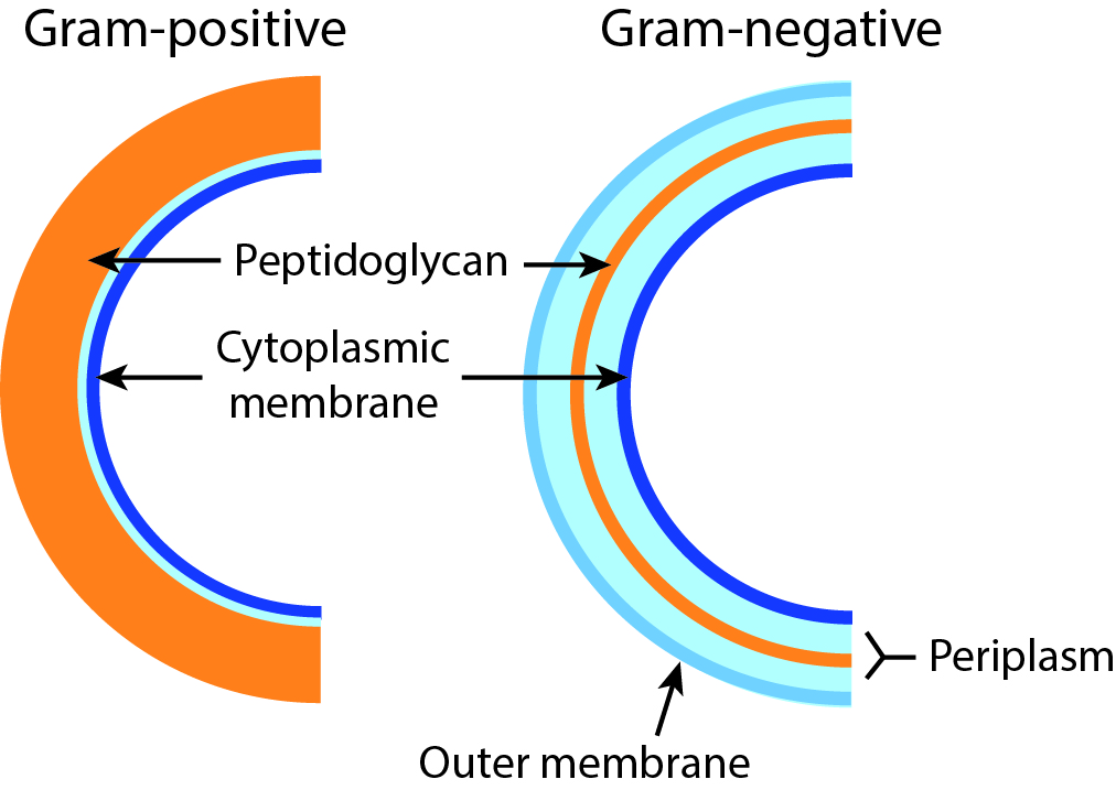
The cell walls of Bacteria have a rigid layer composed of peptidoglycan that is the main source of strength of the wall (Madigan et al., 2003). Peptidoglycan is a polymer consisting of sugars and amino acids. For some Bacteria, peptidoglycan makes up as much as 90% of the cell wall. However, for other Bacteria, it makes up only about 10% of the cell wall (Fig. \(3.3\)).
These two groups of Bacteria can be distinguished based on a staining procedure. By this method, cells are exposed to a stain, crystal violet, that binds to peptidoglycan. Cells with an abundance of peptidoglycan retain more of the stain than those that have little. The cells that retain more stain are referred to as gram-positive cells (i.e., the staining result is positive). Those that bind little stain are referred to as gram-negative cells. This difference in stain retention was traditionally used to help classify Bacteria and was originally developed in the nineteenth century by a Danish scientist, Hans Christian Gram.
Aside from the difference in peptidoglycan thickness, gram-negative cell walls differ from gram-positive cell walls in a few other ways as well. Gram-negative cell walls have an outer membrane. Unlike the cytoplasmic membrane, the outer membrane is not composed entirely of phospholipid but also contains polysaccharide and protein components and can be referred to as the lipopolysaccharide layer (LPS). Moreover, gram-negative cells also contain a thicker periplasm, a gel-like matrix between the inner cytoplasmic membrane and the outer membrane (Fig. \(3.3\)).

Powerpoint Templates
Icon Bundle
Kpi Dashboard
Professional
Business Plans
Swot Analysis
Gantt Chart
Business Proposal
Marketing Plan
Project Management
Business Case
Business Model
Cyber Security
Business PPT
Digital Marketing
Digital Transformation
Human Resources
Product Management
Artificial Intelligence
Company Profile
Acknowledgement PPT
PPT Presentation
Reports Brochures
One Page Pitch
Interview PPT
All Categories

Gram Positive Examples Bacteria In Powerpoint And Google Slides Cpb
Our Gram Positive Examples Bacteria In Powerpoint And Google Slides Cpb are topically designed to provide an attractive backdrop to any subject. Use them to look like a presentation pro.

- Add a user to your subscription for free
You must be logged in to download this presentation.
Do you want to remove this product from your favourites?
PowerPoint presentation slides
Presenting our Gram Positive Examples Bacteria In Powerpoint And Google Slides Cpb PowerPoint template design. This PowerPoint slide showcases four stages. It is useful to share insightful information on Gram Positive Examples Bacteria This PPT slide can be easily accessed in standard screen and widescreen aspect ratios. It is also available in various formats like PDF, PNG, and JPG. Not only this, the PowerPoint slideshow is completely editable and you can effortlessly modify the font size, font type, and shapes according to your wish. Our PPT layout is compatible with Google Slides as well, so download and edit it as per your knowledge.

People who downloaded this PowerPoint presentation also viewed the following :
- Diagrams , Business , Strategy , Icons , Business Slides , Flat Designs , Strategic Planning Analysis , Process Management
- Gram Positive Examples Bacteria
Gram Positive Examples Bacteria In Powerpoint And Google Slides Cpb with all 10 slides:
Use our Gram Positive Examples Bacteria In Powerpoint And Google Slides Cpb to effectively help you save your valuable time. They are readymade to fit into any presentation structure.

Ratings and Reviews
by Chuck James
February 9, 2024
by Edward Nunez
February 8, 2024

GRAM POSITIVE BACTERIA
Nov 14, 2014
961 likes | 2k Views
GRAM POSITIVE BACTERIA. Ross University School of Veterinary Medicine Bacteriology March 2004 Amy Fayette. What is the major source of infection with Listeria monocytogenes?. Silage pH over 5.5. What is the morphology of rhodococcus equi?. Gram + Some rods, some cocci.
Share Presentation
- adult sheep
- necrotic enteritis
- large gram rods
- diamond skin lesions urticaria

Presentation Transcript
GRAM POSITIVE BACTERIA Ross University School of Veterinary Medicine Bacteriology March 2004 Amy Fayette
What is the major source of infection with Listeria monocytogenes? • Silage • pH over 5.5
What is the morphology of rhodococcus equi? • Gram + • Some rods, some cocci
What is a synonym of C. pseudotuberculosis? • C. ovis
What is the most common sign of C. pseudotuberculosis in horses? • Ulcerative lymphangitis • Abscesses and swellings in lnn. That burst and leave ulcers
What are the host factors behind rhodococcus equi? • Age- 6 weeks • Breed lines
What is the bacteria that causes listeriosis? • Listeria monocytogenes
What is the most common sign of C. pseudotuberculosis in sheep? • Pus filled lymph nodes
What are the forms of disease caused by Listeria monocytogenes? • Neural disease- meningoencephalitis • Septicemia • Abortion • Mastitis
What is the morphology of typical corynebacterium spp.? • Small gram + rods • Club-shaped swellings at one or both ends • “chinese lettering”
What is the morphology of Actinomyces? • Gram + • Slender rods
What causes cystitis and pyelonephritis in cattle and “pizzle rot” in sheep and goats? • Corynebacterium renale group • C. renale • C. pilosum • C. cystidis
What is the disease forms associated with Actinomyces suis? • Habitat is prepuce and urethra of boar • Disease only in sow, causing cystitis and pyelonephritis
How is tetanus diagnosed? • Clinical signs • Discovery of appropriate wound
What is the morphology of Erysipelothrix rhusiopathiae? • Gram + • slender rods
What is the main cause of pizzle rot in sheep? • High protein diet
What are the main forms of erysipelas in pigs? • Septicemia • Diamond skin lesions (urticaria) • Endocarditis • arthritis
What is the disease associated with Actinomyces bovis in horses? • Infection of bursae in horses • “poll evil” or “fistulous withers”
What is the bacteria associated with Swine erysipelas? • Erysipelothrix rhusiopathiae
What is the morphology of Listeria monocytogenes? • Small gram + rods • May be coccoid
How is Listeria typically enriched in the laboratory? • “cold enrichment”
What is the morphology of Arcanobacterium pyogenes? • Small gram + rods • Resemble corynebacterium
What is the disease associated with Actinomyces bovis in cattle? • “lumpy jaw” • Caused by erupting teeth or abrasive food
What are the disease forms associated with clostridium sordellii? • Isolated from sudden deaths, and “big head” in rams
What are the disease forms associated with clostridium botulinum in wild birds? • “limberneck”- ingestion of rotting vegetation and dead invertebrates • Ingestion of dead fish
What are the signs associated with rhodococcus equi? • Bronchopenumonia • Extensive anscessation and lymphangitis • Necrotizing enterocolitis with ulceration
What is the disease forms associated with Nocardia asteroides? • Cattle • mastitis • Dogs, cats • aspiration of organisms • Blood stained purulent fluid in pleural cavity
What are the disease forms associated with clostridium perfringes Type A? • Gas gangrene (man and occasionally animals) • Food Poisoning (man • Colitis in horses • Necrotic enteritis in chickens • Enterotoxemic jaundice in lambs
What is another name for Rhodococcus equi? • Corynebacterium equi
What are the predisposing factors of dermatophilus congolensis? • Prolonged rainfall • Humid conditions • Heavy tick infestation
What are the disease forms associated with clostridium perfringes Type B? • Lamb dysentery • 2d-2wks old • Causes severed diarrhea sometimes with blood • Neonatal hemorrhagic enteritis in calves and foals • Enterotoxemia in adult sheep and goats
What is the morphology of dermatophilus congolensis? • Gram + rods • Branching filaments • Motile zoospores
What is the significance of Corynebacterium bovis? • Commensal of bovine udder • Provokes neutrophil response, thought to protect the udder from more serious infections
What are the disease forms associated with clostridium perfringes Type C? • Hemorrhagic enterotoxemia- esp. in neonatal piglets, lambs, foals and calves • Necrotic enteritis- chicks under 2 weeks • Enterotoxemia in post weaning/ adult sheep in Britain and adult goats
What is the general lesion produced by actinomyces spp.? • Pyogranulomatous lesions • Presence of hard “sulfur granules”
What is the morphology of bacillus spp.? • Large gram+ rods • Aerobic or facultative anaerobic • Sporeforming • Catalase +
What are the disease forms associated with clostridium perfringes Type D? • Enterotoxemia (“pulpy kidney”) • Associated with change in diet usually for better • Increases cerebral pressure which causes nervous signs • Also causes hydropericardium, and edema of lungs • Hyperglycemia, and glucosuria • Enterotoxemia in goats and possibly cattle
What are the cultural characteristics of streptococcus? • Facultative anaerobes • Catalase neg (staph catalase pos)
What are the cultural characteristics of Actinomyces? • Anaerobic or microaerophilic- some CO2 prefered
What are the disease forms associated with Staph hyicus? • “Greasy pig disease” • “exudative epidermitis” • Primarily in Suckling piglets • Excessive sebaceous secretion • Subacute disease- thickening and wrinkling of skin
What is the disease forms associated with bovine farcy? • Nocardia sp. • Infection of the lymphatics of the lower limbs or head of cattle
What are the disease forms associated with clostridium perfringes Type E? • Enteritis/enterotoxemia in calves and lambs
What is the morphology of Nocardia spp. • Slender gram + rods and filaments • Modified acid-fast
What are some synonyms of dermatophilus congolensis? • Cattle- dermatophilosis, streptothricosis, senkobo skin disease • Sheep- lumpy wool, mycotic dermatitis, strawberry footrot • Horse- rain scald
What are the disease forms associated with staph intermedius? • Pyoderma in dogs and cats • Lesions in skin folds • Otitis externa
What is the disease forms associated with B. anthracis? • Septicemia with sudden death • Dark, tarry unclotted blood oozing from orifices • Spleen is greatly enlarged
What is the disease forms associated with Actinomyces hordeovulneris? • Injury by grass awns • Local abscesses and serositis
What are the disease forms associated with clostridium piliforme? • Was: bacillus piliformis • Aka: tyzzers disease • Enteritis of lower small intestine and proximal large intestine and focal necrosis of liver • Disease seen in young animals
What are the three most common streptococci associated with bovine mastitis? • S. agalactiae • S. dysgalactiae • S. uberis
What is the disease forms associated with dermatophilus congolensis? • Scab and crust formation with shallow clean wound underneath
- More by User

Agents for Infections Due to Gram Positive Bacteria
Sorting Out Antibiotics: A systematic approach to antibiotic selection Kenneth Alexander, M.D., Ph.D. Associate Professor of Pediatrics and Microbiology Duke University Medical Center Durham, North Carolina [email protected] Streptococci (Group A and Group B) Pseudomonas aeruginosa
648 views • 6 slides

antibacterial agent vs. gram-positive bacteria
New antibiotics, C21. Oxazolidinones: structure / activity. . . N. O. O. . . . N. H. C. . . . . O. CH3. . . . . F. . . N. O. . H. 5. Morpholino enhances pharmacokinetics
1.05k views • 34 slides

Gram Negative Gram Positive
Gram Negative Gram Positive. Two Drug Bugs. Aerobes. S erratia P seudomonas A cinetobactor C itrobactor E nterobactor. Salmonella Shigella. H. Flu Klebsiella E. Coli. MRSA MRSE. VRE. Staph. Strep.
1.09k views • 32 slides

High G + C Gram Positive Bacteria
High G + C Gram Positive Bacteria. Derek Frantz, Kara Sporik, Jessica Teague, Katie Stevenson, Kent Worthington. Phylum: Actinobacteria Typically rod shaped Most are pleomorphic Gram positive High G+C ratio Some are pathogenic, but some are very useful. Nocardia.
1.26k views • 33 slides

Gram Positive Rods
Gram Positive Rods. Listeria. Small Gram positive rods or coccobacilli (<2 μ m ) Tolerate wide temperature and pH range Small haemolytic colonies on blood agar ( β haemolysis ) Facultative anaerobes Catalase positive, oxidase negative Tumbling motility at 25 degrees Aesculin hydrolysed
1.3k views • 56 slides
Overview of Gram-Positive and Other Bacteria
© 2012 Pearson Education, Inc. Overview of Gram-Positive and Other Bacteria. Bacteria has many phyla other than Proteobacteria , including Gram-positive bacteria Large group of mostly chemoorganotrophs Cyanobacteria
850 views • 42 slides
GRAM POSITIVE & GRAM NEGATIVE BACTERIA
GRAM POSITIVE & GRAM NEGATIVE BACTERIA. Dr . Fawzia Al-O tabi. Bacterial cells. GRAM STAIN. Developed in 1884 by the Danish physician Hans Christian Gram
1.61k views • 23 slides
Gram positive bacilli
Gram positive bacilli. Bacillus spp. Corynebacterium diphteria Erysiphelothrix Listeria spp. Gram positive aerobic bacilli (2 hours): Learning Objectives. 1 . Defines ‘’Gram positive aerobic bacilli’’ 1.1 Lists Gram positive aerobic bacilli in normal flora.
1.25k views • 48 slides

Gram Positive
Corynebacterium Spp. Clostridium spp. Listeria spp. Bacillus spp. Gram Positive. Bacilli. Cocci. Catalase negative Streptococcaceae ( strepto - cocay -see-eye). S. viridans , S. Mutans , S. sanguis Optochin resistant Bile-insoluble No capsule. Catalase positive Staphylococcus.
253 views • 5 slides

Gram-positive bacteria
Gram-positive bacteria. Gram-positive bacteria Separated on basis of G + C content of chromosomal DNA Low G + C Gram-positives = Firmicutes High G + C Gram-positives = Actinobacteria. Firmicutes (Low G + C ) Divided into 3 classes: Clostridia Mollicutes Bacilli
1.35k views • 38 slides

Structure of Gram-Positive Bacteria
Antimicrobial Therapy David H. Spach, MD Professor of Medicine Division of Infectious Diseases University of Washington, Seattle. Structure of Gram-Positive Bacteria. Penicillin Binding Proteins. DNA. Cell Wall. Cell Membrane. Structure of Gram -Negative Bacteria. Outer Membrane.
1.05k views • 60 slides

Gram negative bacteria
Gram negative bacteria. PHYLUM Proteobacteria Chlamydiae Spirochaetes Bacteroidetes Fusobacteria. Phylum Proteobacteria. Class Alphaproteobacteria Betaproteobacteria Gamaproteobacteria Deltaproteobacteria Epsilonproteobacteria. C:Alphaproteobacteria. O. Rickettsiales
644 views • 34 slides

Gram-Positive Bacteria
Gram-Positive Bacteria. By: Caleb Martin, Darrell Potter &Kerri Anderson. Gram Stain. Gram-Positive Bacteria retained the color of the crystal violet stain in the Gram stain. Where are they found?. Gram-Positive Bacteria are found in the throat. Also found in the intestines.
574 views • 6 slides

Bergey’s Volume 2 – Gram Positive Bacteria of Importance
Bergey’s Volume 2 – Gram Positive Bacteria of Importance. 1) Gram positive spheres: Micrococcus Staphylococcus Streptococcus. Micrococcus. Demos chromogenicity Isolated from human and animal skin, dairy products and beer Breaks down compounds in sweat to produce odors
287 views • 11 slides
Medically Important Bacteria Gram Positive Cocci
Medically Important Bacteria Gram Positive Cocci. Phase I/ Module VII Dr Ekta Chourasia. Learning Objectives. At the end of the session, you should be able to: Enlist medically important GPCs Compare & contrast the features of staphylococci & streptococci
821 views • 38 slides

Gram positive
Lecture 5: Protein translocation. Gram negative. Gram positive. Why do bacteria need to secrete or translocate proteins?. bacteria move: they have flagella. bacteria secrete proteins into their host during pathogenesis: type III or IV secretion systems.
470 views • 23 slides

GRAM POSITIVE & GRAM NEGATIVE BACTERIA. DR.THAMINA SAYYED M.B.B.S. MD. MICROBIOLOGY REGISTRAR PROF KAMBALMBBS FRCpath. Bacterial cells. GRAM STAIN. Developed in 1884 by the Danish physician Hans Christian Gram
1.67k views • 21 slides
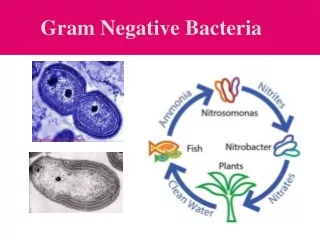
Gram Negative Bacteria
Gram Negative Bacteria. Differences Between Gram Positive and Gram Negative Cell Wall. The Gram positive cell wall is thick and composed of many layers of peptidoglycan that are bound together by teichoic acids and lipoteichoic acids.
1.24k views • 106 slides
Gram-Positive Cocci
Gram-Positive Cocci. Prof. Dr. Asem Shehabi Faculty of Medicine University of Jordan. Gram-psitive cocci. Micrococcaceae family.. Facultative Anaerobic Gram-positive cocci .. includes the following Genera/Groups:
310 views • 12 slides

GRAM POSITIVE & GRAM NEGATIVE BACTERIA. (Foundation Block, Microbiology). Lecturer name: Dr. Fawzia Alotaibi Department of Pathology, Microbiology Unit. Objectives:. By the end of this lecture, the student should able to: Know the general basic characteristics of bacteria
334 views • 24 slides

- My presentations
Auth with social network:
Download presentation
We think you have liked this presentation. If you wish to download it, please recommend it to your friends in any social system. Share buttons are a little bit lower. Thank you!
Presentation is loading. Please wait.
Gram-Positive Bacteria
Published by Alexina McCoy Modified over 6 years ago
Similar presentations
Presentation on theme: "Gram-Positive Bacteria"— Presentation transcript:

Highlights from Section 2 – Bacteria in Your Life

What kind of impact does bacteria have on the world around you?

Wonderful World of Bacteria. The Cell The cell is a unit of organization Cells are classified as prokaryotes or eukaryotes Living things are classified.

Kingdom Eubacteria (True Bacteria) Bacteria are located everywhere – air, water, land, and living organisms including people. General Characteristics:

Viruses, Bacteria, Protists, and Fungi Ch. 7. Section 2: Bacteria The Bacterial Cell A Dutch merchant named Anton van Leeuwenhoek found bacteria in the.

Chapter 10 Section 2 Bacteria’s Role in the World Bellringer

Bacteria Kingdoms Eubacteria & Archaebacteria. Bacteria Single-celled prokaryotes Two kingdoms of bacteria: Archaebacteria Eubacteria.

Bacteria Smallest and simplest organisms on the planet Smallest and simplest organisms on the planet Also the most abundant Also the most abundant 1 gram.

A substance used to kill or slow the growth of bacteria Antibiotics:

10.2. Warm upWarm up What is a prokaryote? Give some examples where you might find bacteria. Which one is more important: prokaryotes or eukaryotes?

BACTERIA Basic structure: –Prokaryotes (no nucleus or membrane-bound organelles) –Single-celled –Single circular piece of DNA –May have pili (attachment)

Chapter 7 Bacteria and Viruses.

Chapter 7 Bacteria.

Domain Bacteria 4 Single Celled 4 Prokaryote - no nucleus or other organelles to 10 micrometers long (100 times smaller than the cells we’ve looked.

Microorganisms Graphic Organizer

Bacteria and Disease A pathogen is a disease-causing agent. Bacteria can damage the tissues of the affected organism directly or release toxins that harm.
About project
© 2024 SlidePlayer.com Inc. All rights reserved.
- Preferences

GRAM POSITIVE BACTERIA - PowerPoint PPT Presentation

GRAM POSITIVE BACTERIA
Scarlet fever. pyoderma - erysipelas. necrotising fascitis. puerperal sepsis ... acute rheumatic fever. acute glomerulonephritis. streptococci. pathogenicity factors ... – powerpoint ppt presentation.
- Classification
- morphology and staining behavior
- structure of murein of cell wall
- metabolic properties
- DNA homologies
- antigenic similarities
- Aerobic / Facultatively anaerobic
- Micrococcaciae
- Staphylococcus spp.
- Streptococcus spp., Enterococcus spp., Lactococcus spp.
- Leuconostoc spp, Pediococcus, spp, Gemella spp., Aerococcus spp..
- e.g. Peptococcaceae
- Peptococcus spp.
- Peptostreptococcus spp.
- Bacillaceae e.g. Bacillus anthracis
- Corynebacteriaceae e.g. Corynebacteria diphtheriae
- Lactobacillaceae e.g Lactobacillus acidophilus
- Other e.g. Listeria monocytogenes, Erysipelothrix spp.
- Anaerobic / Microaerophilic
- Bacillaceae e.g. Clostridium perfringens
- Actinomycetaceae e.g. Actinomyces
- Gram positive cocci in clusters
- Aerobic/facultatively anaerobic
- Catalase positive
- Coagulase enzyme - clots plasma
- Coagulase negative staphylococci e.g.
- S. epidermidis
- S. haemolyticus
- Pathogenicity factors
- Capsule polysaccharide
- Protein A, Peptidoglycan, Clumping factor
- Haemolysins, Leukocidin, Staphylokinase, Protease, Nuclease
- B-lactamase
- Epidermolytic toxins
- Enterotoxins
- Exotoxins e.g. TSST1
- Non-toxin mediated
- Furuncles/carbuncles
- Wound infections
- Bacteraemia
- Osteomyelitis/Endocarditis/Pneumonia
- Toxin-mediated
- Acute staphylococcal enterocolitis
- Staphylococcal scalded skin syndrome
- Toxic shock syndrome
- Normal skin / mucous membrane flora
- Non-pathogenic in normal host
- Pathogenic in immunocompromised host
- adherence to plastic material
- central lines
- artificial heart valves
- endoprosthesis etc.
- Gram positive cocci in chains
- Catalase enzyme - negative
- Classified on basis of haemolysis
- ?-haemolysis
- Streptococcus pneumoniae
- viridans streptococci
- Lancefield group A, B, C, D, E, F, G
- cell wall C-polysaccharide
- non-haemolysis
- Group A ? -haemolytic streptococcus
- Small colonies after 24 hours incubation
- Identified using slide agglutination / latex antigen detection kits
- By invasion
- Pharyngitis, tonsillitis, peritonsillar abscess
- Sinusitis, otitis media
- Scarlet fever
- Pyoderma - erysipelas
- Necrotising fascitis
- Puerperal sepsis
- Long term immunological
- Acute rheumatic fever
- Acute glomerulonephritis
- C carbohydrate
- Lipoteichoic acid
- peptidoglycan
- M-proteins (group A), T-proteins, R-proteins
- Exotoxins and enzymes
- Group B ? -haemolytic streptococcus
- Early neonatal infection
- Late neonatal infection
- Adult disease
- wound infection, sepsis, meningitis, urinary tract infection
- Group D ? -haemolytic streptococcus
- Urinary tract infection
- Endocarditis
- Wound infections (mixed)
- Peritonitis
- Gram positive diplococci
- ?-haemolytic
- Autolysis - depressed central colonies, Optochin sensitive, Bile soluble
- Lobar pneumonia
- Sinusitis, otitis media, conjunctivitis
- S. mutans, S. salivarius, S. angiosus, S. sanguis etc.
- Numerous genospecies
- tooth decay
- oropharyngeal infection
- endocarditis
- septicaemia in immunocompromised
- Peptococcaceae
- Peptostreptococcus spp
- wound infection
- abscess formation
- Corynebacteria
- Gram positive bacillus
- Obligate pathogens
- C. diphtheriae
- Clinical manifestations produced by exotoxin
- inhibits protein synthesis
- Local vs systemic effects (cardiac/neurological)
- Clinical diagnosis
- Rapid treatment
- C. ulcerans, C. pseudotuberculosis etc
- Local effects
- Diphtheroids
- Opportunistic
- Non-spore forming Gram positive bacillus
- Haemolytic, cryophilic
- Food borne outbreaks
- Pregnant women
- Neonatal infection
- Elderly / immunocompromised
- Gram positive bacillus with endospores
- Bacillus spp.
- B. anthracis - toxin composed of 3 proteins
- B. cereus - enterotoxins
- C. tetani - tetanus
- swarming, obligate anaerobe
- produce toxins (metalloproteinase)
- degrades synaptobrevin - prevents transmitter release in inhibitory cells of CNS
- irreversible event
- Clinical diagnosis - laboratory diagnosis too late
- C. botulinum - botulism
- produces toxin (metalloproteinase)
- targets the peripheral neuromuscular junction and autonomic synapses
- classically causes acute cranial neuropathy in association with descending weakness
- C. perfringens
- non-motile, boxcar appearance on gram staining
- produces a number of exo-toxins
- gas gangrene
- food poisoning
PowerShow.com is a leading presentation sharing website. It has millions of presentations already uploaded and available with 1,000s more being uploaded by its users every day. Whatever your area of interest, here you’ll be able to find and view presentations you’ll love and possibly download. And, best of all, it is completely free and easy to use.
You might even have a presentation you’d like to share with others. If so, just upload it to PowerShow.com. We’ll convert it to an HTML5 slideshow that includes all the media types you’ve already added: audio, video, music, pictures, animations and transition effects. Then you can share it with your target audience as well as PowerShow.com’s millions of monthly visitors. And, again, it’s all free.
About the Developers
PowerShow.com is brought to you by CrystalGraphics , the award-winning developer and market-leading publisher of rich-media enhancement products for presentations. Our product offerings include millions of PowerPoint templates, diagrams, animated 3D characters and more.

COMMENTS
Oct 22, 2019 • Download as PPTX, PDF •. 38 likes • 25,829 views. Microbiology. Gram positive bacteria. Health & Medicine. Download now. Gram positive bacteria - Download as a PDF or view online for free.
Gram positive bacteria. Jun 12, 2018 • Download as PPTX, PDF •. 15 likes • 914 views. Ramza Rasheed. Aero tolerant organisms cannot use oxygen for growth but are tolerate its presence. use fermentation to produce ATP. Characteristics: They don't possess cytochromes. The cells are arranged in pairs, chain or tetrads.
Each envelope type possesses peptidoglycan located outside the cytoplasmic membrane (CM) as a major component conferring shape and integrity to the cell wall. In Gram-negative bacteria, a selectively permeable asymmetric outer membrane (OM) usually contains lipopolysaccharides. In Gram-positive bacteria, the much thicker peptidoglycan layer is ...
Download ppt "Lecture: Gram positive and negative bacteria". Objectives: By the end of this lecture, the student should be able to: Know the general basic characteristics of bacteria 5 Differentiate between gram positive and gram negative bacteria's characteristics Know the classes and groups of gram positive bacteria, cocci and bacilli (rods ...
Based on its genome, it is placed into the high G+C gram-positive group. G. vaginalis can cause bacterial vaginosis in women; symptoms are typically mild or even undetectable, but can lead to complications during pregnancy. Table 4.4.1 4.4. 1 summarizes the characteristics of some important genera of Actinobacteria.
4 Bacterial cells 4. 5 GRAM STAIN Developed in 1884 by the Danish physician Hans Christian Gram An important tool in bacterial taxonomy, distinguishing so- called Gram-positive bacteria, which remain coloured after the staining procedure, from Gram- negative bacteria, which do not retain dye and need to be counter-stained.
3 Gram Positive Bacteria Stain blue or violet* Produce exotoxins Have thick layer of peptidoglycan in cell membrane More easily treated with antibiotic therapy than gram negative bacteria Staphyloccus aureus is shown at the right *Using gram staining technique. Note: due to different photography techniques, you may note colors other than expected.
PowerPoint Presentation. Overview of Gram-Positive and Other Bacteria. Bacteria has many phyla other than Proteobacteria, including. Gram-positive bacteria. Large group of mostly chemoorganotrophs. Cyanobacteria. Oxygenic phototrophs that have evolutionary roots near those of gram-positive bacteria. Phylogenetically early-branching phyla.
Sep 10, 2014. 540 likes | 1.35k Views. Gram-positive bacteria. Gram-positive bacteria Separated on basis of G + C content of chromosomal DNA Low G + C Gram-positives = Firmicutes High G + C Gram-positives = Actinobacteria. Firmicutes (Low G + C ) Divided into 3 classes: Clostridia Mollicutes Bacilli. Download Presentation. genus. nonspore forming.
Bacterial cells. GRAM STAIN • Developed in 1884 by the Danish physician Hans Christian Gram • An important tool in bacterial taxonomy, distinguishing so-called Gram-positive bacteria, which remain coloured after the staining procedure, from Gram-negative bacteria, which do not retain dye and need to be counter-stained.
Presentation Transcript. Gram Positive Bacteria • Grouped based on C + G ratio (nitrogen bases cytosine and guanine) • Divided into 2 Phyla • Firmicutes (low C + G ) • Actinobacteria (High C + G) Firmicutes • Low G + C ratio • Includes endospore forming and wall-less bacteria. Phylum: Firmicute • 3 Classes: • Clostridia ...
Health professionals need to understand the important difference between gram-positive and gram-negative bacteria. Gram-positive bacteria are bacteria classified by the color they turn in the staining method. Hans Christian Gram developed the staining method in 1884. The staining method uses crystal violet dye, which is retained by the thick peptidoglycan cell wall found in gram-positive ...
24 Reference book and the relevant page numbers.. Download ppt "GRAM POSITIVE & GRAM NEGATIVE BACTERIA". Objectives: By the end of this lecture, the student should able to: Know the general basic characteristics of bacteria Differentiate between gram positive and gram negative bacteria characteristics. Know the classes and groups of gram ...
This difference in stain retention was traditionally used to help classify Bacteria and was originally developed in the nineteenth century by a Danish scientist, Hans Christian Gram. Aside from the difference in peptidoglycan thickness, gram-negative cell walls differ from gram-positive cell walls in a few other ways as well.
Presentation on theme: "Gram Positive Bacteria"— Presentation transcript: 1 Gram Positive Bacteria 1- Spore-Forming Gram-Positive Bacilli: A- Strict aerobic - Genus Bacillus. B- Strict anaerobic - Genus Clostridium. ... PowerPoint ® Lecture Slides for M ICROBIOLOGY Pathogenic Gram-Positive Bacilli (Bacillus)
PowerPoint presentation slides: Presenting our Gram Positive Examples Bacteria In Powerpoint And Google Slides Cpb PowerPoint template design. This PowerPoint slide showcases four stages. It is useful to share insightful information on Gram Positive Examples Bacteria This PPT slide can be easily accessed in standard screen and widescreen aspect ...
GRAM POSITIVE BACTERIA. Ross University School of Veterinary Medicine Bacteriology March 2004 Amy Fayette. What is the major source of infection with Listeria monocytogenes?. Silage pH over 5.5. What is the morphology of rhodococcus equi?. Gram + Some rods, some cocci.
Presentation on theme: "Gram-Positive Bacteria"— Presentation transcript: 1 Gram-Positive Bacteria Actinomycetes: Gram-positive bacteria that form branching filaments; used to make antibiotics. ... Download ppt "Gram-Positive Bacteria" Similar presentations . Highlights from Section 2 - Bacteria in Your Life.
Transcript and Presenter's Notes. Title: GRAM POSITIVE BACTERIA. 1. GRAM POSITIVE BACTERIA. Classification. Systematic. morphology and staining behavior. structure of murein of cell wall. metabolic properties.