- Biology Article

Cells are the basic, fundamental unit of life. So, if we were to break apart an organism to the cellular level, the smallest independent component that we would find would be the cell.
Explore the cell notes to know what is a cell, cell definition, cell structure, types and functions of cells. These notes have an in-depth description of all the concepts related to cells.
Table of Contents

Cell Definition
What is a cell, characteristics of cells, types of cells, cell structure, cell theory.
- Functions of a Cell
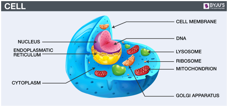
Cells are the fundamental unit of life. They range in size from 0.0001 mm to nearly 150 mm across.
“A cell is defined as the smallest, basic unit of life that is responsible for all of life’s processes.”
Cells are the structural, functional, and biological units of all living beings. A cell can replicate itself independently. Hence, they are known as the building blocks of life .
Each cell contains a fluid called the cytoplasm, which is enclosed by a membrane. Also present in the cytoplasm are several biomolecules like proteins, nucleic acids and lipids. Moreover, cellular structures called cell organelles are suspended in the cytoplasm.
A cell is the structural and fundamental unit of life. The study of cells from its basic structure to the functions of every cell organelle is called Cell Biology. Robert Hooke was the first Biologist who discovered cells.
All organisms are made up of cells. They may be made up of a single cell (unicellular), or many cells (multicellular). Mycoplasmas are the smallest known cells. Cells are the building blocks of all living beings. They provide structure to the body and convert the nutrients taken from the food into energy.
Cells are complex and their components perform various functions in an organism. They are of different shapes and sizes, pretty much like bricks of the buildings. Our body is made up of cells of different shapes and sizes.
Cells are the lowest level of organisation in every life form. From organism to organism, the count of cells may vary. Humans have more number of cells compared to that of bacteria .
Cells comprise several cell organelles that perform specialised functions to carry out life processes. Every organelle has a specific structure. The hereditary material of the organisms is also present in the cells.
Discovery of Cells
Discovery of cells is one of the remarkable advancements in the field of science. It helps us know that all the organisms are made up of cells, and these cells help in carrying out various life processes. The structure and functions of cells helped us to understand life in a better way.
Who discovered cells?
Robert Hooke discovered the cell in 1665. Robert Hooke observed a piece of bottle cork under a compound microscope and noticed minuscule structures that reminded him of small rooms. Consequently, he named these “rooms” as cells. However, his compound microscope had limited magnification, and hence, he could not see any details in the structure. Owing to this limitation, Hooke concluded that these were non-living entities.
Later Anton Van Leeuwenhoek observed cells under another compound microscope with higher magnification. This time, he had noted that the cells exhibited some form of movement (motility). As a result, Leeuwenhoek concluded that these microscopic entities were “alive.” Eventually, after a host of other observations, these entities were named as animalcules.
In 1883, Robert Brown, a Scottish botanist, provided the very first insights into the cell structure. He was able to describe the nucleus present in the cells of orchids.
Following are the various essential characteristics of cells:
- Cells provide structure and support to the body of an organism.
- The cell interior is organised into different individual organelles surrounded by a separate membrane.
- The nucleus (major organelle) holds genetic information necessary for reproduction and cell growth.
- Every cell has one nucleus and membrane-bound organelles in the cytoplasm.
- Mitochondria, a double membrane-bound organelle is mainly responsible for the energy transactions vital for the survival of the cell.
- Lysosomes digest unwanted materials in the cell.
- Endoplasmic reticulum plays a significant role in the internal organisation of the cell by synthesising selective molecules and processing, directing and sorting them to their appropriate locations.
Also Read : Nucleus
Cells are similar to factories with different labourers and departments that work towards a common objective. Various types of cells perform different functions. Based on cellular structure, there are two types of cells:
- Prokaryotes
Explore: Difference Between Prokaryotic and Eukaryotic Cells
Prokaryotic Cells
Main article: Prokaryotic Cells
- Prokaryotic cells have no nucleus. Instead, some prokaryotes such as bacteria have a region within the cell where the genetic material is freely suspended. This region is called the nucleoid.
- They all are single-celled microorganisms. Examples include archaea, bacteria, and cyanobacteria.
- The cell size ranges from 0.1 to 0.5 µm in diameter.
- The hereditary material can either be DNA or RNA.
- Prokaryotes generally reproduce by binary fission, a form of asexual reproduction. They are also known to use conjugation – which is often seen as the prokaryotic equivalent to sexual reproduction (however, it is NOT sexual reproduction).
Eukaryotic Cells
Main article : Eukaryotic Cells
- Eukaryotic cells are characterised by a true nucleus.
- The size of the cells ranges between 10–100 µm in diameter.
- This broad category involves plants, fungi, protozoans, and animals.
- The plasma membrane is responsible for monitoring the transport of nutrients and electrolytes in and out of the cells. It is also responsible for cell to cell communication.
- They reproduce sexually as well as asexually.
- There are some contrasting features between plant and animal cells. For eg., the plant cell contains chloroplast, central vacuoles, and other plastids, whereas the animal cells do not.
The cell structure comprises individual components with specific functions essential to carry out life’s processes. These components include- cell wall, cell membrane, cytoplasm, nucleus, and cell organelles. Read on to explore more insights on cell structure and function.
Cell Membrane
- The cell membrane supports and protects the cell. It controls the movement of substances in and out of the cells. It separates the cell from the external environment. The cell membrane is present in all the cells.
- The cell membrane is the outer covering of a cell within which all other organelles, such as the cytoplasm and nucleus, are enclosed. It is also referred to as the plasma membrane.
- By structure, it is a porous membrane (with pores) which permits the movement of selective substances in and out of the cell. Besides this, the cell membrane also protects the cellular component from damage and leakage.
- It forms the wall-like structure between two cells as well as between the cell and its surroundings.
- Plants are immobile, so their cell structures are well-adapted to protect them from external factors. The cell wall helps to reinforce this function.
- The cell wall is the most prominent part of the plant’s cell structure. It is made up of cellulose, hemicellulose and pectin.
- The cell wall is present exclusively in plant cells. It protects the plasma membrane and other cellular components. The cell wall is also the outermost layer of plant cells.
- It is a rigid and stiff structure surrounding the cell membrane.
- It provides shape and support to the cells and protects them from mechanical shocks and injuries.
- The cytoplasm is a thick, clear, jelly-like substance present inside the cell membrane.
- Most of the chemical reactions within a cell take place in this cytoplasm.
- The cell organelles such as endoplasmic reticulum, vacuoles, mitochondria, ribosomes, are suspended in this cytoplasm.
- The nucleus contains the hereditary material of the cell, the DNA.
- It sends signals to the cells to grow, mature, divide and die.
- The nucleus is surrounded by the nuclear envelope that separates the DNA from the rest of the cell.
- The nucleus protects the DNA and is an integral component of a plant’s cell structure.
Cell Organelles
Cells are composed of various cell organelles that perform certain specific functions to carry out life’s processes. The different cell organelles, along with its principal functions, are as follows:
Cell Theory was proposed by the German scientists, Theodor Schwann, Matthias Schleiden, and Rudolf Virchow. The cell theory states that:
- All living species on Earth are composed of cells.
- A cell is the basic unit of life.
- All cells arise from pre-existing cells.
A modern version of the cell theory was eventually formulated, and it contains the following postulates:
- Energy flows within the cells.
- Genetic information is passed on from one cell to the other.
- The chemical composition of all the cells is the same.
Functions of Cell
A cell performs major functions essential for the growth and development of an organism. Important functions of cell are as follows:
Provides Support and Structure
All the organisms are made up of cells. They form the structural basis of all the organisms. The cell wall and the cell membrane are the main components that function to provide support and structure to the organism. For eg., the skin is made up of a large number of cells. Xylem present in the vascular plants is made of cells that provide structural support to the plants.
Facilitate Growth Mitosis
In the process of mitosis, the parent cell divides into the daughter cells. Thus, the cells multiply and facilitate the growth in an organism.
Allows Transport of Substances
Various nutrients are imported by the cells to carry out various chemical processes going on inside the cells. The waste produced by the chemical processes is eliminated from the cells by active and passive transport. Small molecules such as oxygen, carbon dioxide, and ethanol diffuse across the cell membrane along the concentration gradient. This is known as passive transport. The larger molecules diffuse across the cell membrane through active transport where the cells require a lot of energy to transport the substances.
Energy Production
Cells require energy to carry out various chemical processes. This energy is produced by the cells through a process called photosynthesis in plants and respiration in animals.
Aids in Reproduction
A cell aids in reproduction through the processes called mitosis and meiosis. Mitosis is termed as the asexual reproduction where the parent cell divides to form daughter cells. Meiosis causes the daughter cells to be genetically different from the parent cells.
Thus, we can understand why cells are known as the structural and functional unit of life. This is because they are responsible for providing structure to the organisms and perform several functions necessary for carrying out life’s processes.
Also Read: Difference Between Plant Cell and Animal Cell
To know more about what is a cell, its definition, cell structure, types of cells, the discovery of cells, functions of cells or any other related topics, explore BYJU’S Biology . Alternatively, download BYJU’S app for a personalised learning experience.
Frequently Asked Questions
1. what is a cell, 2. state the characteristics of cells..
- Cells provide the necessary structural support to an organism.
- The genetic information necessary for reproduction is present within the nucleus.
- Structurally, the cell has cell organelles which are suspended in the cytoplasm.
- Mitochondria is the organelle responsible for fulfilling the cell’s energy requirements.
- Lysosomes digest metabolic wastes and foreign particles in the cell.
- Endoplasmic reticulum synthesises selective molecules and processes them, eventually directing them to their appropriate locations.
3. Highlight the cell structure and its components.
The cell structure comprises several individual components which perform specific functions essential to carry out life processes. The components of the cell are as follows:
- Cell membrane
- Nuclear membrane
- Endoplasmic reticulum
- Golgi Bodies
- Mitochondria
- Chloroplast
4. State the types of cells.
Cells are primarily classified into two types, namely
- Prokaryotic cells
- Eukaryotic cells
5. Elaborate Cell Theory.
Cell Theory was proposed by Matthias Schleiden, Theodor Schwann, and Rudolf Virchow, who were German scientists. The cell theory states that:
6. What is the function of mitochondria in the cells?
7. what are the functions of the cell.
The essential functions of the cell include:
- The cell provides support and structure to the body.
- It facilitates growth by mitosis.
- It helps in reproduction.
- Provides energy and allows the transport of substances.
8. What is the function of Golgi bodies?
9. who discovered the cell and how, 10. name the cell organelle that contains hydrolytic enzymes capable of breaking down organic matter., 11. which cellular structure regulates the entry and exit of molecules to and from the cell.
Register at BYJU’S for cell related Biology notes. Refer to these notes for reference.
Further Reading: Cell Biology MCQs

Put your understanding of this concept to test by answering a few MCQs. Click ‘Start Quiz’ to begin!
Select the correct answer and click on the “Finish” button Check your score and answers at the end of the quiz
Visit BYJU’S for all Biology related queries and study materials
Your result is as below
Request OTP on Voice Call
Leave a Comment Cancel reply
Your Mobile number and Email id will not be published. Required fields are marked *
Post My Comment
Nice note given in this context
Yes, it’s really helpful as it is clear and simple to understand
Nice note was given in this context…
useful details thanks
I love the content of the notes
It is very useful notes
ok thank you
Yes its very useful
these notes are very helpful in my preparation .but i also wants mcqs for cell biology kindly help me
Thank you for helping me 👼👼
THIS HELPS ME SO MUCH IN SCHOOL THANK YOU!!!!!!!!!!!!
Nice note and it helps a lot
Awesome explanation. It was really helpful for my exam preparation
It is very useful
easy to understand
These notes are very beautiful for our helping
This is very beautiful notes for our helping
Nice note in the context for our exam preparation
It is very important notes thanks for helping me
I like this upload more so other too can study as well
Really appreciate this it is well understanding and readable
Awesome points for this topic
Excellent lecture / notes
I really appreciate the notes
Amazing points. It’s also very useful.
wonderful material thanks for sharing
Yep they are, I like them
This is really informative
Perfect notes about cell and their component so thanks very much😊
nice work done on these notes
Nice presentation on cells
I must say very helpful insight . Thank you for your contribution to the community.
These notes are really effective and presentation of cell is nicely designed
Thank You Byjus for explaining topics in detail. By this I can understand the concepts easily 😄
That’s great
Thank you for explaining about cells in very simple and understanding way☺️.
very good presentation
REALLY NICE NOTES IT REALLY HELP ME A LOT IN SCHOOL AND EXTRA ACTIVITIES LIKE SO ON. I REALLY GOT TO CATCH UP ON WHAT I MISSED OUT IN SCIENCE ALTHOUGHT I DON’T LIKE SCIENCE
Thanks, it helped in my holiday homework. 🙂
This notes are very usefull to me Its easy to understand and i get evry point related to any topic thank you byju’s !
Really good that help me and I have taken the app also
very good notes very useful
This is realy usefull i like this
Thank you I got all the points for the project from this Byjus
Very useful during the exam. Should study from this app #learning with Byju’s
This helps me to understand the following chapters
This point really helps me to understand the lesson
Good notes, very useful
splendiferous
Nice very nice
Wow, now I understand
very useful notes
IT HELPS TO KNOW EVERYTHING
Very beautiful and useful notes
Awesome explanation
I am very happy with this website
I am grateful for the notes, It was really informative and readable.
Very good explanation
Great notes
Thanks for your help
Thanks alot Really nice notes
A good, perfect, conceptual notes provided by the world’s no. 1 and most loved learning app that is BYJU’S The Learning App. That notes helps me about the cell after pair the app with this notes and understand perfect. Thank you!
Thanks. It’s very useful
Really excited with the explanation
Wow, really I am understanding very well how you explain the lesson
Comfortable and so understanding points, topics and questions are given in Byjus app
Very helpful
Thanks to you all. it’s very helpful 😍😊😃
The diagram and the keynotes were understandable, and it’s useful
Thanks for this note, it is really helpful to me
Thanks for this note, it’s very helpful and interesting
First of all honestly, cell was a chapter completely beyond my understandings. I studied hard but I was just unable to remember the concepts. I was just confused in words like lysozome n rybozome. SER n RER. But today I mastered this chapter completely with the help of Byju’s. I also tried to learn beyond my consepts. I was like asking the same question to my mentor again n again. But she never got frustrated and also explained me each n every consept. For me Byju’s is best way to learn only for those who really wanna learn. From my side:- Dream it- work hard – make it happen. Best wishes to all of you.
Hi students and teachers I love notes
Very useful
- Share Share
Register with BYJU'S & Download Free PDFs
Register with byju's & watch live videos.

- Workshops & Institutes
- Curriculum Index
- Research Opportunities
Sign In with Google
Create an Account
Stay informed! Sign up for our newsletter. We will never send you spam or sell your information.
Please verify that you are a teacher
Sign up with Google
Why should I sign up?
Even without an account, you’ll still have free access to most of the award-winning content on Teach.Genetics. Creating an account will give you access to additional content and tools.
Reset Password Email: Reset Password Email
- Amazing Cells
Here you’ll find a number of multimedia and paper-based classroom resources, featuring dynamic and realistic depictions to help you explore the inner-most workings of cells.
The Cells In Context section describes a sequence of related resources that work together as a middle school cell biology unit.
The Cells Communicate section describes additional resources designed for high school.
Interactive Tools
This magical microscope lets viewers jump between levels of magnification from organ systems to cells.
This dynamic tour features 3 different cell types, each with animated depictions of organelles working together to carry out basic life functions. Explore the functions to learn the name of each cell structure and its role in the cell.
Use an interactive slider to compare the relative sizes of objects, cells, organelles, molecules, and other biological structures.
Image Files
We offer most of the graphics from the print-based materials below as image files. You can download activity-specific bundles of images as ZIP files and use them with your favorite tools. Plug them into digital whiteboards (like Jamboard), slides, documents—anywhere you can drop a jpeg file!
Cells in Context Image Files
CELLS IN CONTEXT - Suggested Lesson Sequence
This middle school unit’s resources are designed to be used in any order, with or without outside lessons. However, we hope you will consider the suggested sequence below. It pulls together the unit’s resources in a way that illuminates the connection between structure and function and examine how cells work together in systems.
The unit’s materials offer an in-depth exploration of specialized cell types. Student pairs can follow one cell type through several activities, or they can learn about multiple cell types. Three cell types (airway, intestine, and leaf) appear in all the key modeling activities: Mystery Cell Model, Teaming with Cells, Hijacked Cells!, and Hijacked Teams! Mystery Cell Model features two additional cell types: neuron and plant root cell.
- Three-dimensional
- US Middle School level (ages 12-14)
- Flexible: use in sequence as a complete unit or integrate with other curriculum materials
- Consistent visual language: structures are depicted similarly throughout
- Uses models to visualize cell structure and function
NGSS Connections
NGSS Phenomena
Is it Alive?
How do you know if something is living or not? Students look at objects on illustrated cards (24 total) and determine whether they are living or non-living. Several tricky examples are included (such as seeds and wood) to encourage discussion about what exactly it is that makes something alive. Regardless of the criteria your students use, you’ll want to underscore that all living things are made of cells.
Have students sort cards independently, then lead a class discussion.
- There are criteria that some use to determine if something is living or not, but some examples are tricky.
- Living things are made of cells.
- Cells are the smallest unit that can be said to be alive.
Printable Object Cards with Teacher Guide (pdf) — Make one set per pair or small group (card sets can be re-used)
Is It Alive? (online version)
Mystery Cell Model
How do cells carry out the basic functions of life? Students label the structure & function of organelles on a cell model—with a slight twist. There are 5 models to distribute, each depicting a specialized cell with some parts that are unique to its function. While labeling the functions of their cell organelles, students compare their cells to find organelles that are: (1) common to all cells, and (2) unique to each cell type. Finally, they deduce their cell’s identity.
Note: The airway, intestine, and leaf cells appear in other modeling activities: Mystery Cell Model, Teaming with Cells, Hijacked Cells!, and Hijacked Teams! You may choose to work with any number of cell types, as appropriate for you students. You may wish to have each student follow the same cell type throughout, as we have found this to be a little quicker.
Have students work individually or in pairs. See Teacher Guide for details.
- Before — to introduce organelles and their function.
- During — as a whole-group or individual reference to help students identify and label common organelles.
- After — as a check to make sure students labeled their organelles correctly.
- Within cells, special structures carry out particular functions.
- All cells have many of the same basic structures, yet they also have differences that allow the cells to perform specialized roles.
Prep time: 30 minutes
Class time: 45 minutes
- Computers with internet and headphones
- Generic cell models and copies of the Most Cells Have These Parts sheet
Teacher Guide (pdf)
Cell Models:
- 8.5 x 11 (pdf) or 11 x 17 (pdf) —
Structure-Function Organizer (fillable pdf)
Most Cells Have These Parts (pdf)
Inside a Cell (interactive)
Coffee to Carbon
How big are cells? Put the relationship between cells, organelles and molecules in to perspective. Using copy-and-cut cards, students place biological structures in order by their relative size from largest to smallest.
Distribute shuffled sets of object cards to student groups and instruct them to arrange the objects pictured in order from largest to smallest. Ask students to compare the order of their cards with another group and discuss any discrepancies. Use Cell Size and Scale to check answers.
- Understand the relative size of microscopic biological structures
Prep time: 10 minutes
Class time: 20 minutes
Cell Size and Scale (interactive)
(Optional) Real Cell Gallery
Cells your textbook never dreamed of
In biology, there is always an exception to the rule. Real and illustrated examples of some interesting prokaryotic and eukaryotic cells underscore the specialized functions of cells as well the things all cells have in common.
20 - 30 minutes
Computers with internet access
Real Cell Gallery (interactive)
Introduce Levels of Organization
Using this online interactive as a demo, zoom in and orient students to cells and their context in higher levels of organization.
Navigate to the Virtual Microscope. Project and orient students to the levels of organization that they will be using throughout the unit: organ system, organ, tissue, and cell.
- Living things are made of many different numbers and types of cells.
5 - 10 minutes
Teaming with Cells
How do groups of cells work together to carry out functions in organisms? Students examine labeled illustrations and construct explanations for how a particular cell type—and the tissue and organ that it is part of—works with others to help an organism function.
Builds on Mystery Cell Model — Model a cell, then learn how it works with other cell types in a tissue and beyond!
Have students use either printed illustrations or the virtual microscope to explore four levels of organization (cell, tissue, organ, organ system).
If students have trouble finding the words for their organizers, you could either point them to the yellow boxes on the cards or provide a word bank.
- Cells form tissues and tissues form organs specialized for particular body functions.
Class time: 45 - 60 minutes
Student Organizer (fillable pdf)
Printable Illustrations (pdf) — Make one set per pair or small group (card sets can be re-used)
Online alternative for viewing illustrations: Virtual Microscope (interactive)

Hijacked Cells!
This and the next activity explore what happens when an organism’s cells are disrupted by pathogens, using the following pathogen/cell type pairs:
- Influenza virus & Airway cell
- E. coli bacteria & Intestine cell
- Tomato spotted wilt virus (TSWV) & Leaf cell
You may choose to work with any number of the infections. If you plan to work with more than one, we have found that it is a little quicker to have students follow the same cell type all the way through.
Hijacked Cells! builds on Mystery Cell Models . Students model the process a specific pathogen uses to infect a cell. They identify which organelles the pathogen uses and how it disrupts the cell’s function.
Have students work individually, in pairs, or in small groups.
- Distribute matching sets of Mystery Cell Models (linked above), Pathogen Cut-Outs, and Modeling Instructions.
- Have students follow the instructions, taping the cut-outs onto the cell models only where the instructions say to (some parts should not be taped down).
- Have students work through the Organizer, using the instructions there.
- Pathogens interrupt the normal function of particular cells.
Prep time: 15 minutes Class time: 45 minutes
Copies, tape, scissors (if students need to prepare their own cut-outs)
- How are the 3 essential functions of a cell (instructions, energy, container) affected in the case of each of the pathogens?
- In the case of E. coli , one of the essential functions is not affected. Which one, and what does the pathogen do instead to spread the infection?
- In the case of a virus, one of the essential functions is not affected. Which one and why?
- Are viruses and bacteria living? Why or why not?
- What are the top reasons the pathogen you modeled needs a host cell?
Use the optional Structure & Harm cards to help students find the right words. Provide one or both sets of cards for students to tape onto their organizers.
If you’re using the fillable pdf, you could use the information from the cards to make a word bank.
Act out a class-wide infection of either Influenza or TSWV. One student/group starts the infection. Their cell becomes the factory. After going through the virus infection cycle, they give their mature virus particles to another group. Now 2 groups are making virus and infecting others. Continue until all groups are infected.
Printable Pathogen Cut-Outs (pdf) —
Printable Modeling Instructions (pdf) —
Student Organizer (fillable pdf) — Make one per student
Structure & Harm Cards (pdf) — optional
Hijacked Teams!
Students follow their infections to the next level. Building on what they learned in Teaming with Cells, students see how pathogens disrupt tissues, organs, and systems. They piece it together to understand how exactly pathogens make you sick.
- Distribute sets of infection cards and organizers. Have students follow the instructions to complete the organizer. It may be useful for students to refer to the healthy structures and functions shown in the Teaming with Cells illustrations or Virtual Microscope
Note: Not all symptoms can be traced back to the cell level, but at least one can for each pathogen/cell type pair (see answer key); students will need to grapple with the information in the infection cards to identify which one it is.
- When cell function is disrupted, tissue function is disrupted.
- The symptoms of a disease or illness are a direct result of disrupted cell, tissue, and organ function.
Copies Student organizers from earlier activities may be useful
- Often it is the immune system that kills an infected cell. Why would it be bad for a pathogen to kill the cell? Why would an organism need to kill its own cells?
- What strategy or strategies does the pathogen use to spread to other individuals?
Have students go back through the Hijacked Teams! cards and look for instances where multiple symptoms can be traced back to one effect at the cellular or tissue level.
Play Pathogen Attacks , a board game where teams of cell specialists apply their new knowledge of pathogens and symptoms.
Hijacked Teams! Cards (pdf) Includes card sets for 3 infections. (can be re-used)
CELLS COMMUNICATE
Designed for high school students, the materials in this section build on the middle-school-level materials above. The lessons here explore cell communication from a molecular perspective.
Build-A-Membrane - Advanced
Cut, fold, and tape biomolecules to create a three-dimensional cell membrane with embedded proteins.
Have students (individually or in pairs) build membrane segments, then put them all together to form a large membrane.
- Membranes have proteins embedded in them.
- Membrane-embedded proteins allow cellular signals and other molecules to pass through the membrane.
Class time: 30 minutes
Copies, scissors, tape
- A cell is enclosed and defined by an outer membrane.
- Integral proteins, which extend through one of both layers of the phospholipid bilayer
- Proteins attached to lipid molecules that anchor them to the membrane
- Receptor proteins, which transmit signals across a membrane
- Transporter and channel proteins, which form pores through the membrane that can open and close to let specific molecules through
- Membranes also organize the interior of a cell. They wrap around compartments / organelles
- Phospholipids spontaneously arrange themselves into membranes
Student Instructions and Cut-Outs (pdf) Make one per student or pair
The Fight or Flight Response - Advanced
Watch how cell communication carried out by molecular signals bring about physiological change during the fight or flight response.
Project to the class or have students explore individually in pairs.
- Cell communication is a multi-step process.
- Cells communicate via signaling pathways made of interacting components.
- Components of cell signaling pathways sometimes change shape as a result of their interaction (conformational change)
15 - 30 minutes
Projector and speakers or individual student computers
The Fight or Flight Response (video)
(optional) Play-By-Play (pdf) - A scene-by-scene guide to the molecular interactions taking place in the video.
Related Resource: How Cells Communicated During Fight or Flight (web page) - An in-depth look at one axis of cell communication during the fight or flight response.
Pathways with Friends - Advanced
Directed by instructional cards, students kinesthetically model cell communication by acting as components in a cell signaling pathway.
- Create a space in which students can move freely.
- Each person will be given a card.
- Do not let others what know what your card says.
- When prompted, follow the instructions on the card to create a cell signaling pathway.
- Distribute one set of Cell Communication Cards to each group, and ask the students to choose a card from their set.
- Once every student has a card, prompt the groups to begin by following instruction #1 on their card.
- Next, instruct your students to follow instruction #2 on their card.
- When each group is finished, project to the class the Cell Signaling Steps diagram, summarizing the steps the students just demonstrated. Discuss the activity and how it models signaling pathways in the cell.
Copies, projector
- What happened?
- How did you recognize where to go?
- How does this model cell communication?
- What effect did joining the pathway have on you? (Looking for something to indicate conformational change.)
- What problems did you encounter?
- What would have happened if someone did not do their job (follow instructions) or were not there?
Instructional Cards (pdf) - includes Cell Signaling Steps diagram
Dropping Signals - Advanced
Students drag and drop to see how various signals affect a selection of cell types.
Have students work individually or in pairs to explore the interactive. Students can record information on an optional student organizer.
- There are different types of cells, and different types of signals.
- Cells respond differently to signals depending on cell and signal type.
Student computers with internet access (optional) copies
Student Sheet (fillable pdf)
Dropping Signals
Troubleshooting
About These Resources
This work was supported by Science Education Partnership Awards (Nos. R25RR023288 and 1R25GM021903) from the National Institute of General Medical Sciences of the National Institutes of Health.
The contents provided here are solely the responsibility of the authors and do not necessarily represent the official views of the National Institutes of Health.

- school Campus Bookshelves
- menu_book Bookshelves
- perm_media Learning Objects
- login Login
- how_to_reg Request Instructor Account
- hub Instructor Commons
- Download Page (PDF)
- Download Full Book (PDF)
- Periodic Table
- Physics Constants
- Scientific Calculator
- Reference & Cite
- Tools expand_more
- Readability
selected template will load here
This action is not available.

4.1: Cell Structure and Function
- Last updated
- Save as PDF
- Page ID 11113
Learning ObjectiveS
- Define a cell, identify the main common components of human cells, and differentiate between intracellular fluid and extracellular fluid
- Describe the structure and functions of the plasma (cell) membrane
- Describe the nucleus and its function
- Identify the structure and function of cytoplasmic organelles
A cell is the smallest living thing in the human organism, and all living structures in the human body are made of cells. There are hundreds of different types of cells in the human body, which vary in shape (e.g. round, flat, long and thin, short and thick) and size (e.g. small granule cells of the cerebellum in the brain (4 micrometers), up to the huge oocytes (eggs) produced in the female reproductive organs (100 micrometers) and function. However, all cells have three main parts, the plasma membrane , the cytoplasm and the nucleus. The plasma membrane (often called the cell membrane) is a thin flexible barrier that separates the inside of the cell from the environment outside the cell and regulates what can pass in and out of the cell. Internally, the cell is divided into the cytoplasm and the nucleus. The cytoplasm ( cyto- = cell; - plasm = “something molded”) is where most functions of the cell are carried out. It looks a bit-like mixed fruit jelly, where the watery jelly is called the cytosol ; and the different fruits in it are called organelles . The cytosol also contains many molecules and ions involved in cell functions. Different organelles also perform different cell functions and many are also separated from the cytosol by membranes. The largest organelle, the nucleus is separated from the cytoplasm by a nuclear envelope (membrane). It contains the DNA (genes) that code for proteins necessary for the cell to function.
Generally speaking, the inside environment of a cell is called the intracellular fluid (ICF) , (intra- = within; referred to all fluid contained in cytosol, organelles and nucleus) while the environment outside a cell is called the extracellular fluid (ECF) (extra- = outside of; referred to all fluid outside cells). Plasma, the fluid part of blood, is the only ECF compartment that links all cells in the body.

Figure \(\PageIndex{1}\) 3-D representation of a simple human cell. The top half of the cell volume was removed. Number 1 shows the nucleus, numbers 3 to 13 show different organelles immersed in the cytosol, and number 14 on the surface of the cell shows the plasma membrane
Concepts, terms, and facts check
Study Questions Write your answer in a sentence form (do not answer using loose words)
1. What is a cell? 2. What is a plasma membrane? 3. What is a cytoplasm? 4. What is the intracellular fluid (ICF)? 5. What is the extracellular fluid (ECF)?
The plasma (cell) membrane separates the inner environment of a cell from the extracellular fluid. It is composed of a fluid phospholipid bilayer (two layers of phospholipids) as shown in figure \(\PageIndex{2}\) below, and other molecules. Not many substances can cross the phospholipid bilayer, so it serves to separate the inside of the cell from the extracellular fluid. Other molecules found in the membrane include cholesterol, proteins, glycolipids and glycoproteins , some of which are shown in figure \(\PageIndex{3}\) below. Cholesterol, a type of lipid, makes the membrane a little stronger. Different proteins found either crossing the bilayer (integral proteins) or on its surface (peripheral proteins) have many important functions. Channel and transporter (carrier) proteins regulate the movement of specific molecules and ions in and out of cells. Receptor proteins in the membrane initiate changes in cell activity by binding and responding to chemical signals, such as hormones (like a lock and key). Other proteins include those that act as structural anchors to bind neighboring cells and enzymes. Glycoproteins and glycolipids in the membrane act as identification markers or labels on the extracellular surface of the membrane. Thus, the plasma membrane has many functions and works as both a gateway and a selective barrier.

Figure \(\PageIndex{2}\) Phospholipids form the basic structure of a cell membrane. Hydrophobic tails of phospholipids are facing the core of the membrane, avoiding contact with the inner and outer watery environment. Hydrophilic heads are facing the surface of the membrane in contact with intracellular fluid and extracellular fluid.

Figure \(\PageIndex{3}\) Small area of the plasma membrane showing lipids (phospholipids and cholesterol), different proteins, glycolipids and glycoproteins.
1. What is the function of the cell membrane? 2. Which are the three types of biomolecules that form the cell membrane?
Almost all human cells contain a nucleus where DNA, the genetic material that ultimately controls all cell processes, is found. The nucleus is the largest cellular organelle, and the only one visible using a light microscope. Much like the cytoplasm of a cell is enclosed by a plasma membrane, the nucleus is surrounded by a nuclear envelope that separates the contents of the nucleus from the contents of the cytoplasm. Nuclear pores in the envelope are small holes that control which ions and molecules (for example, proteins and RNA) can move in and out the nucleus. In addition to DNA, the nucleus contains many nuclear proteins. Together DNA and these proteins are called chromatin . A region inside the nucleus called the nucleolus is related to the production of RNA molecules needed to transmit and express the information coded in DNA. See all these structures below in figure \(\PageIndex{4}\).

Figure \(\PageIndex{4}\) Nucleus of a human cell. Find DNA, nuclear envelope, nucleolus, and nuclear pores. The figure also shows how the outer layer of the nuclear envelope continues as rough endoplasmic reticulum, which will be discussed in the next learning objective.
1. What is the nuclear envelope? 2. What is a nuclear pore? 3. What is the function of the nucleus?
An organelle is any structure inside a cell that carries out a metabolic function. The cytoplasm contains many different organelles, each with a specialized function. (The nucleus discussed above is the largest cellular organelle but is not considered part of the cytoplasm). Many organelles are cellular compartments separated from the cytosol by one or more membranes very similar in structure to the cell membrane, while others such as centrioles and free ribosomes do not have a membrane. See figure \(\PageIndex{5}\) and table \(\PageIndex{1}\) below to learn the structure and functions of different organelles such as mitochondria (which are specialized to produce cellular energy in the form of ATP) and ribosomes (which synthesize the proteins necessary for the cell to function). Membranes of the rough and smooth endoplasmic reticulum form a network of interconnected tubes inside of cells that are continuous with the nuclear envelope. These organelles are also connected to the Golgi apparatus and the plasma membrane by means of vesicles. Different cells contain different amounts of different organelles depending on their function. For example, muscle cells contain many mitochondria while cells in the pancreas that make digestive enzymes contain many ribosomes and secretory vesicles.

Figure \(\PageIndex{5}\) Typical example of a cell containing the primary organelles and internal structures. Table \(\PageIndex{1}\) below describes the functions of mitochondrion, rough and smooth endoplasmic reticulum, Golgi apparatus, secretory vesicles, peroxisomes, lysosomes, microtubules and microfilaments (fibers of the cytoskeleton)
1. What is an organelle? 2. Which are the organelles listed in the module?
Resources: Course Assignments
Module 4 assignment: cell builder.
Create a model of a eukaryotic cell using any material of your choice. In your model be sure to include all the organelles appropriate to your cell (either plant or animal). Once complete, take multiple photographs of your model from all angles. Include these images in a document that also contains the following in table format:
- A detailed key/legend that matches the model;
- Each organelle or part with its basic function;
- A disease or disorder that is associated with the malfunction of each cellular component
- How this organelle is visualized microscopically
Note for the disease information, you can list a disease in either animals or plants, regardless of what type of cell you are modeling. In other words, its okay to discuss a “human” disease even if you are making a plant model, provided the organelle is present in both types of cells.
Some suggestions for 3D models include Legos, a decorated cake with candy toppings, or standard Styrofoam base with appropriate pieces attached. You can also draw or illustrate a model. Here is an example of what you might make.
Basic Requirements (the assignment will not be accepted or assessed unless the follow criteria have been met):
- Assignment has been proofread and does not contain any major spelling or grammatical errors
- Assignment includes appropriate references
- Assignment includes photographs or images of created model from all angles.
- Assignment includes a key documenting how each organelle is represented in the model.
- Assignment includes a completed table such as the one illustrated in the example document.
- Assignment includes a disease caused by malfunction of each identified component in the model.
- Assignment includes at least 7 organelles in the model and table.
- Performance Assessments: Cell Builder. Authored by : Shelli Carter. Provided by : Columbia Basin College. Located at : https://www.columbiabasin.edu/ . License : CC BY: Attribution

Cell Structure, Function and Organisation
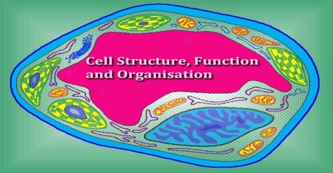
The cell is the structural and functional unit of all living organisms, and is sometimes called the “building block of life.” Some organisms, such as bacteria, are unicellular, consisting of a single cell.
Cell is the basic structural and functional unit of any living thing. Each cell is a small container of chemicals and water wrapped in a membrane. There are 100 trillion cells in a human, and each contains all of the genetic information necessary to manufacture a human being. This information is encoded within the cell nucleus in 6 billion subunits of DNA called base pairs. These base pairs are packaged in 23 pairs of chromosomes, with 1 chromosome in each pair coming from each parent. Each of the 46 human chromosomes contains the DNA for thousands of individual genes.

Cell Theory consists of three principles:
- All living things are composed of one or more cells.
- Cells are the basic units of structure and function in an organism.
- Cells come only from the replication of existing cells.
The cell was discovered by Robert Hooke in 1665, who named the biological unit for its resemblance to cells inhabited by Christian monks in a monastery. Cell theory, first developed in 1839 by Matthias Jakob Schleiden and Theodor Schwann, states that all organisms are composed of one or more cells, that cells are the fundamental unit of structure and function in all living organisms, that all cells come from preexisting cells, and that all cells contain the hereditary information necessary for regulating cell functions and for transmitting information to the next generation of cells. Cells emerged on Earth at least 3.5 billion years ago.
Structure of Cell
There are many cells in an individual, which performs several functions throughout the life. The different types of cell include- prokaryotic cell, plant and animal cell. The size and the shape of the cell range from millimeter to microns, which are generally based on the type of function that it performs. A cell generally varies in their shapes. A few cells are in spherical, rod, flat, concave, curved, rectangular, oval and etc. These cells can only be seen under microscope.
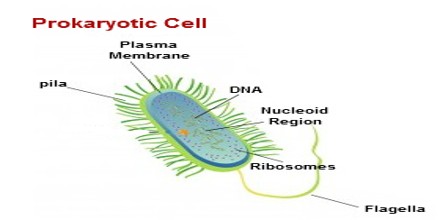
Prokaryotic Cell Structure: They are the first organisms to be present on our planet earth. Organisms, with this cell type are known by the term prokaryotic organisms (or) prokaryotes. Bacteria, blue green algae and E.coli are few examples of this category. Prokaryotic cells are single-celled organisms, with the absence of nucleus and comprises of capsule, cell wall, cell membrane, cytoplasm, nucleiod, ribosome, plasmids, pili and flagella.
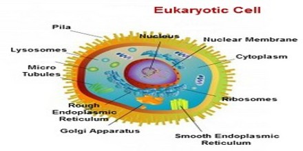
Eukaryotic Cell Structure: They are the cells with the presence of true nucleus. Organisms, with this cell type are known by the term eukaryotic organisms (or) eukaryotes. Animals, plants and other organisms excluding bacteria, blue green algae and E.coli have been grouped into this category. Eukaryotic cells are more complex than prokaryotic cells. These organisms have membrane bound nucleus with many cell organelles to perform several cellular functions within the system.
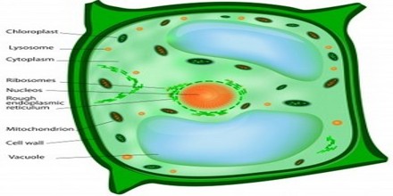
Plant Cell Structure: A plant cells are eukaryotic cells, with the presence of true nucleus, multicellular large and advanced membrane bound organelles. These plant cells are quite different from animal cells like in shape and other few organelles which are only found in animal cells but are absent in plant cells.
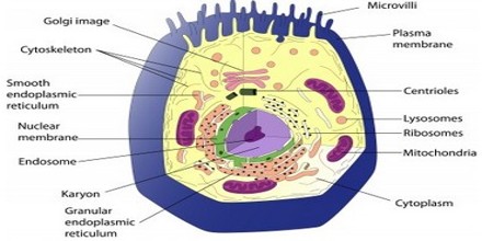
Animal Cell Structure: Animal cells are eukaryotic cells, with the presence of true nucleus; multicellular large and advanced membrane bound organelles. Like plant cells, animal cells have same organelles except the cell wall, chloroplasts, number of vacuoles and many more. Due to the absence of cell wall the shape of an animal cell is irregular.
Function of Cell
- Cell wall: It helps in protecting the plasma membrane and plays a vital role in supporting and protecting the cells. It is a thick outer layer made of cellulose.
- Cell membrane: It is a double layered, thin barrier, surrounding the cell to control the entry and exit of certain substances.
- Cytoplasm: It is a membrane, which protects the cell by keeping the cell organelles separate from each other. This helps to keep a cell in stable. Cytoplasm is the site, where many vital biochemical reactions take place.
- Nucleus: They are the membrane bound organelles, which are found in all eukaryotic cells. It is the very important organelle of a cell as it controls the complete activity of a cell and also plays a vital role in reproduction.
- Nuclear membrane: The bilayer membrane, which protects the nucleus by surrounding around it and acts as a barrier between the cell nucleus and other organs of a cell.
- Nucleolus: It is an important membrane found inside the nucleus. It plays a vital role in the production of cell’s ribosome.
- Chromosomes: It is made up of DNA and stored in the nucleus, which contains the instructions for traits and characteristics.
- Endoplasmic reticulum: It helps in the movement of materials around the cell. It contains an enzyme that helps in building molecules and in manufacturing of proteins. The main function of this organelle is storage and secretion.
- Ribosome: It plays a vital role in protein synthesis.
- Mitochondria: They are double membrane, filamentous organelles, which play a vital role in generating and transforming the energy. Mitochondria play a vital role in various functions of the cell metabolisms including oxidative phosphorylation.
- Golgi Bodies: It helps in the movement of materials within the cell.
- Lysosomes: It is also called as suicidal bags as it helps in cell renewal and break down old cell parts.
- Vacuoles: It helps plants in maintaining its shape and it also stores water, food, wastes, etc.
- Chloroplast: They are the site of photosynthesis, which are present in chlorophyll bacteria, blue-green algae, etc.
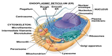
Organisation of Cell
- Cells contain a variety of internal structures called organelles
- An organelle is a cell component that performs a specific function in that cell
- Just as the organs of a multicellular organism carry out the organism’s life functions, the organelles of a cell maintain the life of the cell
- There are many different cells; however, there are certain features common to all cells
- The entire cell is surrounded by a thin cell membrane
- All membranes have the same thickness and basic structure
- Organelles often have their own membranes too – once again, these membranes have a similar structure
- The nucleus, mitochondria and chloroplasts all have double membranes, more correctly called envelopes
- Because membranes are fluid mosaics, the molecules making them up – phospholipids and proteins – move independently. The proteins appear to ‘float’ in the phospholipids bilayer
- Membranes can thus be used to transport molecules within the cell e.g. endoplasmic reticulum.
- Proteins in the membrane can be used to transport substances across the membrane – e.g. by facilitated diffusion or by active transport.
- The proteins on the outside of cell membranes identify us as unique.
Reprogenetics
Environmental Microbiology
Chromosomal Crossover

Searching for Dangerous Viruses now to avoid Future Pandemics

Procurement Contracts

Researchers use DNA-modified Building Blocks to create Colloidal Quasicrystals

Report on Marketing Stategies for real Estate Companies

Fractional-reserve Banking

Requirement Analysis System Boundary
Latest post.

Plasmonic Metamaterial

Split-pi Topology in Electronics

Control Valve

Bioluminescence First Evolved in Animals at least 540 Million Years Ago

Origin of Pluto’s Heart


1. Introduction
- 2. Solving the phase problem for data expanded to space group P1
3. Using phases to find the origin shift and space group
4. assigning chemical elements to the electron-density peaks, 5. isotropic refinement and absolute structure determination, 6. building the structure, 7. examples, 8. program development and distribution.

research papers \(\def\hfill{\hskip 5em}\def\hfil{\hskip 3em}\def\eqno#1{\hfil {#1}}\)

SHELXT – Integrated space-group and crystal-structure determination
a Department of Structural Chemistry, Georg-August Universität Göttingen, Tammannstrasse 4, Göttingen, 37077, Germany * Correspondence e-mail: [email protected]
The new computer program SHELXT employs a novel dual-space algorithm to solve the phase problem for single-crystal reflection data expanded to the space group P 1. Missing data are taken into account and the resolution extended if necessary. All space groups in the specified Laue group are tested to find which are consistent with the P 1 phases. After applying the resulting origin shifts and space-group symmetry, the solutions are subject to further dual-space recycling followed by a peak search and summation of the electron density around each peak. Elements are assigned to give the best fit to the integrated peak densities and if necessary additional elements are considered. An isotropic refinement is followed for non-centrosymmetric space groups by the calculation of a Flack parameter and, if appropriate, inversion of the structure. The structure is assembled to maximize its connectivity and centred optimally in the unit cell. SHELXT has already solved many thousand structures with a high success rate, and is optimized for multiprocessor computers. It is, however, unsuitable for severely disordered and twinned structures because it is based on the assumption that the structure consists of atoms.
Keywords: Patterson superposition ; direct methods ; dual-space recycling ; space-group determination ; element assignment .
Before the phase problem can be solved, the usual procedure is to determine the space group of the crystal with the help of the Laue symmetry of the diffraction pattern, the presence or absence of certain reflections (the systematic absences) and statistical tests ( e.g. to distinguish between centrosymmetric and non-centrosymmetric structures). This space-group determination may be upset by the presence of dominant heavy atoms or by pseudo-symmetry affecting the intensities of certain classes of reflections, and in some cases the space group is ambiguous. For example, the space groups I 222 and I 2 1 2 1 2 1 have the same systematic absences, as do Pmmn and two different orientations of Pmn 2 1 .
2. Solving the phase problem for data expanded to space group P 1
SHELXT reads standard SHELX format . i n s and . h k l files. It extracts the unit cell, Laue group (but not space group) and the elements that are expected to be present (but not how many atoms of each). A number of options, e.g. that all trigonal and hexagonal Laue groups should be considered ( - L 15 ), may be specified by command-line switches. A summary of the possible options is output when no filename is given on the SHELXT command line and further details are available on the SHELX home page.
The data are first merged according to the specified Laue group and then expanded to P 1. In theory, SHELXT could also have been programmed to determine the Laue group, e.g. by calculating the R values or correlation coefficients when the equivalent reflections are merged. However, the Laue group has to be known to scale the data, which is an essential step for the highly focused beams now common for synchrotrons and laboratory microsources, because the effective volume of the crystal irradiated is different for different reflections and needs to be corrected for. So in practice it is best to determine the Laue group first anyway. Even though programs such as XPREP (Bruker AXS, Madison, WI 53711, USA) are no longer required to determine the space group, it is still necessary to identify the correct unit cell and metric symmetry.
2.1. Dual-space iteration starting from a Patterson superposition
2.2. the random omit procedure.
Omit maps are frequently used in macromolecular crystallography to reduce model bias. A small part of the structure is deleted and the rest is refined to reduce memory effects, then a new difference-density map is generated and interpreted. This concept plays an important role in SHELXT , but because no model is available at the P 1 dual-space stage, it is implemented differently. The following density modification is performed unless otherwise specified by the user. A mask M ( x ) is constructed consisting of Gaussian-shaped peaks of unit volume at the positions of the maxima in the electron-density map. A small number of these Gaussian peaks are then deleted from the mask at random, usually every third dual-space cycle, and the new density is obtained by multiplying the original density ρ ( x ) with the mask:
where X is 1.0 unless reset by the user. For organic or organometallic structures, especially for low resolution or incomplete data, the alternative,
is sometimes better, but this is not the default option because it is not appropriate for inorganic and mineral structures. If CFOM is less than a preset threshold, the program refines further sets of starting phases, increasing the number of iterations each time this is done.
The idea of trying all possible space groups in a specified Laue group is also sometimes used in macromolecular crystal structure determination. For example, if the crystal is orthorhombic P , Laue group mmm , and only the Sohncke space groups need to be considered, a molecular-replacement program can be asked to test all eight possibilities. If only one of the eight gives a solution with good figures of merit, both the crystal structure and the space group have been determined! For chemical problems the situation is more interesting, because there are 30 possible orthorhombic P space groups and a total of 120 possibilities when different orientations of the axes are taken into account (as in SHELXT ).
For the correct space group and the correct origin shift Δ x , η should be close to zero. To facilitate comparisons, the figure of merit α is defined as the F 2 -weighted sum of η 2 over all pairs of equivalents for all reflections, normalized so that it should be unity for random phases. α should be as small as possible for the correct combination of space group and origin shift.
Each solution with a reasonable α value is first subject to ten cycles of density modification in the chosen space group after applying the origin shift. This density modification consists only of averaging the phases of equivalent reflections taking the space-group symmetry into account and resetting negative density to zero. A peak search is then performed, and the density inside a sphere (default radius 0.7 Å) about each peak is summed. It is better to use integrated densities rather than peak heights because the atoms may have different atomic displacement parameters. However, these integrated densities are not on an absolute scale, so the problem is how to set the scale so that they correspond to atomic numbers and the elements can be assigned. SHELXT attempts to set the scale as follows, going on to the next test only if the previous tests are negative:
( a ) If carbon is specified as one of the elements present, the program searches for peaks with similar integrated densities separated from each other by typical C—C distances ( i.e. between 1.25 and 1.65 Å). If enough are found, the scale is set so that they will have average atomic numbers of 6.
( b ) If boron is expected, boron cages with distances between 1.65 and 1.8 Å are searched for.
( c ) A search is made for oxyanions. The oxygen atoms should have similar integrated densities to each other and similar distances to a central atom.
( d ) If the above tests are negative, it is assumed that the heaviest atom expected corresponds to the peak with the highest integrated density. This can run into trouble if, for example, there is an unexpected bromide or iodide ion in the structure and it has not been possible to fix the scale by one of the above methods.
When the density scale has been found, it is used to assign elements to the remaining atoms. If it then appears that there are high-density peaks that cannot be assigned because only light atoms were expected, chlorine, bromine or iodine atoms are added. Some rudimentary checks are made to ensure that the element assignments are chemically reasonable.
The following algorithm used to assemble the structure is diabolically simple but almost always builds and clusters the molecules in a way that is instantly recognizable. No covalent radii etc . are used, so the algorithm is independent of the element assignments.
( a ) Generate the SDM (shortest-distance matrix). This is a triangular matrix of the shortest distances between unique atoms, taking symmetry into account.
( b ) Set a flag to -1 for each unique atom, then change it to +1 for one atom (it does not matter which).
( c ) Search the SDM for the shortest distance for which the product of the two flags is -1 . If none, exit.
( d ) Symmetry transform the atom with flag -1 corresponding to this distance so that it is as near as possible to the atom with flag +1 , then set its flag to +1 .
( e ) Go to ( c ).
SHELXT is compiled with the Intel ifort Fortran compiler using the statically linked MKL library and is particularly suitable for multi-CPU computers. It is available free to academics for the 32- or 64-bit Windows, 32- or 64-bit Linux and 64-bit Mac OS X operating systems. The program may be downloaded as part of the SHELX system via the SHELX home page ( https://shelx.uni-ac.gwdg.de/SHELX/ ), which also provides documentation and other useful information. Users are recommended to view the `recent changes' section on the home page from time to time.
The initial development of SHELXT was based on a test databank of about 650 structures, mostly determined in Göttingen, covering a wide range of problems. It has also been tested by more than 200 beta-testers for up to three years, in the course of which several thousand structures were solved (and a few not solved). It is difficult to generalize, but the correct space group was identified in about 97% of cases, and for about half of the structures every atom was located and assigned to the correct element. Most of the remaining structures were basically correct, the most common errors being carbon assigned as nitrogen or vice versa . Poor solutions were sometimes obtained when the heavy atoms corresponded to a centrosymmetric substructure but the full structure possessed a lower symmetry. It is always essential to check the element assignments, especially if the program has added extra elements, and also to check for the presence of disordered solvent molecules that may have been missed. The biggest danger is that inexperienced users may assume that the program is always right!
Acknowledgements
The author is very grateful to the many SHELXT beta-testers for patiently reporting bugs, suggesting improvements and providing interesting data sets for testing. He is particularly grateful to Bruker AXS for their help with the logistics of the three-year beta-test, and for the use of their email list for rapid communication with the beta-testers. He thanks the Volkswagen-Stiftung and the state of Niedersachsen for the award of a Niedersachsen (emeritus) Professorship.
This is an open-access article distributed under the terms of the Creative Commons Attribution (CC-BY) Licence , which permits unrestricted use, distribution, and reproduction in any medium, provided the original authors and source are cited.

- school Campus Bookshelves
- menu_book Bookshelves
- perm_media Learning Objects
- login Login
- how_to_reg Request Instructor Account
- hub Instructor Commons
- Download Page (PDF)
- Download Full Book (PDF)
- Periodic Table
- Physics Constants
- Scientific Calculator
- Reference & Cite
- Tools expand_more
- Readability
selected template will load here
This action is not available.

6.20: Assignment- Cell Builder
- Last updated
- Save as PDF
- Page ID 43571
Create a model of a eukaryotic cell using any material of your choice. In your model be sure to include all the organelles appropriate to your cell (either plant or animal). Once complete, take multiple photographs of your model from all angles. Include these images in a document that also contains the following in table format:
- A detailed key/legend that matches the model;
- Each organelle or part with its basic function;
- A disease or disorder that is associated with the malfunction of each cellular component
- How this organelle is visualized microscopically
Note for the disease information, you can list a disease in either animals or plants, regardless of what type of cell you are modeling. In other words, its okay to discuss a “human” disease even if you are making a plant model, provided the organelle is present in both types of cells.
Some suggestions for 3D models include Legos, a decorated cake with candy toppings, or standard Styrofoam base with appropriate pieces attached. You can also draw or illustrate a model. Here is an example of what you might make.
Basic Requirements (the assignment will not be accepted or assessed unless the follow criteria have been met):
- Assignment has been proofread and does not contain any major spelling or grammatical errors
- Assignment includes appropriate references
- Assignment includes photographs or images of created model from all angles.
- Assignment includes a key documenting how each organelle is represented in the model.
- Assignment includes a completed table such as the one illustrated in the example document.
- Assignment includes a disease caused by malfunction of each identified component in the model.
- Assignment includes at least 7 organelles in the model and table.
Contributors and Attributions
- Performance Assessments: Cell Builder. Authored by : Shelli Carter. Provided by : Columbia Basin College. Located at : https://www.columbiabasin.edu/ . License : CC BY: Attribution

IMAGES
VIDEO
COMMENTS
Learn about the structure and function of cells, the basic units of life, with Khan Academy 's engaging and interactive lessons. Explore how organelles, membranes, and transport systems work together to make cells efficient and diverse. This unit will prepare you for the AP®︎/College Biology exam and beyond.
By structure, it is a porous membrane (with pores) which permits the movement of selective substances in and out of the cell. Besides this, the cell membrane also protects the cellular component from damage and leakage. It forms the wall-like structure between two cells as well as between the cell and its surroundings.
1. Description of Cell Structure and Function. Cells are fundamental to the study of biology. Every living thing is composed of cells, they are the building blocks of life. All cells share similar characteristics and can be defined by the cell theory. Cell Theory. 1. All living things are composed of cells. 2.
Centrioles. Identify these cellular structures. Peroxisome. Identify this cellular structure which neutralizes toxins. Lysosome. Identify this cellular structure which cleans up cellular debris. Mitochondria. Name this cellular structure. biology 141 & 143 Learn with flashcards, games, and more — for free.
Structure of a cell: Quiz 2; Structure of a cell: Unit test; About this unit. This unit is part of the Biology library. Browse videos, articles, and exercises by topic. Introduction to cells. Start your cellular journey the right way: with some history and some microscopy! Here, we'll learn more about how cells were discovered, how they can be ...
Part 1: Cell Structures. 1. Draw an animal cell in the space below. Draw the components of the cell using different colors. Color the parts of an animal cell using a color scheme you developed or on other words, match the color with the cell structure. Use a different color for each of the cell components if possible.
Unlike prokaryotic cells, eukaryotic cells have: 1) a membrane-bound nucleus; 2) numerous membrane-bound organelles such as the endoplasmic reticulum, Golgi apparatus, chloroplasts, mitochondria, and others; and 3) several, rod-shaped chromosomes. Because a eukaryotic cell's nucleus is surrounded by a membrane, it is has "true nucleus.".
This magical microscope lets viewers jump between levels of magnification from organ systems to cells. Inside a Cell. This dynamic tour features 3 different cell types, each with animated depictions of organelles working together to carry out basic life functions. Explore the functions to learn the name of each cell structure and its role in ...
cell, in biology, the basic membrane-bound unit that contains the fundamental molecules of life and of which all living things are composed.A single cell is often a complete organism in itself, such as a bacterium or yeast.Other cells acquire specialized functions as they mature. These cells cooperate with other specialized cells and become the building blocks of large multicellular organisms ...
The plasma (cell) membrane separates the inner environment of a cell from the extracellular fluid. It is composed of a fluid phospholipid bilayer (two layers of phospholipids) as shown in figure 4.1.2 4.1. 2 below, and other molecules. Not many substances can cross the phospholipid bilayer, so it serves to separate the inside of the cell from ...
A cell is the smallest unit of a living thing. A living thing, whether made of one cell (like bacteria) or many cells (like a human), is called an organism. Thus, cells are the basic building blocks of all organisms. There are many types of cells, all grouped into one of two broad categories: prokaryotic and eukaryotic.
The 3 different types of cells that exist in nature are: 1) Prokaryotes. 2) Eukaryotes. 3) Archaeans. Identify the labeled structures of a prokaryotic cell: Arrange the following in order from smallest to largest: Protein, Chloroplast, Atom, Virus, Human egg. 1) Atom. 2) Protein.
Cell Organelles: Definition, Structure, Functions, Diagram. June 6, 2023 by Anupama Sapkota. Edited By: Sagar Aryal. Cell organelles are specialized entities present inside a particular type of cell that performs a specific function. There are various cell organelles, out of which, some are common in most types of cells like cell membranes ...
Module 4 Assignment: Cell Builder. Create a model of a eukaryotic cell using any material of your choice. In your model be sure to include all the organelles appropriate to your cell (either plant or animal). Once complete, take multiple photographs of your model from all angles. Include these images in a document that also contains the ...
OpenStax. Figure 4.1.1 4.1. 1: (a) Nasal sinus cells (viewed with a light microscope), (b) onion cells (viewed with a light microscope), and (c) Vibrio tasmaniensis bacterial cells (seen through a scanning electron microscope) are from very different organisms, yet all share certain characteristics of basic cell structure. (credit a ...
The cell is the structural and functional unit of all living organisms, and is sometimes called the "building block of life.". Some organisms, such as bacteria, are unicellular, consisting of a single cell. Cell is the basic structural and functional unit of any living thing. Each cell is a small container of chemicals and water wrapped in ...
The structure is assembled to maximize its connectivity and centred optimally in the unit cell. SHELXT has already solved many thousand structures with a high success rate, and is optimized for multiprocessor computers. ... With the correct element assignments all the figures of merit would have been very similar for all the space groups. In ...
Cell Builder. Outcome: Identify and explain a variety of cellular components. Criteria. Ratings. Pts. At least 7 membrane-bound organelles found in eukaryotic cells are contained in the model and their functions identified in the table. Organelles and functions are outlined in detail. 5.0 pts.
Quiz 1. Learn for free about math, art, computer programming, economics, physics, chemistry, biology, medicine, finance, history, and more. Khan Academy is a nonprofit with the mission of providing a free, world-class education for anyone, anywhere.