Parts of the Brain: Anatomy, Structure & Functions
Olivia Guy-Evans, MSc
Associate Editor for Simply Psychology
BSc (Hons) Psychology, MSc Psychology of Education
Olivia Guy-Evans is a writer and associate editor for Simply Psychology. She has previously worked in healthcare and educational sectors.
Learn about our Editorial Process
Saul Mcleod, PhD
Editor-in-Chief for Simply Psychology
BSc (Hons) Psychology, MRes, PhD, University of Manchester
Saul Mcleod, PhD., is a qualified psychology teacher with over 18 years of experience in further and higher education. He has been published in peer-reviewed journals, including the Journal of Clinical Psychology.
On This Page:
The brain controls all functions of the body, interprets information from the outside world, and defines who we are as individuals and how we experience the world.
The brain receives information through our senses: sight, touch, taste, smell, and hearing. This information is processed in the brain, allowing us to give meaning to the input it receives.
The brain is part of the central nervous system ( CNS ) along with the spinal cord. There is also a peripheral nervous system (PNS) comprised of 31 pairs of spinal nerves that branch from the spinal cord and cranial nerves that branch from the brain.

Brain Parts
The brain is composed of the cerebrum, cerebellum, and brainstem (Fig. 1).

Figure 1. The brain has three main parts: the cerebrum, cerebellum, and brainstem.
The cerebrum is the largest and most recognizable part of the brain. It consists of grey matter (the cerebral cortex ) and white matter at the center. The cerebrum is divided into two hemispheres, the left and right, and contains the lobes of the brain (frontal, temporal, parietal, and occipital lobes).
The cerebrum produces higher functioning roles such as thinking, learning, memory, language, emotion, movement, and perception.
The Cerebellum
The cerebellum is located under the cerebrum and monitors and regulates motor behaviors, especially automatic movements.
This structure is also important for regulating posture and balance and has recently been suggested for being involved in learning and attention.
Although the cerebellum only accounts for roughly 10% of the brain’s total weight, this area is thought to contain more neurons (nerve cells) than the rest of the brain combined.
The brainstem is located at the base of the brain. This area connects the cerebrum and the cerebellum to the spinal cord, acting as a relay station for these areas.
The brainstem regulates automatic functions such as sleep cycles, breathing, body temperature, digestion, coughing, and sneezing.

Right Brain vs. Left Brain
The cerebrum is divided into two halves: the right and left hemispheres (Fig. 2). The left hemisphere controls the right half of the body, and the right hemisphere controls the left half.
The two hemispheres are connected by a thick band of neural fibers known as the corpus callosum, consisting of about 200 million axons.
The corpus callosum allows the two hemispheres to communicate and allows information being processed on one side of the brain to be shared with the other.

Figure 2. The cerebrum is divided into left and right hemispheres. The nerve fibers corpus callosum connects the two sides.
Hemispheric lateralization is the idea that each hemisphere is responsible for different functions. Each of these functions is localized to either the right or left side.
The left hemisphere is associated with language functions, such as formulating grammar and vocabulary and containing different language centers (Broca’s and Wernicke’s area).
The right hemisphere is associated with more visuospatial functions such as visualization, depth perception, and spatial navigation. These left and right functions are the case in most people, especially those who are right-handed.
Lobes of the Brain
Each cerebral hemisphere can be subdivided into four lobes, each associated with different functions.
The four lobes of the brain are the frontal, parietal, temporal, and occipital lobes (Figure 3).

Figure 3. The cerebrum is divided into four lobes: frontal, parietal, occipital, and temporal.
Frontal lobes
The frontal lobes are located at the front of the brain, behind the forehead (Figure 4).
Their main functions are associated with higher cognitive functions, including problem-solving, decision-making, attention, intelligence, and voluntary behaviors.
The frontal lobes contain the motor cortex responsible for planning and coordinating movements.
It also contains the prefrontal cortex, which is responsible for initiating higher-lever cognitive functioning, and Broca’s Area, which is essential for language production.

Figure 4. Frontal lobe structure.
Temporal lobes
The temporal lobes are located on both sides of the brain, near the temples of the head, hence the name temporal lobes (Figure 5).
The main functions of these lobes include understanding, language, memory acquisition, face recognition, object recognition, perception, and auditory information processing.
There is a temporal lobe in both the left and right hemispheres. The left temporal lobe, which is usually the most dominant in people, is associated with language, learning, memorizing, forming words, and remembering verbal information.
The left lobe also contains a vital language center known as Wernicke’s area, which is essential for language development. The right temporal lobe is usually associated with learning and memorizing non-verbal information and determining facial expressions.

Figure 5. Temporal lobe structure.
Parietal lobes
The parietal lobe is located at the top of the brain, between the frontal and occipital lobes, and above the temporal lobes (Figure 6).
The parietal lobe is essential for integrating information from the body’s senses to allow us to build a coherent picture of the world around us.
These lobes allow us to perceive our bodies through somatosensory information (e.g., through touch, pressure, and temperature). It can also help with visuospatial processing, reading, and number representations (mathematics).
The parietal lobes also contain the somatosensory cortex, which receives and processes sensory information, integrating this into a representational map of the body.
This means it can pinpoint the exact area of the body where a sensation is felt, as well as perceive the weight of objects, shape, and texture.

Figure 6. Parietal lobe structure.
Occipital lobes
The occipital lobes are located at the back of the brain behind the temporal and parietal lobes and below the occipital bone of the skull (Figure 7).
The occipital lobes receive sensory information from the eyes’ retinas, which is then encoded into different visual data. Some of the functions of the occipital lobes include being able to assess the size, depth, and distance, determine color information, object and facial recognition, and mapping the visual world.
The occipital lobes also contain the primary visual cortex, which receives sensory information from the retinas, transmitting this information relating to location, spatial data, motion, and the colors of objects in the field of vision.

Figure 7. Occipital lobe structure.
Cerebral Cortex
The surface of the cerebrum is called the cerebral cortex and has a wrinkled appearance, consisting of bulges, also known as gyri, and deep furrows, known as sulci (Figure 8).
A gyrus (plural: gyri) is the name given to the bumps and ridges on the cerebral cortex (the outermost layer of the brain). A sulcus (plural: sulci) is another name for a groove in the cerebral cortex.

Figure 8. The cortex contains neurons (grey matter) interconnected to other brain areas by axons (white matter). The cortex has a folded appearance. A fold is called a gyrus, and the valley between is a sulcus.
The cerebral cortex is primarily constructed of grey matter (neural tissue made up of neurons), with between 14 and 16 billion neurons found here.
The many folds and wrinkles of the cerebral cortex allow a wider surface area for an increased number of neurons to live there, permitting large amounts of information to be processed.
Deep Structures
The amygdala is a structure deep in the brain that is involved in the processing of emotions and fear learning. The amygdala is a part of the limbic system, a neural network that mediates emotion and memory (Figure 9).
This structure also ties emotional meaning to memories, processes rewards, and helps us make decisions. This structure has also been linked with the fight-or-flight response.

Figure 9. The amygdala in the limbic system plays a key role in how animals assess and respond to environmental threats and challenges by evaluating the emotional importance of sensory information and prompting an appropriate response.
Thalamus and Hypothalamus
The thalamus relays information between the cerebral cortex, brain stem, and other cortical structures (Figure 10).
Because of its interactive role in relaying sensory and motor information, the thalamus contributes to many processes, including attention, perception, timing, and movement. The hypothalamus modulates a range of behavioral and physiological functions.
It controls autonomic functions such as hunger, thirst, body temperature, and sexual activity. To do this, the hypothalamus integrates information from different brain parts and responds to various stimuli such as light, odor, and stress.

Figure 10. The thalamus is often described as the brain’s relay station as a great deal of information that reaches the cerebral cortex first stops in the thalamus before being sent to its destination.
Hippocampus
The hippocampus is a curved-shaped structure in the limbic system associated with learning and memory (Figure 11).
This structure is most strongly associated with the formation of memories, is an early storage system for new long-term memories, and plays a role in the transition of these long-term memories to more permanent memories.

Figure 11. Hippocampus location in the brain
Basal Ganglia
The basal ganglia are a group of structures that regulate the coordination of fine motor movements, balance, and posture alongside the cerebellum.
These structures are connected to other motor areas and link the thalamus with the motor cortex. The basal ganglia are also involved in cognitive and emotional behaviors, as well as playing a role in reward and addiction.

Figure 12. The Basal Ganglia Illustration
Ventricles and Cerebrospinal Fluid
Within the brain, there are fluid-filled interconnected cavities called ventricles , which are extensions of the spinal cord. These are filled with a substance called cerebrospinal fluid, which is a clear and colorless liquid.
The ventricles produce cerebrospinal fluid and transport and remove this fluid. The ventricles do not have a unique function, but they provide cushioning to the brain and are useful for determining the locations of other brain structures.
Cerebrospinal fluid circulates the brain and spinal cord and functions to cushion the brain within the skull. If damage occurs to the skull, the cerebrospinal fluid will act as a shock absorber to help protect the brain from injury.

As well as providing cushioning, the cerebrospinal fluid circulates nutrients and chemicals filtered from the blood and removes waste products from the brain. Cerebrospinal fluid is constantly absorbed and replenished by the ventricles.
If there were a disruption or blockage, this can cause a build-up of cerebrospinal fluid and can cause enlarged ventricles.
Neurons are the nerve cells of the central nervous system that transmit information through electrochemical signals throughout the body. Neurons contain a soma, a cell body from which the axon extends.
Axons are nerve fibers that are the longest part of the neuron, which conduct electrical impulses away from the soma.

There are dendrites at the end of the neuron, which are branch-like structures that send and receive information from other neurons.
A myelin sheath, a fatty insulating layer, forms around the axon, allowing nerve impulses to travel down the axon quickly.
There are different types of neurons. Sensory neurons transmit sensory information, motor neurons transmit motor information, and relay neurons allow sensory and motor neurons to communicate.
The communication between neurons is called synapses. Neurons communicate with each other via synaptic clefts, which are gaps between the endings of neurons.

During synaptic transmission, chemicals, such as neurotransmitters, are released from the endings of the previous neuron (also known as the presynaptic neuron).
These chemicals enter the synaptic cleft to then be transported to receptors on the next neuron (also known as the postsynaptic neuron).
Once transported to the next neuron, the chemical messengers continue traveling down neurons to influence many functions, such as behavior and movement.
Glial Cells
Glial cells are non-neuronal cells in the central nervous system which work to provide the neurons with nourishment, support, and protection.
These are star-shaped cells that function to maintain the environment for neuronal signaling by controlling the levels of neurotransmitters surrounding the synapses.
They also work to clean up what is left behind after synaptic transmission, either recycling any leftover neurotransmitters or cleaning up when a neuron dies.
Oligodendrocytes
These types of glial have the appearance of balls with spikes all around them. They function by wrapping around the axons of neurons to form a protective layer called the myelin sheath.
This is a substance that is rich in fat and provides insulation to the neurons to aid neuronal signaling.
Microglial cells have oval bodies and many branches projecting out of them. The primary function of these cells is to respond to injuries or diseases in the central nervous system.
They respond by clearing away any dead cells or removing any harmful toxins or pathogens that may be present, so they are, therefore, important to the brain’s health.
Ependymal cells
These cells are column-shaped and usually line up together to form a membrane called the ependyma. The ependyma is a thin membrane lining the spinal cord and ventricles of the brain .
In the ventricles, these cells have small hairlike structures called cilia, which help encourage the flow of cerebrospinal fluid.
Cranial Nerves
There are 12 types of cranial nerves which are linked directly to the brain without having to pass through the spinal cord. These allow sensory information to pass from the organs of the face to the brain:

Mnemonic for Order of Cranial Nerves:
S ome S ay M arry M oney B ut M y B rother S ays B ig B rains M atter M ore
- Cranial I: Sensory
- Cranial II: Sensory
- Cranial III: Motor
- Cranial IV: Motor
- Cranial V: Both (sensory & motor)
- Cranial VI: Motor
- Cranial VII: Both (sensory & motor)
- Cranial VIII: Sensory
- Cranial IX: Both (sensory & motor)
- Cranial X: Both (sensory & motor)
- Cranial XI: Motor
- Cranial XII: Motor
Purves, D., Augustine, G., Fitzpatrick, D., Katz, L., LaMantia, A., McNamara, J., & Williams, S. (2001). Neuroscience 2nd edition . sunderland (ma) sinauer associates. Types of Eye Movements and Their Functions.
Mayfield Brain and Spine (n.d.). Anatomy of the Brain. Retrieved July 28, 2021, from: https://mayfieldclinic.com/pe-anatbrain.htm
Robertson, S. (2018, August 23). What is Grey Matter? News Medical Life Sciences. https://www.news-medical.net/health/What-is-Grey-Matter.aspx
Guy-Evans, O. (2021, April 13). Temporal lobe: definition, functions, and location. Simply Psychology. www.www.www.www.www.www.simplypsychology.org/temporal-lobe.html
Guy-Evans, O. (2021, April 15). Parietal lobe: definition, functions, and location. Simply Psychology. www.www.www.www.www.www.simplypsychology.org/parietal-lobe.html
Guy-Evans, O. (2021, April 19). Occipital lobe: definition, functions, and location. Simply Psychology. www.www.www.www.www.www.simplypsychology.org/occipital-lobe.html
Guy-Evans, O. (2021, May 08). Frontal lobe function, location in brain, damage, more. Simply Psychology. www.www.www.www.www.www.simplypsychology.org/frontal-lobe.html
Guy-Evans, O. (2021, June 09). Gyri and sulci of the brain. Simply Psychology. www.www.www.www.www.www.simplypsychology.org/gyri-and-sulci-of-the-brain.html

Essay on Human Brain
Students are often asked to write an essay on Human Brain in their schools and colleges. And if you’re also looking for the same, we have created 100-word, 250-word, and 500-word essays on the topic.
Let’s take a look…
100 Words Essay on Human Brain
The human brain: an overview.
The human brain is a complex organ, responsible for all our thoughts, feelings, and actions. It’s made up of billions of nerve cells, or neurons, which communicate through electrical signals.
Parts of the Brain
The brain is divided into three main parts: the cerebrum, cerebellum, and brainstem. The cerebrum is the largest part and controls thinking, learning, and emotions. The cerebellum manages balance and coordination. The brainstem connects the brain to the spinal cord and controls automatic functions like breathing.
Brain’s Functionality
The brain is always active, even during sleep. It processes information from our senses, helps us understand the world around us, and makes decisions. It’s truly a remarkable organ!
250 Words Essay on Human Brain
Introduction.
The human brain, a marvel of biological engineering, is the most complex organ in the human body. It is the epicenter of human consciousness, responsible for our thoughts, emotions, and actions.
Structure and Function
The brain is divided into three main parts: the cerebrum, cerebellum, and brainstem. The cerebrum, the largest part, is responsible for higher brain functions such as thought, emotion, and sensory processing. The cerebellum coordinates motor functions, while the brainstem controls automatic functions like heart rate and breathing.
Neuroplasticity
A remarkable feature of the brain is its neuroplasticity, the ability to form and reorganize synaptic connections in response to learning, experience, or injury. This adaptability underscores the brain’s capacity for lifelong learning and recovery.
Cognitive Abilities
Cognitive abilities such as memory, attention, and problem-solving are facilitated by the brain’s intricate network of neurons. These abilities enable us to navigate and interpret the world around us, engage in social interactions, and make decisions.
Brain and Technology
Advancements in technology have led to breakthroughs in understanding the brain. Techniques like fMRI and EEG provide detailed insights into brain activity, paving the way for treatments of neurological disorders.
The human brain, with its intricate structure and impressive capabilities, continues to be a subject of fascination and study. Its complexity and adaptability underscore the limitless potential of human cognition, making it a cornerstone of our identity as a species.
500 Words Essay on Human Brain
Introduction to the human brain.
The human brain, a product of millions of years of evolutionary progression, is a marvel of biological engineering. It is a complex organ, responsible for controlling all the functions of the human body, processing sensory information, and coordinating responses. The brain is an intricate network of billions of neurons, which communicate and work together to generate our thoughts, feelings, and actions.
Structural Complexity of the Brain
The human brain is composed of several distinct regions, each with specific functions. The cerebrum, the largest part, is responsible for higher cognitive functions like thinking, learning, and consciousness. It is divided into two hemispheres, each further subdivided into four lobes: the frontal lobe, parietal lobe, occipital lobe, and temporal lobe.
The cerebellum, located beneath the cerebrum, coordinates motor control, balance, and coordination. The brainstem, connecting the brain to the spinal cord, controls automatic functions vital for survival, such as heartbeat, breathing, and digestion.
Neurons: The Building Blocks
Neurons, the fundamental units of the brain, transmit information through electrical and chemical signals. They consist of a cell body, dendrites, and an axon. The dendrites receive signals from other neurons, which are then passed through the cell body and down the axon to the next neuron. This communication forms neural networks, the basis for all brain activity.
Brain Plasticity
One of the most fascinating aspects of the human brain is its plasticity, the ability to reorganize itself by forming new neural connections throughout life. This adaptability allows us to learn new skills, adapt to changes, and recover from brain injuries. Neuroplasticity underscores the brain’s remarkable capacity for resilience and growth.
The Brain and Consciousness
The brain is not only a biological organ but also the seat of consciousness and identity. It is responsible for our thought processes, emotions, memories, and perceptions. The intricate interplay of neural networks generates the rich tapestry of human experience, from the most mundane thoughts to the most profound creative insights.
Future Research Directions
Despite significant advances in neuroscience, much about the brain remains a mystery. Key questions about consciousness, memory formation, and the nature of intelligence are yet to be fully answered. The development of advanced neuroimaging techniques and computational models offers exciting possibilities for future research.
Understanding the brain is not merely an academic exercise but has profound implications for treating neurological disorders, improving education, and even addressing ethical questions about artificial intelligence and brain-computer interfaces.
In conclusion, the human brain, with its intricate architecture and dynamic functionality, is a testament to the complexity and beauty of human life. As we continue to unravel its mysteries, we deepen our understanding of what it means to be human, highlighting the importance of continued research in this fascinating field.
That’s it! I hope the essay helped you.
If you’re looking for more, here are essays on other interesting topics:
- Essay on Honey Bee
- Essay on How I Celebrate Holi
- Essay on Hiroshima Day
Apart from these, you can look at all the essays by clicking here .
Happy studying!
Leave a Reply Cancel reply
Your email address will not be published. Required fields are marked *
Save my name, email, and website in this browser for the next time I comment.

- Science Notes Posts
- Contact Science Notes
- Todd Helmenstine Biography
- Anne Helmenstine Biography
- Free Printable Periodic Tables (PDF and PNG)
- Periodic Table Wallpapers
- Interactive Periodic Table
- Periodic Table Posters
- How to Grow Crystals
- Chemistry Projects
- Fire and Flames Projects
- Holiday Science
- Chemistry Problems With Answers
- Physics Problems
- Unit Conversion Example Problems
- Chemistry Worksheets
- Biology Worksheets
- Periodic Table Worksheets
- Physical Science Worksheets
- Science Lab Worksheets
- My Amazon Books
Parts of the Brain and Their Functions

The human brain is the epicenter of our nervous system and plays a pivotal role in virtually every aspect of our lives. It’s a complex, highly organized organ responsible for thoughts, feelings, actions, and interactions with the world around us. Here is a look at the intricate anatomy of the brain, its functions, and the consequences of damage to different areas.
Introduction to the Brain and Its Functions
The brain is an organ of soft nervous tissue that is protected within the skull of vertebrates. It functions as the coordinating center of sensation and intellectual and nervous activity. The brain consists of billions of neurons (nerve cells) that communicate through intricate networks. The primary functions of the brain include processing sensory information, regulating bodily functions, forming thoughts and emotions, and storing memories.
Main Parts of the Brain – Anatomy
The three main parts of the brain are the cerebrum, cerebellum, and brainstem.
1. Cerebrum
- Location: The cerebellum occupies the upper part of the cranial cavity and is the largest part of the human brain.
- Functions: It’s responsible for higher brain functions, including thought, action, emotion, and interpretation of sensory data.
- Effects of Damage: Depending on the area affected, damage leads to memory loss, impaired cognitive skills, changes in personality, and loss of motor control.
2. Cerebellum
- Location: The cerebellum is at the back of the brain, below the cerebrum.
- Functions: It coordinates voluntary movements such as posture, balance, coordination, and speech.
- Effects of Damage: Damage causes problems with balance, movement, and muscle coordination (ataxia).
3. Brainstem
- Location: The brainstem is lower extension of the brain, connecting to the spinal cord. It includes the midbrain, pons, and medulla oblongata.
- Functions: This part of the brain controls many basic life-sustaining functions, including heart rate, breathing, sleeping, and eating.
- Effects of Damage: Damage results in life-threatening conditions like breathing difficulties, heart problems, and loss of consciousness.
Lobes of the Brain
The four lobes of the brain are regions of the cerebrum:
- Location: This is the anterior or front part of the brain.
- Functions: Decision making, problem solving, control of purposeful behaviors, consciousness, and emotions.
- Location: Sits behind the frontal lobe.
- Functions: Processes sensory information it receives from the outside world, mainly relating to spatial sense and navigation (proprioception).
- Location: Below the lateral fissure, on both cerebral hemispheres.
- Functions: Mainly revolves around auditory perception and is also important for the processing of both speech and vision (reading).
- Location: At the back of the brain.
- Functions: Main center for visual processing.
Left vs. Right Brain Hemispheres
The cerebrum has two halves, called hemispheres. Each half controls functions on the opposite side of the body. So, the left hemisphere controls muscles on the right side of the body, and vice versa. But, the functions of the two hemispheres are not entirely identical:
- Left Hemisphere: It’s dominant in language and speech and plays roles in logical thinking, analysis, and accuracy. .
- Right Hemisphere: This hemisphere is more visual and intuitive and functions in creative and imaginative tasks.
The corpus callosum is a band of nerves that connect the two hemispheres and allow communication between them.
Detailed List of Parts of the Brain
While knowing the three key parts of the brain is a good start, the anatomy is quite a bit more complex. In addition to nervous tissues, the brain also contains key glands:
- Cerebrum: The cerebrum is the largest part of the brain. Divided into lobes, it coordinates thought, movement, memory, senses, speech, and temperature.
- Corpus Callosum : A broad band of nerve fibers joining the two hemispheres of the brain, facilitating interhemispheric communication.
- Cerebellum : Coordinates movement and balance and aids in eye movement.
- Pons : Controls voluntary actions, including swallowing, bladder function, facial expression, posture, and sleep.
- Medulla oblongata : Regulates involuntary actions, including breathing, heart rhythm, as well as oxygen and carbon dioxide levels.
- Limbic System : Includes the amygdala, hippocampus, and parts of the thalamus and hypothalamus.
- Amygdala: Plays a key role in emotional responses, hormonal secretions, and memory formation.
- Hippocampus: Plays a vital role in memory formation and spatial navigation.
- Thalamus : Acts as the brain’s relay station, channeling sensory and motor signals to the cerebral cortex, and regulating consciousness, sleep, and alertness.
- Basal Ganglia : A group of structures involved in processing information related to movement, emotions, and reward. Key structures include the striatum, globus pallidus, substantia nigra, and subthalamic nucleus.
- Ventral Tegmental Area (VTA) : Plays a role in the reward circuit of the brain, releasing dopamine in response to stimuli indicating a reward.
- Optic tectum : Also known as the superior colliculus, it directs eye movements.
- Substantia Nigra : Involved in motor control and contains a large concentration of dopamine-producing neurons.
- Cingulate Gyrus : Plays a role in processing emotions and behavior regulation. It also helps regulate autonomic motor function.
- Olfactory Bulb : Involved in the sense of smell and the integration of olfactory information.
- Mammillary Bodies : Plays a role in recollective memory.
- Function: Regulates emotions, memory, and arousal.
Glands in the Brain
The hypothalamus, pineal gland, and pituitary gland are the three endocrine glands within the brain:
- Hypothalamus : The hypothalamus links the nervous and endocrine systems. It contains many small nuclei. In addition to participating in eating and drinking, sleeping and waking, it regulates the endocrine system via the pituitary gland. It maintains the body’s homeostasis, regulating hunger, thirst, response to pain, levels of pleasure, sexual satisfaction, anger, and aggressive behavior.
- Pituitary Gland : Known as the “master gland,” it controls various other hormone glands in the body, such as the thyroid and adrenals, as well as regulating growth, metabolism, and reproductive processes.
- Pineal Gland : The pineal gland produces and regulates some hormones, including melatonin, which is crucial in regulating sleep patterns and circadian rhythms.
Gray Matter vs. White Matter
The brain and spinal cord consist of gray matter (substantia grisea) and white matter (substantia alba).
- White Matter: Consists mainly of axons and myelin sheaths that send signals between different brain regions and between the brain and spinal cord.
- Gray Matter: Consists of neuronal cell bodies, dendrites, and axon terminals. Gray matter processes information and directs stimuli for muscle control, sensory perception, decision making, and self-control.
Frequently Asked Questions (FAQs) About the Human Brain
- The human brain contains approximately 86 billion neurons. Additionally, it has a similar or slightly higher number of non-neuronal cells (glial cells), making the total number of cells in the brain close to 170 billion.
- There are about 86 billion neurons in the human brain. These neurons are connected by trillions of synapses, forming a complex networks.
- The average adult human brain weighs about 1.3 to 1.4 kilograms (about 3 pounds). This weight represents about 2% of the total body weight.
- The brain is about 73% water.
- The myth that humans only use 10% of their brain is false. Virtually every part gets use, and most of the brain is active all the time, even during sleep.
- The average size of the adult human brain is about 15 centimeters (6 inches) in length, 14 centimeters (5.5 inches) in width, and 9 centimeters (3.5 inches) in height.
- Brain signal speeds vary depending on the type of neuron and the nature of the signal. They travel anywhere from 1 meter per second to over 100 meters per second in the fastest neurons.
- With age, the brain’s volume and/or weight decrease, synaptic connections reduce, and there can be a decline in cognitive functions. However, the brain to continues adapting and forming new connections throughout life.
- The brain has a limited ability to repair itself. Neuroplasticity aids recovery by allowing other parts of the brain to take over functions of the damaged areas.
- The brain consumes about 20% of the body’s total energy , despite only making up about 2% of the body’s total weight . It requires a constant supply of glucose and oxygen.
- Sleep is crucial for brain health. It aids in memory consolidation, learning, brain detoxification, and the regulation of mood and cognitive functions.
- Douglas Fields, R. (2008). “White Matter Matters”. Scientific American . 298 (3): 54–61. doi: 10.1038/scientificamerican0308-54
- Kandel, Eric R.; Schwartz, James Harris; Jessell, Thomas M. (2000). Principles of Neural Science (4th ed.). New York: McGraw-Hill. ISBN 978-0-8385-7701-1.
- Kolb, B.; Whishaw, I.Q. (2003). Fundamentals of Human Neuropsychology (5th ed.). New York: Worth Publishing. ISBN 978-0-7167-5300-1.
- Rajmohan, V.; Mohandas, E. (2007). “The limbic system”. Indian Journal of Psychiatry . 49 (2): 132–139. doi: 10.4103/0019-5545.33264
- Shepherd, G.M. (1994). Neurobiology . Oxford University Press. ISBN 978-0-19-508843-4.
Related Posts
- Bipolar Disorder
- Therapy Center
- When To See a Therapist
- Types of Therapy
- Best Online Therapy
- Best Couples Therapy
- Best Family Therapy
- Managing Stress
- Sleep and Dreaming
- Understanding Emotions
- Self-Improvement
- Healthy Relationships
- Student Resources
- Personality Types
- Guided Meditations
- Verywell Mind Insights
- 2023 Verywell Mind 25
- Mental Health in the Classroom
- Editorial Process
- Meet Our Review Board
- Crisis Support
Parts of the Brain
Anatomy, Functions, and Conditions
Kendra Cherry, MS, is a psychosocial rehabilitation specialist, psychology educator, and author of the "Everything Psychology Book."
:max_bytes(150000):strip_icc():format(webp)/IMG_9791-89504ab694d54b66bbd72cb84ffb860e.jpg "parts of the brain essay")
Steven Gans, MD is board-certified in psychiatry and is an active supervisor, teacher, and mentor at Massachusetts General Hospital.
:max_bytes(150000):strip_icc():format(webp)/steven-gans-1000-51582b7f23b6462f8713961deb74959f.jpg "parts of the brain essay")
The Cerebral Cortex
The four lobes, the brain stem, the cerebellum, the limbic system, other parts of the brain, brain conditions, protecting your brain.
The human brain is not only one of the most important organs in the human body; it is also the most complex. The brain is made up of billions of neurons and it also has a number of specialized parts that are each involved in important functions.
While there is still a great deal that researchers do not yet know about the brain, they have learned a great deal about the anatomy and function of the brain. Understanding these parts can help give people a better idea of how disease and damage may affect the brain and its ability to function.
The cerebral cortex is the part of the brain that makes human beings unique. Functions that originate in the cerebral cortex include:
- Consciousness
- Higher-order thinking
- Imagination
- Information processing
- Voluntary physical action
The cerebral cortex is what we see when we look at the brain. It is the outermost portion that can be divided into four lobes. Each bump on the surface of the brain is known as a gyrus, while each groove is known as a sulcus.
The cerebral cortex is the part of the brain that is responsible for a number of complex functions including information processing, language, and memory.
The cerebral cortex can be divided into four sections, which are known as lobes. The frontal lobe, parietal lobe, occipital lobe, and temporal lobe have been associated with different functions ranging from reasoning to auditory perception.
Frontal Lobe
This lobe is located at the front of the brain and is associated with reasoning, motor skills, higher level cognition, and expressive language. At the back of the frontal lobe, near the central sulcus, lies the motor cortex.
The motor cortex receives information from various lobes of the brain and uses this information to carry out body movements. Damage to the frontal lobe can lead to changes in sexual habits, socialization, and attention as well as increased risk-taking .
Parietal Lobe
The parietal lobe is located in the middle section of the brain and is associated with processing tactile sensory information such as pressure, touch, and pain . A portion of the brain known as the somatosensory cortex is located in this lobe and is essential to the processing of the body's senses.
Temporal Lobe
The temporal lobe is located on the bottom section of the brain. This lobe is also the location of the primary auditory cortex, which is important for interpreting sounds and the language we hear.
The hippocampus is also located in the temporal lobe, which is why this portion of the brain is also heavily associated with the formation of memories . Damage to the temporal lobe can lead to problems with memory, speech perception, and language skills.
Occipital Lobe
The occipital lobe is located at the back portion of the brain and is associated with interpreting visual stimuli and information. The primary visual cortex, which receives and interprets information from the retinas of the eyes, is located in the occipital lobe.
Damage to this lobe can cause visual problems such as difficulty recognizing objects, an inability to identify colors, and trouble recognizing words.
The brain comprises four lobes, each associated with different functions. The frontal lobe is found at the front of the brain; the parietal lobe is behind the frontal lobe; the temporal lobe is located at the sides of the head; and the occipital lobe is found at the back of the head.
The brainstem is an area located at the base of the brain that contains structures vital for involuntary functions such as the heartbeat and breathing. The brain stem is comprised of the midbrain, pons, and medulla.
The midbrain is often considered the smallest region of the brain. It acts as a sort of relay station for auditory and visual information. The midbrain controls many important functions such as the visual and auditory systems as well as eye movement.
Portions of the midbrain called the red nucleus and the substantia nigra are involved in the control of body movement. The darkly pigmented substantia nigra contains a large number of dopamine-producing neurons.
The degeneration of neurons in the substantia nigra is associated with Parkinson’s disease.
The medulla is located directly above the spinal cord in the lower part of the brain stem and controls many vital autonomic functions such as heart rate, breathing, and blood pressure.
The pons connects the cerebral cortex to the medulla and to the cerebellum and serves a number of important functions. It plays a role in several autonomic processes, such as stimulating breathing and controlling sleep cycles.
The brainstem, which includes the midbrain, medulla, and pons, is responsible for involuntary processes, including breathing, heartbeat, and blood pressure.
Sometimes referred to as the "little brain," the cerebellum lies on top of the pons behind the brain stem. The cerebellum makes up approximately 10% of the brain's total size , but it accounts for more than 50% of the total number of neurons located in the entire brain .
The cerebellum is comprised of small lobes and serves several functions.
- It receives information from the inner ear's balance system, sensory nerves, and auditory and visual systems. It is involved in the coordination of movements as well as motor learning.
- It is also associated with motor movement and control, but this is not because the motor commands originate here. Instead, the cerebellum modifies these signals and makes motor movements accurate and useful.
- The cerebellum helps control posture, balance, and the coordination of voluntary movements. This allows different muscle groups to act together and produce coordinated fluid movement.
- In addition to playing an essential role in motor control, the cerebellum is also important in certain cognitive functions, including speech.
The cerebrum is the largest part of the brain and is responsible for managing conscious thought, the coordination of movement, learning, speech, behavior, and personality.
Although there is no totally agreed-upon list of the structures that make up the limbic system, four of the main regions include:
The Hypothalamus
The hypothalamus is a grouping of nuclei that lie along the base of the brain near the pituitary gland. The hypothalamus connects with many other regions of the brain and is responsible for controlling hunger, thirst, emotions , body temperature regulation, and circadian rhythms.
The hypothalamus also controls the pituitary gland by secreting hormones. This gives the hypothalamus a great deal of control over many body functions.
The Amygdala
The amygdala is a cluster of nuclei located close to the base of the brain. It is primarily involved in functions including memory, emotion, and the body's fight-or-flight response . The structure processes external stimuli and then relays that information to the hippocampus, which can then prompt a response to deal with outside threats.
The Thalamus
Located above the brainstem, the thalamus processes and transmits movement and sensory information . It is essentially a relay station, taking in sensory information and then passing it on to the cerebral cortex. The cerebral cortex also sends information to the thalamus, which then sends this information to other systems.
The Hippocampus
The hippocampus is a structure located in the temporal lobe. It is important in memory and learning and is sometimes considered to be part of the limbic system because it plays an important part in the control of emotional responses . It plays a role in the body's fight-or-flight response and in the recall and regulation of emotional memories.
The limbic system controls behaviors essential for survival, including the fight or flight response, feeding behavior, and reproduction.
Other important structures play an essential role in supporting the structure and function of the brain. Some of these parts of the brain include:
The meninges are the layers that surround the brain and spinal cord and provide protection. There are three layers of meninges:
- The dura mater : This is the thick, outmost layer located directly under the skull and vertebral column.
- The arachnoid mater : This is a thin layer of web-like connective tissue. Under this layer is cerebrospinal fluid that helps cushion the brain and spinal cord.
- The pia mater : This layer contains veins and arteries and is found directly atop the brain and spinal cord.
The brain also contains 12 cranial nerves. Each nerve plays a vital role in relaying essential information to the brain. These nerves include:
- The olfactory nerve : Essential for the sense of smell
- The optic nerve : Controls eyesight
- The oculomotor nerve : Controls the motions of the eye and the response of the pupil
- The trochlea nerve : Controls the muscles of the ey
- The trigeminal nerve : Carries sensory and motor information to and from the face, jaw, teeth, and scalp
- Abducens nerve : Associated with specific movements of the eye
- Facial nerve : Responsible for sensory and motor functions controlling the face, tongue, tear glands, and parts of the ear
- The vestibulocochlear nerve , which regulates hearing and balance
- The glossopharyngeal nerve : Important for sensory information from parts of the tongue and stimulating specific throat muscles
- The vagus nerve : Plays many important roles, including carrying sensory information from the ear, heart, intestines
- The accessory nerve : Controls the muscles of the neck
- The hypoglossal nerve : Responsible for the muscle movements of the tongue
In addition to the main parts of the brain, there are also other important structures that are important for normal functioning. This includes the protective meninges and the cranial nerves that transmit signals to and from the brain.
The brain can also be affected by a number of conditions and by damage. According to the National Institute of Neurological Disorders and Stroke, there are more than 600 types of neurological diseases. Some conditions that can affect the brain and its function include:
- Brain tumors
- Cerebrovascular diseases such as stroke and vascular dementia
- Convulsive disorders such as epilepsy
- Degenerative diseases such as Alzheimer's disease and Parkinson's disease
- Developmental disorders such as cerebral palsy
- Infectious diseases such as AIDS dementia
- Metabolic diseases such as Gaucher's disease
- Neurogenetic diseases including Huntington's disease and muscular dystrophy
- Trauma such as head injury and spinal cord injury
By studying the brain and learning more about its anatomy and function, researchers are able to develop new treatments and preventative strategies for conditions that affect the brain.
Disease and damage can affect the brain's ability to function. Tumors, strokes, degenerative conditions, trauma, and infectious diseases are just a few of the conditions that can damage the brain.
You can't change your genetics or some other risk factors. But it's important to take steps to help protect the health of your brain.
Diet and Exercise
Research suggests that regular physical activity is essential for brain health. For example, that exercise can help delay brain aging as well as degenerative diseases such as Alzheimer's, diabetes, and multiple sclerosis. It is also associated with improvements in cognitive abilities and memory.
Similarly, a nutritious, balanced diet that includes omega-3 fatty acids, vitamins, and antioxidants is important for brain function (as well as overall health).
It's also essential to protect your brain from injury by, for example, wearing a helmet when participating in physical activities that pose a risk for collision or falls, and always wearing a seatbelt when driving or riding in a car.
Sleep can also play a pivotal role in brain health and mental well-being . Studies have found that sleep can actually play a role in the development and maintenance of some psychiatric conditions including anxiety, depression, and bipolar disorder.
Mental Activity
Evidence also suggests that staying mentally engaged can also play an important role in protecting your brain from some degenerative conditions. Activities that may help include learning new things and staying socially active.
A Word From Verywell
The human brain is remarkably complex and researchers are still discovering many of the mysteries of how the mind works. By better understanding how different parts of the brain function, you can also better appreciate how disease or injury may impact it. If you think that you are experiencing symptoms of a brain condition, talk to your doctor for further evaluation.
Boly M, Massimini M, Tsuchiya N, Postle BR, Koch C, Tononi G. Are the neural correlates of consciousness in the front or in the back of the cerebral cortex? Clinical and neuroimaging evidence . J Neurosci . 2017;37(40):9603-9613. doi:10.1523/JNEUROSCI.3218-16.2017
Jawabri KH, Sharma S. Physiology, cerebral cortex functions . In: StatPearls [Internet]. StatPearls Publishing.
Hurley RA, Flashman LA, Chow TW, Taber KH. The brainstem: anatomy, assessment, and clinical syndromes . J Neuropsychiatry Clin Neurosci . 2010;22(1):iv-7. doi:10.1176/jnp.2010.22.1.iv
Basinger H, Hogg JP. Neuroanatomy, brainstem . In: StatPearls [Internet]. StatPearls Publishing.
Wagner MJ, Kim TH, Savall J, Schnitzer MJ, Luo L. Cerebellar granule cells encode the expectation of reward . Nature . 2017;544(7648):96-100. doi:10.1038/nature21726
Biran J, Tahor M, Wircer E, Levkowitz G. Role of developmental factors in hypothalamic function . Front Neuroanat . 2015;9:47. doi:10.3389/fnana.2015.00047
Baxter MG, Croxson PL. Facing the role of the amygdala in emotional information processing . Proc Nat Acad Sci . 2012;109(52):21180-21181. doi:10.1073/pnas.1219167110
Fama R, Sullivan EV. Thalamic structures and associated cognitive functions: Relations with age and aging . Neurosci Biobehav Rev . 2015;54:29-37. doi:10.1016/j.neubiorev.2015.03.008
Anand KS, Dhikav V. Hippocampus in health and disease: An overview . Ann Indian Acad Neurol . 2012;15(4):239-46. doi:10.4103/0972-2327.104323
Zhu Y, Gao H, Tong L, et al. Emotion regulation of hippocampus using real-time fmri neurofeedback in healthy human . Front Hum Neurosci . 2019;13:242. doi:10.3389/fnhum.2019.00242
National Institute of Neurological Disorders and Stroke. Brain basics: know your brain .
Di Liegro CM, Schiera G, Proia P, Di Liegro I. Physical activity and brain health . Genes (Basel) . 2019;10(9):720. doi:10.3390/genes10090720
Scott AJ, Webb TL, Rowse G. Does improving sleep lead to better mental health?. A protocol for a meta-analytic review of randomised controlled trials . BMJ Open . 2017;7(9):e016873. doi:10.1136/bmjopen-2017-016873
Sommerlad A, Sabia S, Singh-manoux A, Lewis G, Livingston G. Association of social contact with dementia and cognition: 28-year follow-up of the Whitehall II cohort study . PLoS Med . 2019;16(8):e1002862. doi:10.1371/journal.pmed.1002862
Carter R. The Human Brain Book . Penguin; 2014.
Kalat JW. Biological Psychology . Cengage Learning; 2016.
By Kendra Cherry, MSEd Kendra Cherry, MS, is a psychosocial rehabilitation specialist, psychology educator, and author of the "Everything Psychology Book."
Choose Your Test
Sat / act prep online guides and tips, the 3 major parts of the brain and what they do.
General Education

Mission control. Command center. Control tower. No, I'm not talking about space or your laptop hard drive, or even airport flight control. I'm talking about the human brain—the most complex and essential organ our bodies have. What is the brain structure? What part of the brain controls emotions?
Whether you're studying it in class, preparing for an AP exam, or just curious about brain structure, in this article, you'll learn about the main parts of brain anatomy and their functions and as well as get a general overview of the brain's supporting cast.
What Is the Brain and Why Does It Matter?
The brain is a three-pound organ that serves as headquarters for our bodies. Without it, we wouldn't be able to process information, move our limbs, or even breathe . Together with the spinal cord, brain structure and function helps control the central nervous system—the main part of two that make up the human nervous system. (The other part, the peripheral nervous system, is made up of nerves and neurons that connect the central nervous system to the body's limbs and organs.) The human nervous system is responsible for helping us think, breathe, move, react and feel.

Like any good command center, there is a structure to the brain and its operations that help it carry out its basic functions.
What Are the Main Parts of the Brain?
There are three main parts of the brain: the cerebrum, cerebellum and the brain stem.

Image source: Denise Wawrzyniak , used under CC BY-NC 4.0
What Is the Cerebrum?
Was I A Bee /Wikimedia Commons
The cerebrum is the largest part of the brain. Located in the front and middle part of the brain, it accounts for 85% of the brain's weight. Of the three main parts of the brain, the cerebrum is considered the most recent to develop in human evolution. The cerebrum is responsible for all voluntary actions (e.g.: motor skills), communication, emotions, creativity, intelligence and personality.
What Are the Main Parts of the Cerebrum?
The cerebrum's structure is made up of:

What Are the Layers of the Cerebrum?
The cerebrum has two layers: one inner and one outer . The outer layer is known as the cerebral cortex (represented in red in the spinning image above). Most times, whenever you see photos of the brain, you are looking at the cerebral cortex. This area houses the brain's "gray matter," and is considered the "seat" of human consciousness. Higher brain functions such as thinking, reasoning, planning, emotion, memory, the processing of sensory information and speech all happen in the cerebral cortex. In other words, the cerebral cortex is what sets humans apart from other species.

The cerebral cortex is referred to as "gray matter," due to its color and is responsible for several vital functions, such as those listed above.
What Is the Corpus Callosum?
The cerebrum's inner core houses the brain's "white matter." The major part of the inner core is known as the corpus callosum. The corpus callosum is a thick tract of fibrous nerves that serve as a kind of switchboard enabling the brain's hemispheres to communicate with one another. Whereas the cerebral cortex is the cerebrum's outer layer made up of gray matter, and is responsible for thinking, motor function and information processing; the corpus callosum is the cerebrum's inner core, made up of white matter, with four parts of nerve tracts connecting to different parts of the hemispheres.
Home of the white matter: corpus callosum./ Life Sciences Database /Wikimedia Commons
The corpus callosum's nerve fibers (or axons) are coated with myelin. This fatty substance helps increase the transmission of information between the next part of the cerebrum: the two hemispheres.
The Cerebrum's Left and Right Hemispheres
In addition to two layers, the cerebrum also has two halves, or hemispheres: the left hemisphere and the right hemisphere. Although each hemisphere is known for managing different functions, it is important to note that both handle most processes of the brain structure.
The existence of the hemispheres is vital to our body's functions. The relationship between our brain and body is contralateral. This means, generally speaking, that the left side of the brain (left hemisphere) controls the right side of the body, and the right side of the brain (right hemisphere) controls the left side of the body. Because the hemispheres carry out different tasks, they need to "talk" to one another in real time to coordinate our movements, thoughts, etc.
The left hemisphere is responsible for controlling the right side of the body. It handles language, reasoning, logic and speech. If the left hemisphere were a set of classes in school, it would be your math, science, and English classes.
The right hemisphere is responsible for controlling the left side of the body. It handles spatially-related tasks and visual understanding. In terms of classes, the right hemisphere would be your arts, music, and creative writing classes.

The deep groove separating the hemispheres is called the longitudinal fissure (or, cerebral fissure).
The longitudinal fissure is prevented from completely splitting the cerebrum in half by the corpus callosum. Thanks to the corpus callosum (our brain's speedy switchboard), the left side of your brain can chat instantaneously with the right side of your brain.
The red line down the center of the cerebrum is the longitudinal fissure. Life Sciences Database /Wikimedia Commons
All this hemisphere talk brings us to the final part of the cerebrum: the four lobes.
What Are the 4 Lobes of the Brain?
Database Center for Life Sciences /Wikimedia Commons
The cerebrum's left and right hemispheres are each divided into four lobes: the frontal, parietal, occipital and temporal lobes. The lobes generally handle different functions, but much like the hemispheres, the lobes don't function alone. The lobes are separated from each other by depressions in the cortex known as sulcus (or sulci) and are protected by the skull with bones named after their corresponding lobes.

Cancer Research UK /Wikimedia Commons
The frontal lobe is located in the front of the brain, running from your forehead to your ears. It is responsible for problem-solving and planning, thought, behavior, speech, memory and movement. The frontal lobe is separated from the parietal lobe by the central sulcus and is protected by a singular frontal skull bone.
The parietal lobe picks up where the frontal lobe ends and goes until the mid-back part of the brain (about where a ponytail would be). It is responsible for processing information from the senses (touch, sight, hearing, smelling and sight), as well as language interpretation and spatial perception. It is separated from the other lobes on all four sides: from the frontal lobe by central sulcus; from the opposite hemisphere by the longitudinal fissure; from the occipital lobe by parieto-occipital sulcus; and from the temporal lobe below by a depression known as the lateral sulcus, or lateral fissure. Because each hemisphere has a parietal lobe, there are two parietal skull bones—one on the external side of each hemisphere.
The occipital lobe is located in the back of the brain. It is considered the brain's "visual processing center" because it's where the bulk of information our eyes take in gets analyzed and sorted. It is separated from the parietal lobe by the parieto-occipital sulcus; from the temporal lobe by the lateral occipital sulcus; and from the cerebellum (the second part of the brain, coming up soon) by what is called the cerebellar tentorium (or tentorium cerebelli). It is protected by the skull's singular occipital bone.
The temporal lobe is located in behind and below the frontal lobe (and beneath the parietal lobe), under the lateral fissure. It is responsible for our memory, emotions, language and speech, and auditory and visual processing. It is separated from the parietal and frontal lobe by the lateral sulcus (lateral fissure); from the occipital lobe by the lateral occipital sulcus and occipitotemporal sulcus; and is adjacent to the corpus callosum. Similar to its parietal neighbor, the temporal lobe is protected by two bones—one temporal bone on the external side of each hemisphere.

The brain's lobes serve as a map for understanding where brain functions happen. Image source: Denise Wawrzyniak , used under CC BY-NC 4.0
What Is the Cerebellum?
The cerebellum stands for "little brain" in Latin. It looks like a separate mini-brain behind and underneath the cerebrum (beneath the temporal and occipital lobes) and above the brain stem. The cerebellum (along with the brain stem) is considered evolutionarily to be the oldest part of the brain.
Where the cerebrum makes up 85% of the brain's mass, the cerebellum takes up only 10%. However, the cerebellum accounts for more than half of the brain's neurons. The cerebellum is responsible for voluntary movements, coordination, balance, posture, muscle tone, and cognitive functions.
What Are the Main Parts of the Cerebellum?
The cerebellum's structure is made up of:

The Cerebellum's Inner and Outer Layers
Like the cerebrum, the cerebellum has two layers: one inner and one outer. The outer layer is called the cerebellar cortex. Like the cerebral cortex, it is full of gray matter. Functions such as movement, motor learning, balance and posture happen here.
Underneath the cortex lies the cerebellum's white matter. Called "arbor vitae" ("tree of life") for its appearance, the cerebellum's white matter contains cerebellar nuclei. These neurons are vital because they relay information between the cerebral cortex and the peripheral nervous system to assist in learning and cognitive functions, motor control, balance and coordination. (In a very loose parallel, both the cerebellar nuclei of the cerebellum and the corpus callosum of the cerebrum are responsible for internal communications: The corpus callosum relays messages between there cerebrum's hemispheres, and the cerebellar nuclei relays messages between the body and cerebrum.)
The Cerebellum's Left and Right Hemispheres
The cerebellum also has two hemispheres: the left cerebellar hemisphere and the right cerebellar hemisphere. Just as the longitudinal fissure divides the cerebrum's hemispheres, the "vermis" (Latin for "worm") separates the cerebellum's hemispheres.
The Database Center for Life Science /Wikimedia Commons

Cerebellar hemispheres seen from front (r) and back (l)/ The Database Center for Life Science/ Wikimedia Commons
What Are the Three Lobes of the Cerebellum?
The cerebellum's hemispheres are each divided into three lobes: the anterior lobe, posterior lobe, and the flocculonodular lobe. These lobes are split up by two fissures (grooves), called the primary fissure and the posterolateral fissure.

The three lobes of the Cerebellum, where purple is the anterior lobe, green is the posterior lobe and orange is the Flocculonodular lobe. /Database Center for Life Science /Wikimedia Commons
Unlike the cerebral cortex, there are no clear separation of functions in the cerebellar cortex. The best way to identify the tasks are by the information each section processes.
Spinocerebellum
The anterior lobe and the vermis together are known as the spinocerebellum. The spinocerebellum helps regulate muscle tone and body movement. It's also responsible for our sense of our body's position in relation to our surroundings, and in relation to other parts of our body (a.k.a.: proprioceptive information). This area receives input from our spinal cord, auditory and visual systems.
Cerebrocerebellum
The posterior lobe (the cerebellar hemispheres at large, not including the vermis and anterior lobe) is called the cerebrocerebellum. This area is responsible for planning movements that are about to happen, managing sensory information to determine action and motor learning. It receives information from the cerebral cortex (the parietal lobe).
Vestibulocerebellum

The flocculonodular lobe is referred to as the vestibulocerebellum. This area is responsible for managing our eye movement and balance. Unlike the other two areas, this one gets data directly from a sensory nerve, the vestibular nerve. (The vestibular nerve is connected to the vestibular system, which is responsible for our sense of balance and spatial orientation.) This area receives information from the visual cortex.
What Are the Four Nuclei of the Cerebellum?
As the three lobes take in information from the cerebrum, spinal cord and body, the cerebellum also has a way of sending out information. This is done through what are called nuclei—a bundle or neurons embedded deep in the cerebellum's white matter.
Rounding out cerebellum's composition are the four nuclei that pass information between the cerebrum and the body. These nuclei are: dentate, emboliform, globose, and fastcgi. They receive on the body and give information from the cerebellum through Purkinje cells (neurons) and mossy fibers.
What Is the Brain Stem?
Life Sciences Database /Wikimedia Commons
The final section of the brain is a mass of tissue and nerves called the brain stem. Located underneath the cerebrum and cerebellum, the brain stem connects the brain to the spinal cord. All information that goes from the brain to the body (or vice versa), must pass through the brain stem to reach its destination. The brain stem accounts for the remaining 5% of the brain's mass, and is (along with the cerebellum), the oldest part of the brain. The brain stem is responsible for regulating the heart and lungs, communications between the brain and the peripheral nervous system (the nerves of the body), our sleep cyc le, and coordinating reflexes.

The brain stem plugs the brain (central nervous system) into the rest of the body through the spinal cord (peripheral nervous system).
Running throughout the brain stem is an area known as the "reticular formation." This collection of nuclei plays a vital role in managing our consciousness (e.x.: sleep and alertness) and connecting with the various motor nerves to help us move our heads and faces, regulate our involuntary actions, and to help us chew, eat, breathe and see.
What Are the Main Parts of the Brain Stem?
The brain stem is made up of three parts: the midbrain, the pons and the medulla.

BruceBlaus /Wikimedia Commons
Life Sciences Database/ Wikimedia Commons
The midbrain is located underneath the cerebral cortex, near the top of the brain stem. It connects the cerebrum to the brain stem. The midbrain helps process visual and auditory information, such as controlling the eyes and eyelids. It also plays a role in regulating our body temperature and motor movements.
Main Parts of the Midbrain

Pons is the Latin word for "bridge." The pons is responsible for connecting the brain stem to the cerebral cortex and the cerebrum to the cerebellum. It can be found right underneath the midbrain and above the medulla oblongata. Although it is the largest section of the brain stem, the pons is only about 2.5 centimeters long. The pons is responsible for assisting in motor functions, particularly for nerves in the face, ears, and eyes. It also plays a role in regulating the intensity and frequency of breathing. It has both gray and white matter, but it does share gray matter with the midbrain. The reticular formation of the pons' gray matter plays a vital role in dreaming and REM (deep) sleep.
Medulla Oblongata
The medulla oblongata is located behind and partially underneath the cerebellum. It is responsible for our life-sustaining involuntary (autonomic) actions such as breathing, regulating the heartbeat and blood pressure, and reflexes such as sneezing, vomiting and coughing.
Like the pons, the medulla also has gray and white matter. Some of its white matter is shared with the spinal cord, while its gray matter processes cranial nerve information. The reticular formation in the medulla's gray matter assists with breathing and controlling the heart rate.
The Cerebellar Peduncles
Earlier, we learned how four nuclei are responsible for connecting the cerebellum to the body. To connect the cerebellum to the brain stem, the brain depends on nerve tracts called cerebellar peduncles. The cerebellar peduncles help process and analyze motor and sensory information, such as the position of our joints and limbs. There are six cerebellar peduncles (three for each hemisphere) with both white and gray matter. The six cerebellar peduncles are: superior (2), middle (2) and inferior (2).
What Are the Regions of the Brain and How Do They Fit Into the Brain Structure?
The three main parts of the brain are split amongst three regions developed during the embryonic period: the forebrain, midbrain and hindbrain. Together, these regions act as a useful map to understanding the various parts of the brain's structure and functions.

The forebrain, midbrain and hindbrain serve as regions that make finding the various parts of the brain easier./ BruceBlaus /Wikimedia Commons
To better understand the roles of the forebrain, midbrain and hindbrain within the brain, check out the short video below:
The Brain's Support System
While we've covered the basics of brain anatomy, there are a few other supporting players that assist the brain in its role as our command center.
Protective cushioning
Arielinson /Wikimedia Commons
The brain is surrounded by the cranium, or skull. The skull's job is to house the brain and protect its soft tissue from trauma and the elements. The skull is made up of 22 bones, along with fibrous joints called sutures, that keep the brain safe from external injury.
Cushioning the brain from the skull are the meninges. The meninges are three layers of tissue known as the dura mater, arachnoid & pia mater. These layers protect the brain from being displaced; separate the cerebrum from the cerebellum; transfer food and waste from the brain to the body; and clean the brain's fluid to keep it running.

Food and Waste Transport
The cerebrospinal fluid is responsible for bringing in nutrients and removing waste in the brain and spinal cord. It is found in the meninges layers and is moved through the brain by ventricles.
The brain's four main ventricles (spaces) help the cerebrospinal fluid nourish and cleanse the brain. They also cushion the brain from injury.
Information Transport and Boundary Assistants
The gyrus (plural: gyri) and sulcus (sulci) are what give the brain its wrinkly appearance. The grooves (or fissures) of the brain are known as the sulci, while the bumps (or ridges) are called the gyri. These folds and ridges help increase how much of the cerebral cortex can fit into the skull. They also create boundaries between the different sections of the brain, such as the two hemispheres and four lobes of the cerebrum.

Albert Kok /Wikimedia Commons

The gyri and sulci create the wrinkles we traditionally associate with the brain./ Bruce Blaus /Wikimedia Commons
The heart pumps blood to the brain through two arteries: the carotid and vertebral. Because of the brain's importance to the body and the fact that brain cells die without constant blood flow, the heart sends about 20% of the body's bloody supply to the brain. The blood brings oxygen and other nutrients the brain needs to regulate itself and function properly.

The heart pumps blood in and out of the body through the carotid and vertebral arteries.
Neurons and Glial Cells
The human brain has about 80-100 billion neurons, and roughly the same of glial cells. Neurons and glial cells help coordinate and transport signals within the human nervous system. While neurons communicate and receive information with cells, glial cells protect and support neurons in completing their mission.

Cranial Nerves
Twelve cranial nerves help transport information from the brain and body. These motor and sensory nerves are part of the peripheral nervous system and are responsible for controlling muscles and processing information from the organs and bringing it to the brain. These include our senses of sight and smell, as well as our balance and hearing.
The twelve nerves are named for their function and include: the olfactory nerve, optic nerve, oculomotor nerve, trochlear nerve, trigeminal nerve, abducens nerve, facial nerve, vestibulocochlear nerve, glossopharyngeal nerve, vagus nerve, spinal accessory nerve, and hypoglossal nerve.
In Conclusion: Brain Anatomy
The human brain is an incredibly complex, hardworking organ. As one-half of the human nervous system, the brain structure oversees nearly all of the body's operations, including how we move, think, feel and understand ourselves and the world around us. And knowing all this brain anatomy is important. From the cerebrum, cerebellum and the brain stem, to all the parts in between: this three-pound organ is what makes us humans, well, human.
What's Next?
Want to gain medical experience before starting college ? Check out 59 medical programs available for high school students .
Prepping for an AP Biology exam and need some help? Find out what books you need with our Best AP Biology Books of 2019 .
Interested in medical school? Learn what it takes to become a medical student with our guide to pre-med and the main requirements for medical school .

Brittany Logan graduated from Columbia University Graduate School of Journalism with a Master of Science with Honors. She has a dual-degree Master's from Sciences Po School of Journalism in Paris, and earned her Bachelor’s in Global Studies from the University of California, Santa Barbara. She has spent several years working in higher education- including as an English teacher abroad and as a teaching assistant in science writing at Columbia University’s Earth Institute.
Student and Parent Forum
Our new student and parent forum, at ExpertHub.PrepScholar.com , allow you to interact with your peers and the PrepScholar staff. See how other students and parents are navigating high school, college, and the college admissions process. Ask questions; get answers.

Ask a Question Below
Have any questions about this article or other topics? Ask below and we'll reply!
Improve With Our Famous Guides
- For All Students
The 5 Strategies You Must Be Using to Improve 160+ SAT Points
How to Get a Perfect 1600, by a Perfect Scorer
Series: How to Get 800 on Each SAT Section:
Score 800 on SAT Math
Score 800 on SAT Reading
Score 800 on SAT Writing
Series: How to Get to 600 on Each SAT Section:
Score 600 on SAT Math
Score 600 on SAT Reading
Score 600 on SAT Writing
Free Complete Official SAT Practice Tests
What SAT Target Score Should You Be Aiming For?
15 Strategies to Improve Your SAT Essay
The 5 Strategies You Must Be Using to Improve 4+ ACT Points
How to Get a Perfect 36 ACT, by a Perfect Scorer
Series: How to Get 36 on Each ACT Section:
36 on ACT English
36 on ACT Math
36 on ACT Reading
36 on ACT Science
Series: How to Get to 24 on Each ACT Section:
24 on ACT English
24 on ACT Math
24 on ACT Reading
24 on ACT Science
What ACT target score should you be aiming for?
ACT Vocabulary You Must Know
ACT Writing: 15 Tips to Raise Your Essay Score
How to Get Into Harvard and the Ivy League
How to Get a Perfect 4.0 GPA
How to Write an Amazing College Essay
What Exactly Are Colleges Looking For?
Is the ACT easier than the SAT? A Comprehensive Guide
Should you retake your SAT or ACT?
When should you take the SAT or ACT?
Stay Informed
Get the latest articles and test prep tips!
Looking for Graduate School Test Prep?
Check out our top-rated graduate blogs here:
GRE Online Prep Blog
GMAT Online Prep Blog
TOEFL Online Prep Blog
Holly R. "I am absolutely overjoyed and cannot thank you enough for helping me!”
Advertisement
Introduction: The Human Brain
By Helen Phillips
4 September 2006

A false-colour Magnetic Resonance Image (MRI) of a mid-sagittal section through the head of a normal 42 year-old woman, showing structures of the brain, spine and facial tissues
(Image: Mehau Kulyk / Science Photo Library)
The brain is the most complex organ in the human body. It produces our every thought, action , memory , feeling and experience of the world. This jelly-like mass of tissue, weighing in at around 1.4 kilograms, contains a staggering one hundred billion nerve cells, or neurons .
The complexity of the connectivity between these cells is mind-boggling. Each neuron can make contact with thousands or even tens of thousands of others, via tiny structures called synapses . Our brains form a million new connections for every second of our lives. The pattern and strength of the connections is constantly changing and no two brains are alike.
It is in these changing connections that memories are stored, habits learned and personalities shaped , by reinforcing certain patterns of brain activity, and losing others.
Grey matter
While people often speak of their “ grey matter “, the brain also contains white matter . The grey matter is the cell bodies of the neurons, while the white matter is the branching network of thread-like tendrils – called dendrites and axons – that spread out from the cell bodies to connect to other neurons.
But the brain also has another, even more numerous type of cell, called glial cells . These outnumber neurons ten times over. Once thought to be support cells, they are now known to amplify neural signals and to be as important as neurons in mental calculations. There are many different types of neuron, only one of which is unique to humans and the other great apes, the so called spindle cells .
Brain structure is shaped partly by genes , but largely by experience . Only relatively recently it was discovered that new brain cells are being born throughout our lives – a process called neurogenesis . The brain has bursts of growth and then periods of consolidation , when excess connections are pruned. The most notable bursts are in the first two or three years of life, during puberty , and also a final burst in young adulthood.
How a brain ages also depends on genes and lifestyle too. Exercising the brain and giving it the right diet can be just as important as it is for the rest of the body.
Chemical messengers
The neurons in our brains communicate in a variety of ways. Signals pass between them by the release and capture of neurotransmitter and neuromodulator chemicals, such as glutamate , dopamine , acetylcholine , noradrenalin , serotonin and endorphins .
Some neurochemicals work in the synapse , passing specific messages from release sites to collection sites, called receptors. Others also spread their influence more widely, like a radio signal , making whole brain regions more or less sensitive.
These neurochemicals are so important that deficiencies in them are linked to certain diseases. For example, a loss of dopamine in the basal ganglia, which control movements, leads to Parkinson’s disease . It can also increase susceptibility to addiction because it mediates our sensations of reward and pleasure.
Similarly, a deficiency in serotonin , used by regions involved in emotion, can be linked to depression or mood disorders, and the loss of acetylcholine in the cerebral cortex is characteristic of Alzheimer’s disease .
Brain scanning
Within individual neurons, signals are formed by electrochemical pulses. Collectively, this electrical activity can be detected outside the scalp by an electroencephalogram (EEG).
These signals have wave-like patterns , which scientists classify from alpha (common while we are relaxing or sleeping ), through to gamma (active thought). When this activity goes awry, it is called a seizure . Some researchers think that synchronising the activity in different brain regions is important in perception .
Other ways of imaging brain activity are indirect. Functional magnetic resonance imaging ( fMRI ) or positron emission tomography ( PET ) monitor blood flow. MRI scans, computed tomography ( CT ) scans and diffusion tensor images (DTI) use the magnetic signatures of different tissues, X-ray absorption, or the movement of water molecules in those tissues, to image the brain.
These scanning techniques have revealed which parts of the brain are associated with which functions . Examples include activity related to sensations , movement, libido , choices , regrets , motivations and even racism . However, some experts argue that we put too much trust in these results and that they raise privacy issues .
Before scanning techniques were common, researchers relied on patients with brain damage caused by strokes , head injuries or illnesses, to determine which brain areas are required for certain functions . This approach exposed the regions connected to emotions , dreams , memory , language and perception and to even more enigmatic events, such as religious or “ paranormal ” experiences.
One famous example was the case of Phineas Gage , a 19 th century railroad worker who lost part of the front of his brain when a 1-metre-long iron pole was blasted through his head during an explosion. He recovered physically, but was left with permanent changes to his personality , showing for the first time that specific brain regions are linked to different processes.
Structure in mind
The most obvious anatomical feature of our brains is the undulating surfac of the cerebrum – the deep clefts are known as sulci and its folds are gyri. The cerebrum is the largest part of our brain and is largely made up of the two cerebral hemispheres . It is the most evolutionarily recent brain structure, dealing with more complex cognitive brain activities.
It is often said that the right hemisphere is more creative and emotional and the left deals with logic, but the reality is more complex . Nonetheless, the sides do have some specialisations , with the left dealing with speech and language , the right with spatial and body awareness.
See our Interactive Graphic for more on brain structure
Further anatomical divisions of the cerebral hemispheres are the occipital lobe at the back, devoted to vision , and the parietal lobe above that, dealing with movement , position, orientation and calculation .
Behind the ears and temples lie the temporal lobes , dealing with sound and speech comprehension and some aspects of memory . And to the fore are the frontal and prefrontal lobes , often considered the most highly developed and most “human” of regions, dealing with the most complex thought, decision making , planning, conceptualising, attention control and working memory. They also deal with complex social emotions such as regret , morality and empathy .
Another way to classify the regions is as sensory cortex and motor cortex , controlling incoming information, and outgoing behaviour respectively.
Below the cerebral hemispheres, but still referred to as part of the forebrain, is the cingulate cortex , which deals with directing behaviour and pain . And beneath this lies the corpus callosum , which connects the two sides of the brain. Other important areas of the forebrain are the basal ganglia , responsible for movement , motivation and reward.
Urges and appetites
Beneath the forebrain lie more primitive brain regions. The limbic system , common to all mammals, deals with urges and appetites. Emotions are most closely linked with structures called the amygdala , caudate nucleus and putamen . Also in the limbic brain are the hippocampus – vital for forming new memories; the thalamus – a kind of sensory relay station; and the hypothalamus , which regulates bodily functions via hormone release from the pituitary gland .
The back of the brain has a highly convoluted and folded swelling called the cerebellum , which stores patterns of movement, habits and repeated tasks – things we can do without thinking about them.
The most primitive parts, the midbrain and brain stem , control the bodily functions we have no conscious control of, such as breathing , heart rate, blood pressure, sleep patterns , and so on. They also control signals that pass between the brain and the rest of the body, through the spinal cord.
Though we have discovered an enormous amount about the brain, huge and crucial mysteries remain. One of the most important is how does the brain produces our conscious experiences ?
The vast majority of the brain’s activity is subconscious . But our conscious thoughts, sensations and perceptions – what define us as humans – cannot yet be explained in terms of brain activity.
- psychology /
Sign up to our weekly newsletter
Receive a weekly dose of discovery in your inbox! We'll also keep you up to date with New Scientist events and special offers.
More from New Scientist
Explore the latest news, articles and features
Why do some people experience anxiety more intensely than others?
Subscriber-only
How neuroscience can help you make tough decisions - with no regrets
Ai can tell where a mouse is by reading its brain activity, the hidden evolutionary advantages of the teenage brain, popular articles.
Trending New Scientist articles
Introductory essay
Written by the educators who created Mapping and Manipulating the Brain, a brief look at the key facts, tough questions and big ideas in their field. Begin this TED Study with a fascinating read that gives context and clarity to the material.
Here is this mass of jelly, three-pound mass of jelly you can hold in the palm of your hand, and it can contemplate the vastness of interstellar space. It can contemplate the meaning of infinity and it can contemplate itself contemplating on the meaning of infinity. VS Ramachandran
The brain may well be our body's most mysterious organ. Unbelievably complex, utterly fascinating, and notoriously difficult to study, we're left wondering: What exactly does the brain do and how does it do it?
Despite two centuries of intensive research, supported in recent decades by impressive technological advances, answers to many of our questions about the brain are still distant. The reason is easy to appreciate: the brain contains more than ten billion cells — a number equivalent to the total human population on Earth — interacting with each other through about 1,000 times as many connections. Imagine that what's going on in your brain is like a shrunk-down version of the global human population interacting through the Internet. The Internet is hard enough to understand even though we created it; now imagine trying to understand a process of similar complexity without the benefit of knowing how it was generated!
As you listen to these TEDTalks and expand your study of neuroscience through other sources, remember that although we might now know a great deal more about the brain than we did at the start of the 19th century, it's a tiny fraction of what there is to know. Bear in mind that many current ideas may prove wrong. Indeed, it's the excitement of generating and testing, and trying to prove or disprove ideas that might explain the great unknown inside our heads that motivates many research neuroscientists around the world.
A brief history of brain science
The Egyptians wrote the first known descriptions of the brain and its anatomy about 3700 years ago, but another 1200 years elapsed before Greek philosophers of the Hippocratic School identified the brain as the organ responsible for our cognitive functions. Around 400 B.C., Hippocrates declared, "Men ought to know that from the brain, and from the brain only, arise our pleasures, joy, laughter and jests, as well as our sorrows, pains, griefs, and tears." However, not everyone agreed: although Plato and Hippocrates thought that the brain was responsible for sensation, intelligence and mental processes, Aristotle believed it was the heart.
Over the next 2500 years, the work of great European intellectuals including Galen of Bergama, Leonardo da Vinci and Rene Descartes improved our understanding of the brain. By the start of the 19th century, the brain's importance as the organ of perception and higher mental function was beyond doubt.
In the early 1800s, scientists made an important conceptual breakthrough when they hypothesized that different brain functions are carried out in specific and distinct brain regions. Brain regionalization, a concept central to several of the TEDTalks we'll watch, remains an important though controversial component of modern neuroscience.
Some of the initial models of brain regionalization were severely misguided, mainly because they were built on little or no evidence. For example, the Viennese physician Franz Joseph Gall (1758-1828) became convinced for the flimsiest of reasons that each of mankind's mental faculties, including our moral and intellectual capabilities, are each controlled by a separate "organ" within the cerebral hemispheres of the brain. The pseudo-science of phrenology that grew out of Gall's claims gained an enormous popular following in the 19th century; advocates believed that skilled practitioners could feel the lumps and bumps on an individual's skull to gain information about the underlying "organs" and thus fully describe the individual's personality and mental abilities.
Although phrenology is now discredited, the fundamental idea that different functions are localized to different areas of the brain turned out to have merit — even if Gall got the details wrong. The story of phrenology also provides a salutary lesson on the dangers of accepting popular beliefs about aspects of brain function and dysfunction that are difficult to critically evaluate through scientific experimentation. Even today, it's common to find that people think they know more than it's currently possible to know about how and why brains work or go wrong; for example, the causes and cures for various types of mental illness, which may contribute to the social stigma that surrounds these conditions.
Through the late 19th and early 20th centuries, scientists including Pierre Paul Broca, Carl Wernicke, Korbinian Brodmann and Wilder Penfield found credible scientific evidence supporting the subdivision of the brain into discrete areas with different specific functions. Their work was based on studies of patients with localized lesions of the brain, of the anatomical differences between different parts of the brain and of the effects of stimulating discrete brain regions on bodily actions. Together, scientists such as these laid the foundations of modern neuroscience. As you watch the TEDTalks in Mapping and Manipulating the Brain , notice how the speakers reference some of the same approaches used by Broca, Wernicke, Brodmann and Penfield, and how they apply the concepts of brain regionalization and localization of function . Bear in mind, however, that although these concepts are useful, they're also controversial -- more on this below.
How brains are built
Spanish scientist Santiago Ramón Y Cajal (1852-1934) is often thought of as the father of modern neuroscience. Through his extensive and beautiful studies of the microscopic structure of the brain, he discovered that the neuron is the fundamental unit of the nervous system. Since Ramón Y Cajal's breakthrough, scientists have sought to understand how the billions of neurons in the brain are organized to support so many complex functions.
This daunting task would likely be easier if we could follow the process by which the brain is generated, but following brain development is very difficult to do in humans. Thus, we often have to infer how the human brain develops by studying the developing brains of other species, so-called "model organisms" selected for their particular advantages in certain experimental procedures. Aside from helping us to work out how the adult brain functions, research on brain development is a major area in neuroscience for other reasons as well. For example, many conditions like schizophrenia and autism can be traced back to abnormalities in earlier brain development.
The great molecular, structural and functional diversity of brain cells, along with their specializations and precise interactions, are acquired in an organized way through processes that build on differences between the relatively small numbers of cells in the early embryo. As more and more cells are generated in a growing organism, new cells diversify in specific ways as a result of interactions with pre-existing cells, continually adding to the organism's complexity in a highly regulated manner. To understand how brains develop we need to know how their cells develop in specific and reproducible ways as a result of their own internal mechanisms interacting with an expanding array of stimuli from outside the cell.
Since, as discussed above, regionalization is a prominent organizing feature in mature brains, when and how is it established during brain development? Some of the most exciting research on brain development in recent years has focused on this question.
For neurons to develop regional identities, they must possess or acquire information on where they are located within the brain so that they can take on the appropriate specializations. How neurons gain positional information has been one of the most prominent themes in developmental neuroscience in the last 50 years or so, as indeed it has in the broader field of developmental biology (positional identity is required not only by brain cells).
The model that has dominated current thinking was famously elaborated in the 1960s by Lewis Wolpert in his French flag analogy. Here, a signal produced by a group of organizer cells diffuses from its source through a surrounding field of cells. In so doing, it forms a concentration gradient with more of the signal present in areas closer to the source. Cells respond to the concentration of this signal. In Wolpert's French Flag analogy, they become blue, white or red (in reality, they would become cells of different types, not different colors). Close to the source, cells receive signals above the highest threshold (to become blue, or type 1). Beyond this, cells respond to a lower dose (to become white, or type 2) while farther still cells do not receive enough of the signal to respond (and become red, or type 3). Here the model is expressed in terms of three outcomes, but there might be a different number of outcomes depending on the locations and/ or stages of development. The important point is that cells can work out where they are based on the level of signal they receive and they respond accordingly by developing different attributes.
Beyond Wolpert's basic model, the issue of how brain regionalization develops is an important question and we have relatively few answers. Regional specification is a prerequisite for the development of the connections that must link each region of the brain in a stereotypical and highly precise way (but allowing room for plasticity at a fine level). How these trillions of connections are made is another of life's great mysteries.
The connectome and connectionism
Since Ramón Y Cajal's first description of the neuron, scientists have vastly expanded our understanding of the structure and function of these individual building blocks of the brain. However, as Tim Berners-Lee comments, this is just the first step in understanding how our brains really work: "There are billions of neurons in our brains, but what are neurons? Just cells. The brain has no knowledge until connections are made between neurons. All that we know, all that we are, comes from the way our neurons are connected."
You'll hear about the "connectome" in Sebastian Seung's TEDTalk. The suffix "–ome" is used with increasing frequency to indicate a complete collection of whatever units are specified in the first part of the word, such as genes (hence genome), proteins (proteome) or connections (connectome). The connectome of the human brain is bewildering in its complexity, but the development of new brain imaging methods has catalyzed the first serious attempts to map it in living brains. At present, the resolution of imaging methods that can be applied to living brains isn't sufficient to follow individual connections (called axons). In these TEDTalks you'll hear about an attempt to come at the problem from the other direction, using very high resolution imaging of non-living brain tissue to reconstruct the ultramicroscopic anatomy of connections around individual cells. The extent to which these approaches are likely to succeed remains controversial.
The theory known as connectionism addresses a somewhat different matter within the field of brain organization: the relationship between connectivity and function. Essentially, the idea is that higher mental processes such as object recognition, memory and language result from the activity of the connections between areas of the brain rather than the activity of specific discrete regions. Whereas connectionists would agree that primary sensory and motor functions (i.e. responses to sensory stimuli and the activation of movements) are strongly localized to defined areas within the brain, they argue that this applies less clearly at higher cognitive levels. The theory emphasizes the relationship between connected brain areas and the function of the brain as a whole, with all parts having the potential to contribute to cognitive function. You should appreciate, therefore, that there is as yet no accepted view of the extent to which our higher mental functions are localized to particular parts of the brain. It is worth remembering this as you listen to the TEDTalks; keep an open mind on these truly fascinating issues.
Ways of studying brain function
In these TEDTalks, you're going to hear about some of the ways in which we can work out what the human brain does and how it does it. One longstanding approach is to examine what happens when people suffer brain lesions. Phineas Gage, a Vermont railroad worker, provides one spectacular historical example from 1848. Gage was packing gunpowder into a hole when it exploded, blowing the tamping rod through the front of his brain. Astonishingly, he survived and recovered, but those closest to him claimed that he had a very different personality. From this example, scientists hypothesized that elements of human personality are localized to the frontal lobes.
In Jill Bolte Taylor's TEDTalk, you'll hear how Taylor's own stroke provides further evidence for localization of brain function. A few words of caution, however: when we study the effects of a lesion on the brain, we're really learning about what the rest of the brain does without the damaged part, which is not quite the same as what the damaged structure itself does. Maybe this seems rather subtle, but in some cases it becomes important, for example if a lesion causes other parts of the brain to alter what they do.
You'll also hear about powerful techniques for observing the activity of living brains, for example using functional magnetic resonance imaging (FMRI; see the TEDTalk by Oliver Sacks). And you'll hear about methods for looking at the fine structure of neurons in post-mortem material, as in Sebastian Seung's TEDTalk. All have advantages and limitations, but together they give ever- increasing insight into the workings of the human mind.
Let's begin the TEDTalks with neuroanatomist Jill Bolte Taylor, who provides a basic overview of the brain and describes what she learned firsthand about its structure and function when at age 37 she suffered a massive hemorrhage in the left hemisphere of her brain.
Jill Bolte Taylor
My stroke of insight, relevant talks.
VS Ramachandran
3 clues to understanding your brain.

Oliver Sacks
What hallucination reveals about our minds.
Sebastian Seung
I am my connectome.

Christopher deCharms
A look inside the brain in real time.

A light switch for neurons

Rebecca Saxe
How we read each other's minds.
Home — Essay Samples — Nursing & Health — Human Brain — Anatomy of the Brain
Anatomy of The Brain
- Categories: Human Brain
About this sample

Words: 586 |
Published: Jan 4, 2019
Words: 586 | Page: 1 | 3 min read

Cite this Essay
Let us write you an essay from scratch
- 450+ experts on 30 subjects ready to help
- Custom essay delivered in as few as 3 hours
Get high-quality help

Prof Ernest (PhD)
Verified writer
- Expert in: Nursing & Health

+ 120 experts online
By clicking “Check Writers’ Offers”, you agree to our terms of service and privacy policy . We’ll occasionally send you promo and account related email
No need to pay just yet!
Related Essays
2 pages / 1119 words
7 pages / 3233 words
4 pages / 1752 words
2 pages / 858 words
Remember! This is just a sample.
You can get your custom paper by one of our expert writers.
121 writers online

Still can’t find what you need?
Browse our vast selection of original essay samples, each expertly formatted and styled
Related Essays on Human Brain
My impression of Dennett’s location throughout the story is that he is where he is told. When the experiment started, he was supposed to be in Houston, then he was sent to Tulsa for the radioactive device, and at last he [...]
Working memory is a part of the human memory system with a limited capacity. Working Memory’s job is holding temporarily information for processing. Working memory refers to information storage without manipulation. About [...]
“Memory is the diary we all carry about with us,” Oscar Wilde once said. Now for a second imagine a life without any memories! One wouldn’t be able to remember his/her name, how to look after themselves or to even recognize [...]
The idea that people could be left-brained and right-brained is ubiquitous—there are 200 million results on Google, a best-selling book by Daniel Pink, a BuzzFeed quiz, even Oprah describes herself as a “right-brained” person. [...]
Two or three meningeal sinuses may join to form a vestibule just before reaching the superior sagittal sinus. There is a tendency for the veins draining the lateral surface of the anterior frontal and posterior parietal regions [...]
The question of what sets humans apart from animals has intrigued philosophers, scientists, and thinkers for centuries. This essay delves into the differences that distinguish humans from animals, considering cognitive [...]
Related Topics
By clicking “Send”, you agree to our Terms of service and Privacy statement . We will occasionally send you account related emails.
Where do you want us to send this sample?
By clicking “Continue”, you agree to our terms of service and privacy policy.
Be careful. This essay is not unique
This essay was donated by a student and is likely to have been used and submitted before
Download this Sample
Free samples may contain mistakes and not unique parts
Sorry, we could not paraphrase this essay. Our professional writers can rewrite it and get you a unique paper.
Please check your inbox.
We can write you a custom essay that will follow your exact instructions and meet the deadlines. Let's fix your grades together!
Get Your Personalized Essay in 3 Hours or Less!
We use cookies to personalyze your web-site experience. By continuing we’ll assume you board with our cookie policy .
- Instructions Followed To The Letter
- Deadlines Met At Every Stage
- Unique And Plagiarism Free
- - Google Chrome
Intended for healthcare professionals
- Access provided by Google Indexer
- My email alerts
- BMA member login
- Username * Password * Forgot your log in details? Need to activate BMA Member Log In Log in via OpenAthens Log in via your institution
Search form
- Advanced search
- Search responses
- Search blogs
- News & Views
- What is brain health...
What is brain health and why is it important?
Read our brain health collection.
- Related content
- Peer review
- Yongjun Wang , professor 1 2 ,
- Yuesong Pan , associate professor 1 2 ,
- Hao Li , professor 1 2
- 1 Department of Neurology, Beijing Tiantan Hospital, Capital Medical University, Beijing, China
- 2 China National Clinical Research Center for Neurological Diseases, Beijing, China
- Correspondence to: Y Wang yongjunwang{at}ncrcnd.org.cn
Yongjun Wang and colleagues discuss the definition of brain health and the opportunities and challenges of future research
The human brain is the command centre for the nervous system and enables thoughts, memory, movement, and emotions by a complex function that is the highest product of biological evolution. Maintaining a healthy brain during one’s life is the uppermost goal in pursuing health and longevity. As the population ages, the burden of neurological disorders and challenges for the preservation of brain health increase. It is therefore vital to understand what brain health is and why it is important. This article is the first in a series that aims to define brain health, analyse the effect of major neurological disorders on brain health, and discuss how these disorders might be treated and prevented.
Definition of brain health
Currently, there is no universally recognised definition of brain health. Most existing definitions have only a general description of normal brain function or emphasise one or two dimensions of brain health. The US Centers for Disease Control and Prevention defined brain health as an ability to perform all the mental processes of cognition, including the ability to learn and judge, use language, and remember. 1 The American Heart Association/American Stroke Association (AHA/ASA) presidential advisory defined optimal brain health as “average performance levels among all people at that age who are free of known brain or other organ system diseases in terms of decline from function levels, or as adequacy to perform all activities that the individual wishes to undertake.” 2
The brain is a complex organ and has at least three levels of functions that affect all aspects of our daily lives: interpretation of senses and control of movement; maintenance of cognitive, mental, and emotional processes; and maintenance of normal behaviour and social cognition. Brain health may therefore be defined as the preservation of optimal brain integrity and mental and cognitive function at a given age in the absence of overt brain diseases that affect normal brain function.
Effect of major neurological disorders on brain health
Several neurological disorders may disrupt brain function and affect humans’ health. Medically, neurological disorders that cause brain dysfunction can be classified into three groups:
Brain diseases with overt damage to brain structures, such as cerebrovascular diseases, traumatic brain injury, brain tumours, meningitis, and communication and sensory disorders
Functional brain disorders with detectable destruction of brain connections or networks, such as neurodegenerative diseases (eg, Parkinson’s disease, Alzheimer’s disease, and other dementias) and mental disorders (eg, schizophrenia, depression, bipolar disorder, alcoholism, and drug abuse)
Other brain disorders without detectable structural or functional impairment, such as migraine and sleep disorders.
These neurological disorders may have different or common effects on brain health and function. For instance, Alzheimer’s disease is the main type of dementia, with a decline in different domains of cognitive function. Mood disorders may cause dysfunction in execution, reward processing, and emotional regulations. In addition to physical disability, aphasia, gait and balance problems, and cerebrovascular diseases may lead to cognitive impairment and dementia, which are neglected by both patients and physicians.
Ageing and burden of neurological disorders
Human ageing is mainly reflected in the aspects of brain ageing and degradation of brain function. The number of people aged 60 years and over worldwide was around 900 million in 2015 and is expected to grow to two billion by 2050. 3 With the increases in population ageing and growth, the burden of neurological disorders and challenges to the preservation of brain health steeply increase. People with neurological disorders will have physical disability, cognitive or mental disorders, and social dysfunction and be a large economic burden.
Globally, neurological disorders were the leading cause of disability adjusted life years (276 million) and the second leading cause of death (9 million) in 2016, according to the Global Burden of Diseases study. 4 Stroke, migraine, Alzheimer’s disease and other dementias, and meningitis are the largest contributors to neurological disability adjusted life years. 4 About one in four adults will have a stroke in their lifetime, from the age of 25 years onwards. 5 Roughly 50 million people worldwide were living with dementia in 2018, and the number will more than triple to 152 million by 2050. 6 In the following decades, governments will face increasing demand for treatment, rehabilitation, and support services for neurological disorders.
Opportunities and challenges of future research on brain health
Opportunities and challenges exist in the assessment of brain health, the mechanism of brain function and dysfunction, and approaches to promote brain health ( box 1 ).
Lack of metrics or tools to comprehensively assess or quantify brain health
Little knowledge about the mechanisms of brain function and dysfunction
Few effective approaches to prevent and treat brain dysfunction in some major neurological disorders, such as dementia
Need to precisely preserve brain functions for people with neurosurgical diseases
Defining and promoting optimal brain health require the scientific evaluation of brain health. However, it is difficult to comprehensively evaluate or quantify brain health through one metric owing to the multidimensional aspects of brain health. Many structured or semistructured questionnaires have been developed to test brain health by self-assessments or close family member assessments of daily function or abilities. In recent decades new structural and functional neuroimaging techniques have been applied to evaluate brain network integrity and functional connectivity. 7 However, these subjective or objective measures have both strengths and weaknesses. For instance, scales such as the mini-mental state examination and Montreal cognitive assessment are simple and easy to implement but are used only as global screening tools for cognitive impairment; tests such as the digit span, Rey-Osterrieth complex figure test, trail making A and B, Stroop task, verbal fluency test, Boston naming test, and clock drawing test are used mainly to assess one or two specific domains of memory, language, visuospatial, attention, and executive function; and neuroimaging techniques, although non-invasive and objective, still have disadvantages of test contraindications, insufficient temporal or spatial resolution, motion artefact, and high false discovery rates, which limit their clinical transformation.
Another difficulty in measuring brain health is that age, culture, ethnicity, and geography specific variations exist in the perception of optimal brain health. Patient centred assessment of brain function, such as self-perception of cognitive function and quality of life, should also be considered when measuring brain health. 8 Universal acceptable, age appropriate, multidimensional, multidisciplinary, and sensitive metrics or tools are required to comprehensively measure and monitor brain function and brain health.
To promote optimal brain health, we need a better understanding of the mechanisms of brain function and dysfunction. Unfortunately, little is known about the working mechanism of the brain. Although we have made considerable developments in neuroscience in recent decades, we still cannot totally decipher the relations between spatiotemporal patterns of activity across the interconnected networks of neurons and thoughts or the cognitive and mental state of a person. 9 Recent progress in brain simulation and artificial intelligence provides a vital tool to understand biological brains, and vice versa. 10 11 The development of brain inspired computation, brain simulation, and intelligent machines was emphasised in the European Union and China Brain Project. 9 12
Meanwhile, the mechanisms behind the brain dysfunction in some neurological disorders are still not well understood, especially for mental and neurodegenerative disorders. Further investigation of the mechanisms of brain diseases may indicate approaches to treatment and improve brain function. Brain imaging based cognitive neuroscience may unravel the underlying brain mechanism of cognitive dysfunction and provide an avenue to develop a biological framework for precision biomarkers of mood disorders. 13
Most common neurological diseases, such as cerebrovascular diseases and Alzheimer’s disease, have complex aetiopathologies, typically involving spatial-temporal interactions of genetic and environmental factors. However, a single genetic factor could account for the disease progression of monogenic neurological disorders. These diseases could be more readily investigated by simplified cross species modelling, leading to better understanding of their mechanisms and greater efficiency in testing innovative therapies. Such research may provide a window to promote the investigation of common neurological disorders and general brain health, as discussed by Chen and colleagues elsewhere in this series. 14
Few effective approaches are available to prevent and treat brain dysfunction in some major neurological disorders, such as dementia. Neurons are not renewable, and brain dysfunction is always irreversible. Recent trials targeting amyloid clearance and the selective inhibition of tau protein aggregation failed to improve cognition or modify disease progression in patients with mild Alzheimer’s disease. 15 16 More attention has focused on other potential therapeutic targets, such as vascular dysfunction, inflammation, and the gut microbiome, as discussed by Shi and colleagues. 17 In particular, recent studies showed that the early impairment of cognition was induced by the disruption of neurovascular unit integrity, which may cause hypoperfusion and the breakdown of the blood-brain barrier and subsequent impairment in the clearance of proteins in the brain. 18 19 Physical activity, mental exercise, a healthy diet and nutrition, social interaction, ample sleep and relaxation, and control of vascular risk factors are considered six pillars of brain health. The AHA/ASA presidential advisory recommended the AHA’s Life’s Simple 7 (non-smoking, physical activity, healthy diet, appropriate body mass index, blood pressure, total cholesterol, and blood glucose) to maintain optimal brain health. 2 Pan and colleagues discuss how this may indicate a new dawn of preventing some cognitive impairment and brain dysfunction by preventing vascular risk factors or cerebrovascular diseases. 20
For other neurological disorders with potential therapeutic approaches, the main aim is to preserve brain function. Impaired brain function due to anatomical structural damage is underestimated in patients with neurosurgical diseases such as brain tumours, trauma, and epilepsy. In recent years, treatment targets for neurosurgical diseases have changed from focusing on survival or life expectancy to balancing brain structures and functions. Precise preservation of brain function requires an understanding of the exquisite relation between brain structure and function and advanced technologies to visualise brain structure-function relations. 21
Another example of the predicament associated with protection of brain function is uncertainty in the treatment response in epilepsy management. Current standard care for epilepsy relies on a trial and error approach of sequential regimens of antiseizure medications. The time delay due to this treatment approach means that such treatments may be less effective and irreversible damage may occur. Chen and colleagues 22 describe how recent advances in personalised epilepsy management based on artificial intelligence, genomics, and patient derived stem cells are bringing some hope to overcome this predicament in epilepsy management and promise a more effective strategy. 23 24
Brain health is the maintenance of multidimensional aspects of brain function. However, several neurological disorders may affect brain health in one or more aspects of brain function. Deciphering and promoting the function and health of the brain, the most mysterious organ in the human body, will have a dramatic impact on science, medicine, and society. 25 In the past seven years, a number of large scale brain health initiatives have been launched in several countries to promote the development of neuroscience, brain simulation, and brain protection. 9 However, further challenges are raised by the different key research directions of brain projects in different countries. In the face of these challenges, Liu and colleagues argue that collaboration on brain health research is urgently needed. 26 As the other articles in this series describe, coordinated research has enormous potential to improve the prognosis of brain disorders.
Key messages
Brain health is the preservation of optimal brain integrity and mental and cognitive function and the absence of overt neurological disorders
Human ageing increases the burden of brain dysfunction and neurological diseases and the demands for medical resources
Further studies are required to assess brain health, understand the mechanism of brain function and dysfunction, and explore effective approaches to promote brain health.
Contributors and sources: YW proposed the idea for this series on brain health. YW and YP drafted the first manuscript. All the authors critically reviewed and revised the manuscript. YP and HL expertise is in the area of clinical research methods and clinical research on stroke. YW is an expert in clinical research on stroke and neurological diseases. YW is the guarantor.
Competing interests We have read and understood BMJ policy on declaration of interests and declare that the study was supported by grants from the National Science and Technology Major Project (2017ZX09304018), National Key R&D Program of China (2018YFC1312903, 2017YFC1310902, 2018YFC1311700, and 2018YFC1311706), National Natural Science Foundation of China (81971091), Beijing Hospitals Authority Youth Programme (QML20190501), and Beijing Municipal Science and Technology Commission (D171100003017002).
Provenance and peer review: Commissioned; externally peer reviewed.
This article is part of a series launched at the Chinese Stroke Association annual conference on 10 October 2020, Beijing, China. Open access fees were funded by the National Science and Technology Major Project. The BMJ peer reviewed, edited, and made the decision to publish these articles.
This is an Open Access article distributed in accordance with the Creative Commons Attribution Non Commercial (CC BY-NC 4.0) license, which permits others to distribute, remix, adapt, build upon this work non-commercially, and license their derivative works on different terms, provided the original work is properly cited and the use is non-commercial. See: http://creativecommons.org/licenses/by-nc/4.0/ .
- ↵ Centers for Disease Control and Prevention. Healthy aging. What is a healthy brain? New research explores perceptions of cognitive health among diverse older adults. https://www.cdc.gov/aging/pdf/perceptions_of_cog_hlth_factsheet.pdf
- Gorelick PB ,
- Iadecola C ,
- American Heart Association/American Stroke Association
- ↵ WHO Global Health Ethics team. Ageing. https://www.who.int/ethics/topics/ageing/en/ . 2019
- GBD 2016 Neurology Collaborators.
- Feigin VL ,
- GBD 2016 Lifetime Risk of Stroke Collaborators
- ↵ Alzheimer’s Disease International. World Alzheimer report 2018. The state of the art of dementia research: new frontiers. https://www.alz.co.uk/research/world-report-2018
- Gordon MF ,
- Lenderking WR ,
- Patient-Reported Outcome Consortium’s Cognition Working Group
- Ullman TD ,
- Tenenbaum JB ,
- Gershman SJ
- Hassabis D ,
- Kumaran D ,
- Summerfield C ,
- Botvinick M
- Capitão L ,
- Satterthwaite TD ,
- Thomas RG ,
- Alzheimer’s Disease Cooperative Study Steering Committee ,
- Solanezumab Study Group
- Gauthier S ,
- Feldman HH ,
- Schneider LS ,
- Sabbagh MN ,
- Henstridge CM ,
- Spires-Jones TL
- Sweeney MD ,
- Montagne A ,
- Zlokovic BV
- Wardlaw JM ,
- Couldwell WT ,
- Antonic-Baker A ,
- Kuhlmann L ,
- Lehnertz K ,
- Richardson MP ,
- Schelter B ,
- Epi4K Consortium
- Schwamm LH ,
- Koroshetz WJ
The Role of the Brain in Cognition Essay
Introduction, role of the brain in cognition, the case of phineas gage.
Cognition refers to the process through which information is processed, stored, and recovered for use (Glees, 2005). This process involves several mental processes that play different roles in order to enhance functions such as memory, comprehension, learning, problem solving, thinking, and decision making. The brain plays a pivotal role in cognition.
It is a faculty that helps to process sensory information, apply knowledge, and make important decisions (Glees, 2005). Cognition comprises mental functions and processes, as well as intelligent entities. The brain uses its various parts to process information.
For example, certain parts are concerned with memory while other parts deal with learning. The case of Phineas Gage is widely used to demonstrate the role of the brain in cognition. His brain injury is used in the field of psychology to understand and explain the functioning of the human brain with regard to cognition.
The brain plays a pivotal role in supporting cognitive functions. Examples of cognitive functions include learning, memory, and perception (Glees, 2005). The brain has several parts that play different roles in the execution of cognitive functions. Parts of the brain involved in cognition include prefrontal cortex, frontal and parietal lobes, temporal lobes, and occipital lobe (Roizman, 2010).
The prefrontal cortex is the latest part of the brain to be discovered in the field of psychology. It executes high-priority cognitive functions that include planning, assessment of the outcomes of actions, and expression of personality traits (Roizman, 2010). In addition, this area expresses the aptness of various behaviors in different social contexts.
The frontal lobes deal with two main cognitive functions that include language comprehension and memory (Roizman, 2010). The left and right frontal lobes perform different functions. The left lobe deals with language comprehension while the right lobe processes information. Damage to these lobes is characterized by poor decisions and inability to make good plans.
Parietal lobes aid in the processing of sensory information. For example, it converts and consolidates sensory input into memories that are stored in the brain. Temporal lobes serve the role of processing auditory sensory information mainly for speech recognition (Glees, 2005).
In addition, they aid in memory and recognition of physical objects. For example, the brain’s role of identifying sounds and odors is executed by the temporal lobes. Finally, the occipital lobe plays the role of processing visual information (Roizman, 2010). Damage to the occipital lobe causes a condition that is characterized by reduced functionality of sight, and inability to recognize apparent deficits.
Phineas Gage was a railroad construction worker who is an important figure in psychology. Gage survived an accident in which his brain’s frontal lobe was damaged by an iron bar that passed through his head (Flesichman, 2004). The injury had far-reaching effects on his behavior and personality for the 12 years that he lived after the accident.
This incident is widely used in the field of psychology to explain how certain brain areas support cognitive functions. After the injury, Gage showed certain changes in behavior that characterized changes in behavior due to damage to certain brain areas. The frontal lobe plays roles such as problem solving, planning, and decision-making. The prefrontal cortex expresses personality.
After the accident, studies of Gage’s behavior revealed several changes. For example, he could make plans and fail to execute them to completion (Flesichman, 2004). His friends also reported that his personality had changed significantly. Before the accident, Gage was a shrewd, persistent, and energetic businessperson. However, after the accident, these traits were replaced by destructive qualities that affected his life negatively.
He neither made good plans nor completed his projects due to lack of persistence. His personality traits after the accident included irreverence, impatience, and irresponsibility. Other behaviors that resulted from the injury included irresponsible sexual behavior, domestic violence against his wife and children, lying, gambling, bullying, and lack of foresight (Flesichman, 2004).
These behaviors mainly resulted from poor judgment and planning. One of the most common signs of frontal lobe damage is change in behavior. An individual who suffers damage to their frontal lobes does not behave as they used to before the damage.
People who are close to the individual can observe these changes in behavior. After the accident, Gage showed a decrease in the efficiency of functions that included planning, judgment, inhibition, and decision making (Flesichman, 2004).
The brain serves several roles, one of which is supporting cognitive functions. Parts of the brain that support cognition include prefrontal cortex, frontal and parietal lobes, temporal lobes, and occipital lobe. Each of these parts performs a different role. Examples of cognitive functions performed by these parts include judgment, memory, and decision-making, problem solving, and planning.
The brain injury of Phineas Gage is used by psychologists to demonstrate the role of the brain with regard to cognition. After the accident, Gage’s personality changed tremendously after damage to his brain’s frontal lobe. Behavioral changes included irresponsible sexual behavior, domestic violence against his wife and children, lying, gambling, bullying, and lack of foresight.
Flesichman, J. (2004). Phineas Gage: A Gruesome but True Story about Brain Science . New York: Houghton Mifflin Harcourt.
Glees, P. (2005). The Human Brain . London: Cambridge University Press.
Roizman, T. (2010). The Brain Functions Involved in Cognitive Functions . Web.
- Chicago (A-D)
- Chicago (N-B)
IvyPanda. (2024, February 3). The Role of the Brain in Cognition. https://ivypanda.com/essays/the-role-of-the-brain-in-cognition-essay/
"The Role of the Brain in Cognition." IvyPanda , 3 Feb. 2024, ivypanda.com/essays/the-role-of-the-brain-in-cognition-essay/.
IvyPanda . (2024) 'The Role of the Brain in Cognition'. 3 February.
IvyPanda . 2024. "The Role of the Brain in Cognition." February 3, 2024. https://ivypanda.com/essays/the-role-of-the-brain-in-cognition-essay/.
1. IvyPanda . "The Role of the Brain in Cognition." February 3, 2024. https://ivypanda.com/essays/the-role-of-the-brain-in-cognition-essay/.
Bibliography
IvyPanda . "The Role of the Brain in Cognition." February 3, 2024. https://ivypanda.com/essays/the-role-of-the-brain-in-cognition-essay/.
- Cognitive Processing: Phineas Gage’s Accident
- Brain Functions: The Case of Phineas Gage
- Cognitive Psychology: Phineas Gage's Brain Injury
- Medicine Issues: the Phineas Gage Concept
- Insights From Phineas Gage's Story and the Brain's Role in Decision-Making
- Control of Cognitive Processes
- Cognitive Functions
- Cognitive Functions of the Brain - Psychology
- Frontal Lobe Functioning and Behavior
- Anatomy of the Brain: Key Issues
- The Relationship Between Emotion and Cognition: Imposed Emotions and Brain Imaging Investigation
- Hypnosis Therapy Issues
- Teacher Using Cooking Activity
- Senses’ Development Problem: Psychological and Philosophical Perspectives
- Sensory Perceptions and Its Role in People’s Lives

An official website of the United States government
The .gov means it's official. Federal government websites often end in .gov or .mil. Before sharing sensitive information, make sure you're on a federal government site.
The site is secure. The https:// ensures that you are connecting to the official website and that any information you provide is encrypted and transmitted securely.
- Publications
- Account settings
- Browse Titles
NCBI Bookshelf. A service of the National Library of Medicine, National Institutes of Health.
Ackerman S. Discovering the Brain. Washington (DC): National Academies Press (US); 1992.

Discovering the Brain.
- Hardcopy Version at National Academies Press
2 Major Structures and Functions of the Brain
Outside the specialized world of neuroanatomy and for most of the uses of daily life, the brain is more or less an abstract entity. We do not experience our brain as an assembly of physical structures (nor would we wish to, perhaps); if we envision it at all, we are likely to see it as a large, rounded walnut, grayish in color.
This schematic image refers mainly to the cerebral cortex, the outermost layer that overlies most of the other brain structures like a fantastically wrinkled tissue wrapped around an orange. The preponderance of the cerebral cortex (which, with its supporting structures, makes up approximately 80 percent of the brain's total volume) is actually a recent development in the course of evolution. The cortex contains the physical structures responsible for most of what we call ''brainwork": cognition, mental imagery, the highly sophisticated processing of visual information, and the ability to produce and understand language. But underneath this layer reside many other specialized structures that are essential for movement, consciousness, sexuality, the action of our five senses, and more—all equally valuable to human existence. Indeed, in strictly biological terms, these structures can claim priority over the cerebral cortex. In the growth of the individual embryo, as well as in evolutionary history, the brain develops roughly from the base of the skull up and outward. The human brain actually has its beginnings, in the four-week-old embryo, as a simple series of bulges at one end of the neural tube.
FIGURE 2.1.
The brain owes its outer appearance of a walnut to the wrinkled and deeply folded cerebral cortex, which handles the innumerable signals responsible for perception and movement and also for mental processes. Below the surface of the cortex are packed (more...)
The bulges in the neural tube of the embryo develop into the hindbrain, midbrain, and forebrain—divisions common to all vertebrates, from sharks to squirrels to humans. The original hollow structure is commemorated in the form of the ventricles, which are cavities containing cerebrospinal fluid. During the course of development, the three bulges become four ventricles. In the hindbrain is the fourth ventricle, continuous with the central canal of the spinal cord. A cavity in the forebrain becomes the third ventricle, which leads further forward into the two lateral ventricles, one in each cerebral hemisphere.
The hindbrain contains several structures that regulate autonomic functions, which are essential to survival and not under our conscious control. The brainstem, at the top of the spinal cord, controls breathing, the beating of the heart, and the diameter of blood vessels. This region is also an important junction for the control of deliberate movement. Through the medulla, at the lower end of the brainstem, pass all the nerves running between the spinal cord and the brain; in the pyramids of the medulla, many of these nerve tracts for motor signals cross over from one side of the body to the other. Thus, the left brain controls movement of the right side of the body, and the right brain controls movement of the left side.
In addition to being the major site of crossover for nerve tracts running to and from the brain, the medulla is the seat of several pairs of nerves for organs of the chest and abdomen, for movements of the shoulder and head, for swallowing, salivation, and taste, and for hearing and equilibrium.
At the top of the brainstem is the pons—literally, a bridge—between the lower brainstem and the midbrain. Nerve impulses traversing the pons pass on to the cerebellum (or "little brain"), which is concerned primarily with the coordination of complex muscular movement. In addition, nerve fibers running through the pons relay sensations of touch from the spinal cord to the upper brain centers.
Many nerves for the face and head have their origin in the pons, and these nerves regulate some movements of the eyeball, facial expression, salivation, and taste. Together with nerves of the medulla, nerves from the pons also control breathing and the body's sense of equilibrium.
What had been the middle bulge in the neural tube develops into the midbrain, which functions mainly as a relay center for sensory and motor nerve impulses between the pons and spinal cord and the thalamus and cerebral cortex. Nerves in the midbrain also control some movements of the eyeball, pupil, and lens and reflexes of the eyes, head, and trunk.
- Thalamus And Hypothalamus
Deep in the core area of the brain, just above the top of the brainstem, are structures that have a great deal to do with perception, movement, and the body's vital functions. The thalamus consists of two oval masses, each embedded in a cerebral hemisphere, that are joined by a bridge. The masses contain nerve cell bodies that sort information from four of the senses—sight, hearing, taste, and touch—and relay it to the cerebral cortex. (Only the sense of smell sends signals directly to the cortex, bypassing the thalamus.) Sensations of pain, temperature, and pressure are also relayed through the thalamus, as are the nerve impulses from the cerebral hemispheres that initiate voluntary movement.
The hypothalamus, despite its relatively small size (roughly that of a thumbnail), controls a number of drives essential for the functioning of a wide-ranging omnivorous social mammal. At the autonomic level, the hypothalamus stimulates smooth muscle (which lines the blood vessels, stomach, and intestines) and receives sensory impulses from these areas. Thus it controls the rate of the heart, the passage of food through the alimentary canal, and contraction of the bladder.
The hypothalamus is the main point of interaction for the body's two physical control systems: the nervous system, which transmits information in the form of minute electrical impulses, and the endocrine system, which brings about changes of state through the release of chemical factors. It is the hypothalamus that first detects crucial changes in the body and responds by stimulating various glands and organs to release hormones.
The hypothalamus is also the brain's intermediary for translating emotion into physical response. When strong feelings (rage, fear, pleasure, excitement) are generated in the mind, whether by external stimuli or by the action of thoughts, the cerebral cortex transmits impulses to the hypothalamus; the hypothalamus may then send signals for physiological changes through the autonomic nervous system and through the release of hormones from the pituitary. Physical signs of fear or excitement, such as a racing heartbeat, shallow breathing, and perhaps a clenched "gut feeling," all originate here.
Also in the hypothalamus are neurons that monitor body temperature at the surface through nerve endings in the skin, and other neurons that monitor the blood flowing through this part of the brain itself, as an indicator of core body temperature. The front part of the hypothalamus contains neurons that act to lower body temperature by relaxing smooth muscle in the blood vessels, which causes them to dilate and increases the rate of heat loss from the skin. Through its neurons associated with the sweat glands of the skin, the hypothalamus can also promote heat loss by increasing the rate of perspiration. In opposite conditions, when the body's temperature falls below the (rather narrow) ideal range, a portion of the hypothalamus directs the contraction of blood vessels, slows the rate of heat loss, and causes the onset of shivering (which produces a small amount of heat).
The hypothalamus is the control center for the stimuli that underlie eating and drinking. The sensations that we interpret as hunger arise partly from a degree of emptiness in the stomach and partly from a drop in the level of two substances: glucose circulating in the blood and a hormone that the intestine produces shortly after the intake of food. (Receptors for this hormone gauge how far digestion has proceeded since the last meal.) This system is not a simple "on" switch for hunger, however: another portion of the hypothalamus, when stimulated, actively inhibits eating by promoting a feeling of satiety. In experimental animals, damage to this portion of the brain is associated with continued excessive eating, eventually leading to obesity.
In addition to these numerous functions, there is evidence that the hypothalamus plays a role in the induction of sleep. For one thing, it forms part of the reticular activating system, the physical basis for that hard-to-define state known as consciousness (about which more later); for another, electrical stimulation of a portion of the hypothalamus has been shown to induce sleep in experimental animals, although the mechanism by which this works is not yet known. In all, the hypothalamus is a richly complex cubic centimeter of vital connections, which will continue to reward close study for some time to come. Because of its unique position as a midpost between thought and feeling and between conscious act and autonomic function, a thorough understanding of its workings should tell us much about the earliest history and development of the human animal.
- Pituitary And Pineal Glands
The pituitary and the pineal glands function in close association with the hypothalamus. The pituitary responds to signals from the hypothalamus by producing an array of hormones, many of which regulate the activities of other glands: thyroid-stimulating hormone, adrenocorticotropic hormone (which stimulates an outpouring of epinephrine in response to stress), prolactin (involved in the production of milk), and the sex hormones follicle-stimulating hormone and luteinizing hormone, which promote the development of eggs and sperm and regulate the timing of ovulation. The pituitary gland also produces several hormones with more general effects: human growth hormone, melanocyte-stimulating hormone (which plays a role in the pigmentation of skin), and dopamine, which inhibits the release of prolactin but is better known as a neurotransmitter (see Chapter 5 ).
The pineal gland produces melatonin, the hormone associated with skin pigmentation. The secretion of melatonin varies significantly over a 24-hour cycle, from low levels during the day to a peak at night, and the pineal gland has been called a "third eye" because it is controlled by neurons sensitive to light, which originate in the retina of each eye and end in the hypothalamus. In animals with a clear-cut breeding season, the pineal gland is a link between the shifting hours of daylight and the hormonal responses of the hypothalamus, which in turn guide reproductive functions. In humans, who can conceive and give birth throughout the year, the pineal gland plays no known role in reproduction, although there is evidence that melatonin has a share in regulating ovulation.
- The "Little Brain" At The Back Of The Head
While autonomic and endocrine functions are being maintained by structures deep inside the brain, another specialized area is sorting and processing the signals required to maintain balance and posture and to carry out coordinated movement. The cerebellum (the term in Latin means "little brain") is actually a derived form of the hindbrain—as suggested by its position at the back of the head, partly tucked under the cerebral hemispheres. In humans, with our almost unlimited repertoire of movement, the cerebellum is accordingly large; in fact, it is the second-largest portion of the brain, exceeded only by the cerebral cortex. Its great surface area is accommodated within the skull by elaborate folding, which gives it an irregular, pleated look. In relative terms, the cerebellum is actually largest in the brain of birds, where it is responsible for the constant streams of information between brain and body that are required for flight.
In humans, the cerebellum relays impulses for movement from the motor area of the cerebral cortex to the spinal cord; from there, they pass to their designated muscle groups. At the same time, the cerebellum receives impulses from the muscles and joints that are being activated and in some sense compares them with the instructions issued from the motor cortex, so that adjustments can be made (this time by way of the thalamus). The cerebellum thus is neither the sole initiator of movement nor a simple link in the chain of nerve impulses, but a site for the rerouting and in some cases refining of instructions for movement. There is evidence, too, that the cerebellum can store a sequence of instructions for frequently performed movements and for skilled repetitive movements—those that we think of as learned "by rote."
The right and left hemispheres of the cerebellum each connect with the nerve tracts from the spinal cord on the same side of the body, and with the opposite cerebral hemisphere. For example, nerve impulses concerned with movement of the left arm originate in the right cerebral hemisphere, and information about the orientation, speed, and force of the movement is fed back to the right cerebral hemisphere, through the left half of the cerebellum. The nerves responsible for movement at the ends of the arms and legs tend to have their origin near the outer edges of the cerebellum. By contrast, nerves that have their origin near the center of the cerebellum serve to monitor the body's overall orientation in space and to maintain upright posture, in response to information about balance that is transmitted by nerve impulses from the inner ear, among other sources.
- Reticular Network
Some nerve fibers from the cerebellum also contribute to the reticular formation, a widespread network of neurons ("reticular" is derived from the Latin word for "net"). This formation and some neurons in the thalamus, together with others from various sensory systems of the brain, make up the reticular activating system—the means by which we maintain consciousness. The reticular activating system also comes into play when we deliberately focus our attention, "tuning out" distractions to some degree. At the midline of the brainstem are the raphe nuclei, whose axons extend down into the spinal cord and up to the cerebral cortex—a reach that makes it possible for many areas of the nervous system to be contacted simultaneously. The reticular formation plays a role in movement, particularly those forms of movement that do not call for conscious attention: it is also involved in transmitting or inhibiting sensations of pain, temperature, and touch. Less tangibly, the reticular activating system appears to work as a filter for the countless stimuli that can act on the nervous system both from within and from outside the body. It is this filtering of signals that allows a passenger on an airplane, for example, to doze off undisturbed by sounds of nearby conversation and steady jet engines, but to awake and become alert when the pitch of the engines changes and the plane tilts into its descent.
- The "Emotional Brain"
The limbic system (from the Latin limbus , for "hem" or "border") is another assembly of linked structures that form a loose circuit throughout the brain. This system is a fairly old part of the brain and one that humans share with many other vertebrates; in reptiles, it is known as the rhinencephalon, or "smell-brain," because it reacts primarily to signals of odor. In humans, of course, the stimuli that can affect the emotional brain are just about limitless in their variety.
The limbic system is responsible for most of the basic drives and emotions and the associated involuntary behavior that are important for an animal's survival: pain and pleasure, fear, anger, sexual feelings, and even docility and affection. As with the rhinencephalon, the sense of smell is a powerful factor. Nerves from the olfactory bulb, by which all odor is perceived, track directly into the limbic system at several points and are then connected through it to other parts of the brain; hence the ability of pheromones, and perhaps of other odors as well, to influence behavior in quite complex ways without necessarily reaching our conscious awareness.
Also feeding into the limbic system are the thalamus and hypothalamus, as well as the amygdala, a small, almond-shaped complex of nerve cells that receive input from both the olfactory system and the cerebral cortex. These connections are illustrated in an unusual way in the context of epilepsy. Perhaps because the amygdala is located near a common site of origin of epileptic seizures—that is, in the temporal lobe of the cerebral hemispheres—epileptics sometimes experience unidentifiable or unpleasant odors or changes of mood as part of the aura preceding a seizure. The limbic system is not thought to be involved in the causes of epilepsy, but it is indirectly stimulated by the electric discharge in the brain that sets off a seizure and gives evidence of the stimulation in its own characteristic ways.
- Hippocampus
The hippocampus is another major structure of the limbic system. Named for its fanciful resemblance to the shape of a sea horse, the hippocampus is located at the base of the temporal lobe near several sets of association fibers. These are bundles of nerve fibers that connect one region of the cerebral cortex with another, so that the hippocampus, as well as other parts of the limbic system, exchanges signals with the entire cerebral cortex. The hippocampus has been shown to be important for the consolidation of recently acquired information. (In contrast, long-term memory is thought to be stored throughout the cerebral cortex. The means by which short-term memory is converted into long-term memory has posed a particularly challenging riddle that only now is beginning to yield to investigation; see Chapter 8 .)
Recent work with a variety of animals has found dense clusters of receptor sites for tetrahydrocannabinol, the active ingredient of marijuana and related drugs, in the hippocampus and other nearby structures of the limbic system. This localization helps explain the effects of marijuana, which range from mild euphoria to wavering attention to temporarily weakened short-term memory. A loss of short-term memory is also seen in certain syndromes of alcoholism and in Alzheimer's disease, which involves some degeneration of the hippocampus and other limbic structures.
- Cerebral Cortex
The cerebral cortex occupies by far the greatest surface area of the human brain and presents its most striking aspect. Also known as the neocortex, this is the most recently evolved area of the brain. In fact, the enormous expansion in the area of the cerebral cortex is hypothesized to have begun only about 2 million years ago, in the earliest members of the genus Homo ; the result today is a brain weighing approximately three times more than would be expected for a mammal our size. The cortex is named for its resemblance to the bark of a tree, because it covers the surface of the cerebral hemispheres in a similar way. Its wrinkled convoluted appearance is due to a growth spurt during the fourth and fifth months of embryonic development, when the gray matter of the cortex is expanding greatly as its cells grow in size. The supporting white matter, meanwhile, grows less rapidly; as a result, the brain takes on the dense folds and fissures characteristic of an object with great surface area crowded into a small space.
FIGURE 2.2.
The brain is divided into a left and a right hemisphere by a deep groove that runs from the front of the head (at left) to the back (at right). In each hemisphere, the cerebral cortex falls into four main divisions, or lobes, set off from one another (more...)
FIGURE 2.3.
Two miniature ''maps" represent the body on the cerebral cortex. One of these, in the motor area, assigns a specific portion of the cortex to each part of the body that calls for muscular control; the portions assigned to the fingers, lips, and tongue (more...)
Although the folds in the cerebral cortex appear at first to be random, they include several prominent bulges, or gyri, and grooves, or sulci, that act as landmarks in what is in fact a highly ordered structure (the finer details of which are still not completely known). The deepest groove extends from the front to the back of the head, dividing the brain into the left and right hemispheres. The central sulcus, which runs from the middle of the brain outward to both left and right, and the lateral sulcus, another left-to-right groove somewhat lower on the hemispheres and toward the back of the head, further divide each hemisphere into four lobes: frontal, parietal, temporal, and occipital. A fifth lobe, known as the insula, is located deep within the parietal and temporal lobes and is not apparent as a separate structure on the outer surface of the cerebral hemispheres.
Two noticeable bulges, the precentral gyrus and the postcentral gyrus, are named for their positions just in front of and just behind the central sulcus, respectively. The precentral gyrus is the site of the primary motor area, responsible for conscious movement. From eyebrows to toes, the movable parts of the body are "mapped" on this area of the cortex, with each muscle group or limb represented here by a population of neurons. In complementary fashion, the job of receiving sensations from all parts of the body is managed by the primary somatosensory area, which is located in the postcentral gyrus. Here, too, the human form is mapped, and, as with the precentral gyrus, the areas devoted to the hand and the mouth are disproportionately large. Their size reflects the elaborate brain circuitry that makes possible the precision grip of the human hand, the fine motor and sensory signals required for striking up a violin arpeggio or sharpening a tool, and the coordination of the lips, tongue, and vocal apparatus to produce the highly arbitrary and significant sounds of human language.
Close observations of animals and humans after injury to particular sites of the brain indicate that many areas of the cortex control quite specific functions. Additional findings have come from stimulating sites on the cortex with a small electrical charge in experimental procedures or during surgery; the result might be an action in some part of the body (if the motor cortex is involved) or (for a sensory function) a pattern of electrical discharges in other parts of the cortex. Careful exploration has established, for example, that the auditory area in the temporal lobe is made up of smaller regions, each attuned to different sound frequencies.
But for much of the cortex, no such direct functions have been found, and for a time these areas were known as "silent" cortex. It is now clear that "association" cortex is a better name for them because they fill the crucial role of making sense of received stimuli, piecing together the signals from various sensory pathways and making the synthesis available as felt experience. For instance, if there is to be not merely perception but conscious understanding of sounds, the auditory association area (just behind the auditory area proper) must be active. In the hemisphere that houses speech and other verbal abilities—the left hemisphere, for most people—the auditory association area blends into the receptive language area (which also receives signals from the visual association area, thereby providing a neural basis for reading as well as for the comprehension of speech in most languages).
A large portion of the association cortex is found in the frontal lobes, which have expanded most rapidly over the past 20,000 or so generations (about 500,000 years) of human evolution. Medical imaging shows increased activity in the association cortex after other areas of the brain have received electrical stimulation and also before the initiation of movement. On present evidence, it is in the association cortex that we locate long-term planning, interpretation, and the organization of ideas—perhaps the most recently developed elements of the modern human brain.
Visual functions occupy the occipital lobe, the bulge at the back end of the brain. The primary area for visual perception is almost surrounded by the much larger visual association area. Nearby, extending into the lower part of the temporal lobe, is the association area for visual memory —a specialized area in the cortex. Clearly, this function has been important for an omnivorous foraging primate that probably spent a long evolutionary period ranging among scattered food sources. (For an account of the intricate mechanisms that underlie depth perception and color vision, see Chapter 7 .)
A less specific kind of function has been attributed to the prefrontal cortex, located on the forward-facing part of the frontal lobes. This area is connected by association fibers with all other regions of the cortex and also with the amygdala and the thalamus, which means that it, too, makes up part of the "emotional brain," the limbic system. Injury to the prefrontal cortex or its underlying white matter results in a curious disability: the patient suffers from a reduced intensity of emotion and can no longer foretell the consequences of things that are said or done. (The injury must be bilateral to produce such an effect; if only one hemisphere is injured, the other can compensate and avert this strange, potentially crippling social deficit.) Among its other functions, the prefrontal cortex is responsible for inhibiting inappropriate behavior, for keeping the mind focused on goals, and for providing continuity in the thought process.
Long-term memory has not yet been found to reside in any exclusive part of the brain, but experimental findings indicate that the temporal lobes contribute to this function. Electrical stimulation of the cerebral cortex in this area gives rise to sensations of déjà vu ("already seen") and its opposite, jamais vu ("never seen"); it also conjures up images of scenes witnessed or speech heard in the past. That the association areas for vision and hearing and the language areas are all nearby may suggest pathways for the storage and retrieval of memories that include several types of stimuli.
The function of language itself is housed in the left hemisphere (in most cases), in several discrete sites on the cortex.
The expressive language area, responsible for the production of speech, is found toward the center of the frontal lobe; this is also called Broca's area, after the French anatomist and anthropologist of the mid-1800s who was among the first to observe differences in function between the left and right hemispheres. The receptive language area, which is located near the junction of the parietal and temporal lobes, allows us to comprehend both spoken and written language, as described above. This is often called Wernicke's area, after the German neurologist Karl Wernicke, who in the late 1800s laid the basis for much of our current understanding of how the brain encodes and decodes language. A bundle of nerve fibers connects Wernicke's area directly to Broca's area. This tight linkage is important, since before any speech at all can be uttered, its form and appropriate words must first be assembled in Wernicke's area and then relayed to Broca's area to be mentally translated into the requisite sounds; only then can it pass to the supplementary motor cortex for vocal production.
For nine of ten right-handed people and almost two-thirds of all left-handers, language abilities are sited in the left hemisphere. No one knows why there should be this asymmetrical distribution rather than an even balance or, for that matter, a consistent location of language in the left brain. What is clear is that in all cases, the hemisphere that does not contain language abilities holds the key to other functions of a less distinct, more holistic nature. The appreciation of forms and textures, the recognition of the timbre of a voice, and the ability to orient oneself in space all appear to lodge here, as do musical talent and appreciation—a host of perceptions that do not lend themselves well to analysis in words.
The limited specialization of the two hemispheres is efficient in terms of the use of space: it increases the functional abilities of the brain without adding to its volume. (The skull of the human infant, it is calculated, is already as large as can be accommodated through the birth canal, which in turn is constrained by the skeletal requirements for upright walking.) Moreover, the bilateral arrangement allows for some flexibility if one hemisphere is injured; often the other hemisphere can compensate to some degree, depending on the age at which injury occurs (a young, still-developing brain readjusts more readily).
The two hemispheres are connected mainly by a thick bundle of nerve fibers called the corpus callosum, or "hard body," because of its tough consistency. A smaller bundle, the anterior commissure, connects just the two temporal lobes. Although the corpus callosum is a good landmark for students of brain anatomy, its contribution to behavior has been difficult to pin down. Patients in whom the corpus callosum has been severed (a way of ameliorating epilepsy by restricting seizures to one side of the brain) go about their everyday business without impairment. Careful testing does turn up a gap between sensations processed by the right brain and the language centers of the left brain—for instance, a person with a severed corpus callosum is unable to name an object placed unseen in the left hand (because stimuli perceived by the left half of the body are processed in the right hemisphere). On the whole, though, it appears that the massive crossing-over of nerve fibers that takes place in the brainstem is quite adequate for most purposes, at least those related to survival.
Although the cerebral cortex is quite thin, ranging from 1.5 to 4 millimeters deep (less than 3/8 inch), it contains no fewer than six layers. From the outer surface inward, these are the molecular layer, made up for the most part of junctures between neurons for the exchange of signals; the external granular layer, mainly interneurons, which serve as communicating nerve bodies within a region; an external pyramidal layer, with large-bodied "principal" cells whose axons extend into other regions; an internal granular layer, the main termination point for fibers from the thalamus; a second, internal pyramidal layer, whose cells project their axons mostly to structures below the cortex; and a multiform layer, again containing principal cells, which in this case project to the thalamus. The layers vary in thickness at different sites on the cortex; for example, the granular layers (layers 2 and 4) are more prominent in the primary sensory area and less so in the primary motor area.
- Building Blocks Of The Brain
Extensive and intricate as the human brain is, and with the almost limitless variation of which it is capable, it is built from relatively few basic units. The fundamental building block of the human brain, like that of nervous systems throughout the animal kingdom, is the neuron, or nerve cell. The neuron conducts signals by means of an axon, which extends outward from the soma, or body of the cell, like a single long arm. Numerous shorter arms, the dendrites ("little branches"), conduct signals back to the soma.
The ability of the axon to conduct nerve impulses is greatly enhanced by the myelin sheath that surrounds it, interrupted at intervals by nodes. Myelin is a fatty substance, a natural electrical insulator, that protects the axon from interference by other nearby nerve impulses. The arrangement of nodes increases the speed of conductivity, so that an electrical impulse sent along the axon can literally jump from node to node, reaching velocities as high as 120 meters per second.
The site of communication between any two neurons—actually not a physical contact but an infinitesimal cleft across which signals are transmitted—is called a synapse, from the Greek word for "conjunction." An axon may extend over a variable distance to make contact with other neurons at a synapse. The end of an axon near a synapse widens out into a bouton, or button; the bouton contains mitochondria, which supply energy, and a number of synaptic vesicles. It is these vesicles, each less than 200 billionths of a meter in diameter, that contain the chemical neurotransmitters to be released into the synaptic cleft. On the other side of the synapse is usually a dendrite, sometimes with a dendritic spine—a small protuberance that expands the surface area of the dendrite and provides a receptive site for incoming signals.
A completely different arrangement for transmitting signals is the electrical synapse, at which the cell membranes of two neurons are extremely close together and are linked by a bridge of tubular protein molecules. This bridge allows passage of water and electrically charged small molecules; any change in electrical charge in one neuron is instantaneously transmitted to the other. Hence this mechanism for relaying signals relies entirely on direct electrical coupling; an electrical synapse is about 3 nanometers (nm), or billionths of a meter, wide, as compared with the 25-nm gap of a chemical synapse. Outside of nervous tissue, electrical synapses (and other, similar gap junctions) are the messengers of choice.
The brain is sometimes said to be full of "gray matter," which is supposed to be the stuff of intelligence. The material referred to is actually grayish pink in living brain, and only gray in specimens that have been chemically preserved; it consists of nerve cell bodies and dendrites and the origins and boutons of axons. It is gray matter that forms sheets of cortex on the surface of the cerebral hemispheres. White matter receives its name from the appearance of the myelin enclosing the elongated region of axons. The third main form of matter in the brain is the neuroglia, or "glue" cells. These cells do not connect the neurons, as their name implies; connections are already far from scarce, with the vast system of neural soma, axons, and dendrites packed so densely into the brain. Rather, the neuroglia provide structural support and a source of metabolic energy for the roughly 100 billion nerve cells of the human brain.
- Chemical And Electrical Signals
The actual signals transmitted throughout the brain come in two forms, electrical and chemical. The two forms are interdependent and meet at the synapse, where chemical substances can alter the electrical conditions within and outside the cell membrane.
A nerve cell at rest holds a slight negative charge (about -70 millivolts, or thousandths of a volt, mV) with respect to the exterior; the cell membrane is said to be polarized. The negative charge, the resting potential of the membrane, arises from a very slight excess of negatively charged molecules inside the cell.
A membrane at rest is more or less impermeable to positively charged sodium ions (Na + ), but when stimulated it is transiently open to their passage. The Na + ions thus flow in, attracted by the negative charge inside, and the membrane temporarily reverses its polarity, with a higher positive charge inside than out. This stage lasts less than a millisecond, and then the sodium channels close again. Potassium channels (K + ) open, and K + ions move out through the membrane, reversing the flow of positively charged ions. (Both these channels are known as voltage-gated, meaning that they open or close in response to changes in electrical charge occurring across the membrane.) Over the next 3 milliseconds, the membrane becomes slightly hyperpolarized, with a charge of about -80 mV, and then returns to its resting potential. During this time the sodium channels remain closed; the membrane is in a refractory phase.
An action potential—the very brief pulse of positive membrane voltage—is transmitted forward along the axon; it is prevented from propagating backward as long as the sodium channels remain closed. After the membrane has returned to its resting potential, however, a new impulse may arrive to evoke an action potential, and the cycle can begin again.
Gated channels, and the concomitant movement of ions in and out of the cell membrane, are widespread throughout the nervous system, with sodium, potassium, and chlorine being the most common ions involved. Calcium channels are also important, particularly at the presynaptic boutons of axons. When the membrane is at its resting potential, positively charged calcium ions (Ca 2+ ) outside the cell far outnumber those inside. With the advent of an action potential, however, calcium ions rush into the cell. The influx of calcium ions leads to the release of neurotransmitter into the synaptic cleft; this passes the signal to a neighboring nerve cell.
Having taken a close look at the electrical side of the picture, we are in a better position to see where the chemistry comes in. Molecules of neurotransmitter are released into a synaptic cleft and bind to specific receptor sites on the postsynaptic side (the dendrite or dendrite spine), thereby altering the ion channels in the postsynaptic membrane. Some neurotransmitters cause sodium channels to open, allowing the influx of Na + ions and thus a lessening of negative charge inside the cell membrane. If a considerable number of these potentials are received within a short interval, they can depolarize the membrane enough to trigger an action potential; the result is the transmission of a nerve impulse. The substances that can cause this to occur are the excitatory neurotransmitters. By contrast, other chemical compounds cause potassium channels to open, increasing the outflow of K + ions from the cell and making excitation less likely; the neurotransmitters that bring about this state are considered inhibitory.
A given neuron has a great quantity of sites available on its dendrites and cell body and receives signals from many synapses simultaneously, both excitatory and inhibitory. These signals often amount to a rough balance; it is only when the net potential of the membrane in one region shifts significantly up or down from the resting level that a particular neurotransmitter can be said to be exerting an effect. Interestingly, in the membrane's overall balance sheet, the importance of a particular synapse varies with its proximity to where the axon leaves the nerve cell body, so that numerous excitatory potentials out at the ends of the dendrites may be overruled by several inhibitory potentials closer to the soma. Other kinds of synapse regulate the release of neurotransmitters into the synaptic cleft, where they go on to affect the postsynaptic channels as described above.
The list of known neurotransmitters, once thought to be quite short, continues to grow as more substances are found to be synthesized by neurons, contained in presynaptic boutons, and bound on the postsynaptic membrane by specific receptors. Despite stringent requirements for identifying a substance as a neurotransmitter (see Chapter 5 ), well over two dozen have been so named, and another several dozen strong candidates are under review.
The most cursory look at the human brain can excite awe at its complex functions, the intricacy of its structure, and the innumerable connections all maintained on microscopic fibers a few millionths of a meter in diameter. But a slightly more intimate acquaintance with this 3-pound organ inside our heads, an acquaintance that builds on observation of the brain in action and discovery of the principles by which it works, can yield something more satisfying than awe: the sense of mastery and of rewarded curiosity that comes with understanding. With the rewarding of curiosity as our goal, let us take a closer look at a few aspects of the functioning brain.
- Cite this Page Ackerman S. Discovering the Brain. Washington (DC): National Academies Press (US); 1992. 2, Major Structures and Functions of the Brain.
- PDF version of this title (3.2M)
In this Page
Recent activity.
- Major Structures and Functions of the Brain - Discovering the Brain Major Structures and Functions of the Brain - Discovering the Brain
Your browsing activity is empty.
Activity recording is turned off.
Turn recording back on
Connect with NLM
National Library of Medicine 8600 Rockville Pike Bethesda, MD 20894
Web Policies FOIA HHS Vulnerability Disclosure
Help Accessibility Careers

Dagmar Turner, a violinist, during surgery to remove her brain tumour, January 2020. Photo courtesy King’s College Hospital, London
Rethinking the homunculus
When we discovered that the brain contained a map of the body it revolutionised neuroscience. but it’s time for an update.
by Moheb Costandi + BIO
The homunculus is one of the most iconic images in neurology and neuroscience. Usually visualised as a series of disproportionately sized body parts splayed across a section of the brain, it shows how the body is systematically mapped onto the sensory and motor cortices, representing the proportion of brain tissue devoted to each part of the body.
This image has not only had a long-lasting impact on neurosurgical practice and basic brain research, but has also entered the public imagination, with three-dimensional clay models consisting of an enormous head and outsized hands attached to a tiny torso, on display at the Natural History Museum in London, and elsewhere.
The groundbreaking work that led to the homunculus was a major advance in our understanding of the structure and function of the brain, and the homunculus itself revolutionised the art of medical illustration. Yet modern research suggests that the homunculus is far more complex than originally thought, and some argue that it is incorrect and needs to be radically revised.
T he homunculus – meaning little man – is the brainchild of the Canadian neurosurgeon Wilder Penfield (1891-1976), who co-founded the Montreal Neurological Institute at McGill University in 1934 and became its first director. There, he developed a pioneering technique for identifying, and then surgically removing, abnormal brain tissue causing epileptic seizures. Using this method over the course of his career, he and his colleagues produced early detailed maps of the functions of various regions of the cerebral cortex.
Most epileptic patients respond well to anti-convulsant drugs, but for those who do not, and whose seizures become frequent, severe and debilitating, brain surgery is a last-resort treatment. Penfield’s technique involved using an electrode to electrically stimulate the surface of the patient’s brain; crucially, they remained fully conscious on the operating table during the procedure, so that the patient could describe the effects of the stimulation. This enabled Penfield to cut out, or resect , the tissue causing the seizures without damaging neighbouring tissue involved in functions such as movement and language.
With the patient’s scalp anaesthetised and their skull opened, Penfield applied small electrical currents to the exposed surface of his patient’s brain. Because the patient remained fully conscious, Penfield could not only observe the movements evoked by stimulation of a specific area, but also ask them about the sensations and perceptions they experienced.
Stimulation of the top of the brain evoked movement or sensation in the hip and torso
Penfield operated on more than 1,000 patients throughout the 1930s and ’40s, and thus comprehensively ‘mapped’ the function of each area of the cerebral cortex. Electrical stimulation of some regions elicited the recall of long-lost memories; others triggered musical or olfactory hallucinations, famously causing one patient to report: ‘I smell burnt toast!’
His most important discovery, however, was the organisation of the sensory and motor cortices, two narrow, adjacent strips of tissue that run down from the top to the bottom of the brain on either side of the central sulcus, a deep fissure separating the frontal and parietal lobes.
Here, stimulation in front of the fissure evoked small movements or muscle twitches in specific parts of the body, and stimulation just behind it evoked sensations instead. Importantly, the body appeared to be mapped in a highly organised manner in both of these regions, such that stimulation of adjacent patches in either evoked movements or sensations in adjacent body parts on the opposite side of the body.
Thus, stimulation of the top of the brain evoked movement or sensation in the hip and torso, and stimulation progressively further down along the outer surface elicited responses first in the shoulder, arm, elbow, forearm, and then the wrist. Finally, there was a large patch of both strips of tissue devoted to the hand, with each finger represented individually, and another large patch devoted to the face, tongue and throat. Crucially, although the precise size and location of the tissue devoted to each body part differed between patients, the sequence of responses elicited by progressive stimulations from the top to the bottom of the brain was always the same.
During each procedure, Penfield would place small numbered stickers on the patient’s brain, and take note of the response evoked by electrical stimulation of that particular patch of tissue (see figure below):
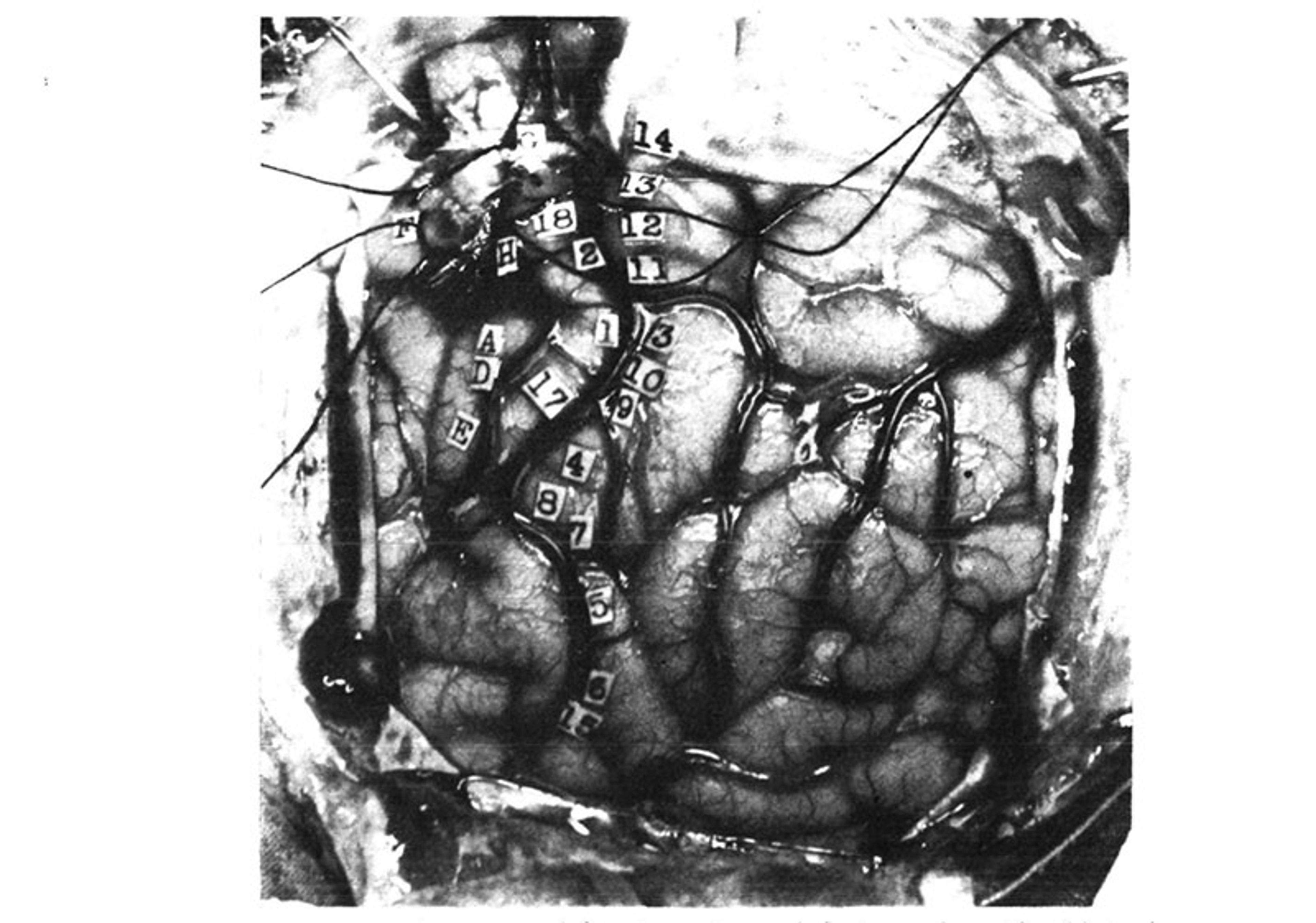
From Wilder Penfield and Edwin Boldrey’s 1937 paper . American Neurological Association
14. Tingling from the knee down to the right foot, no numbness. 13. Numbness all down the right leg, did not include the foot. 12. Numbness over the wrist, lower border, right side. 11. Numbness in the right shoulder. 3. Numb feeling in hand and forearm up to just above the forearm. 10. Tingling feeling in the fifth or little finger. 9. Tingling in first three fingers. 4. Felt like a shock and numbness in all four fingers but not in the thumb. 8. Felt sensation of movement in the thumb; no evidence of movement could be seen. 7. Same as 8. 5. Numbness in the right side of the tongue. 6. Tingling feeling in the right side of the tongue, more at the tip. 15. Tingling in the tongue, associated with up and down vibratory movements. 16. Numbness, back of tongue, mid-line. Precentral gyrus from above down: – (G) Flexion of knee. 18. Slight twitching of arm and hand like a shock, and felt as if he wanted to move them. 2. Shrugged shoulders upwards; did not feel like an attack. (H) Clonic movement of right arm, shoulders, forearm, no movement in trunk. (A) Extreme flexion of wrist, elbow and hand. (D) Closure of hand and flexion of his wrist, like an attack. 17. Felt as if he were going to have an attack, flexion of arms and forearms, extension of wrist. (E) Slight closure of hand; stimulation followed by local flushing of brain; this was repeated with the strength at 24. Flushing was followed by pallor for a few seconds. (B) Patient states that he could not help closing his right eye but he actually closed both. (C) Made a little noise; vocalisation. This was repeated twice. Patient says he could not help it. It was associated with movement of the upper and lower lips, equal on the two sides …
It is these findings that are immortalised in visual form as the homunculus, which first appeared in Penfield’s paper ‘Somatic Motor and Sensory Representation in the Cerebral Cortex of Man as Studied by Electrical Stimulation’ (1937), co-authored with Edwin Boldrey. The findings reveal that the motor and sensory cortices are organised in such a way that there is a point-for-point correspondence of body parts to specific regions of brain tissue, with adjacent body parts ‘represented’ by adjacent patches of tissue.
This organisation is referred to as ‘somatotopy’, and it is widely considered to be a fundamental principle of brain structure and function. Furthermore, Penfield’s technique, which came to be known as ‘the Montreal Procedure’, is still used today. Several years ago, for example, the violinist Dagmar Turner played her instrument throughout a neurosurgical procedure, so that the team performing the operation could remove a brain tumour without damaging the motor cortex.
I t’s also worth discussing what’s been called the ‘ her monculus’. The homunculus is a composite of localisation data that Penfield obtained from the presurgical evaluation of some 400 patients. Yet, while the homunculus clearly shows the cortical representation of male genitalia, female anatomical parts are conspicuously absent. The reasons for this are unclear. It may be because Penfield worked at a time when it was considered inappropriate to ask about or report certain sensations experienced by his female patients; because female patients felt embarrassed reporting genital sensations to male authority figures; or because Hortense Cantlie, the medical illustrator who drew the homunculus, may have been uncomfortable incorporating female genitalia into her illustrations.
Another possibility is that Penfield simply did not have enough data – just nine of the patients on which the homunculus is based were confirmed as female, only one of whom reported any genital sensations during presurgical evaluation. This was a 27-year-old woman referred to as ‘EC’, who had a tumour removed from her right sensory cortex. Before her surgery, the tumour caused spontaneous seizures that produced a tingling sensation that shifted between her left buttock, labium and breast, and, on the operating table, electrical stimulation of the sensory cortex produced a sensation in her left buttock and a twitching of her left foot.
Penfield and his colleagues thus assumed that the female genitals and breasts are represented in the same region as the male genitalia: adjacent to the representation of the foot, on the inner wall of the cortex, deep inside the longitudinal fissure separating the left and right hemispheres.
We need further investigation into the her munculus, and to fill in the rest of the female map
We still know very little about the neural representation of the female body. In Penfield’s time, there was only one other case study hinting at how the female genitalia map onto the cortex, that of an epileptic woman diagnosed with ‘erotomania’ (nymphomania) because she experienced vaginal sensations during her seizures; removal of the tumour causing the seizures relieved the patient of those symptoms.
Between then and 2011, there were only 10 other studies investigating the somatotopic organisation of female anatomical parts. These provided conflicting results, hinting at alternative locations for females: some scientists mapped sensations related to female anatomy onto the inner wall of the cortex, consistent with Penfield, but others mapped them further up, at the brain’s apex. Among some researchers, the call is on to resolve the matter with further, active investigation into the her munculus, and to fill in the rest of the female map. ‘What happens to bodily sensation during pregnancy, menopause … or after surgeries such as oophorectomy[?]’ the neuroscientist Paula Di Noto and colleagues asked in the journal Cerebral Cortex in 2012.
In the most recent such study to address the issue, published in 2022, Andrea Knop of Charité–Universitätsmedizin Berlin and colleagues used functional magnetic resonance (fMRI) to scan 20 women’s brains while stimulation was applied to their clitorises with an air-controlled vibrating membrane placed over disposable underwear just below the pubic mound, to show that the representation of the clitoris in the brain lies adjacent to that of the hips and upper legs, results that ‘provide independent confirmation for the revision of the original homunculus’.
Furthermore, the researchers found that the frequency of sexual intercourse within the 12 months prior to the scan was linked to the thickness of that particular area of the sensory cortex, with the more sexually active participants exhibiting thicker tissue. By contrast, the phase of the menstrual cycle was not associated with differences in thickness of the ‘genital field’.
T he sensory and motor strips of the cortex work together to control and coordinate limb movements. The sensory cortex contains cells that process touch and pain information, and the motor cortex contains cells that execute movements by sending signals down the spinal cord to ‘secondary’ cells that activate specific muscles.
But both regions also contain neurons that exhibit properties associated with spatial navigation. These navigation cells, called ‘place cells’, are located in a deep brain structure called the hippocampus. They were first identified in the 1970s in experiments performed on rats, which showed that individual place cells are activated only when the animal enters a specific place in its environment. Since then, researchers have discovered several other navigational cells in and around the hippocampus: head direction cells, which fire when the animal is moving in a specific direction, and grid cells, which fire periodically as the animal moves through an open space.
Two monkeys navigated a small room in a wheelchair controlled by a brain-machine interface to get food
These cells make up the brain’s global positioning system, working together to generate maps of the environment and contributing to formation of the spatial memories we use to find our way around. Recently, two groups of researchers have independently shown that this same spatial navigation system is also found in the brain’s sensory and motor regions.
In a study published in 2018, researchers at Duke University in North Carolina trained two rhesus monkeys to navigate a small room in a wheelchair controlled by a brain-machine interface in order to get food, while recording the activity of hundreds of cells with microelectrode arrays implanted into the animals’ sensory and motor cortices. Unexpectedly, they found that significant numbers of them exhibited place cell-like activity, firing only when the wheelchair was moved into a specific location.
These findings were confirmed in a 2021 study by researchers at Xinqiao Hospital in China, who recorded from the sensory cortex in foraging rats and identified neurons with the properties of place cells, grid cells and head-direction cells.
Although unexpected, the discovery of navigational cells in the sensory and motor cortices is not entirely surprising. Whereas in the hippocampus they function to generate maps and aid navigation, here they are likely to encode the position and orientation of the body within its surroundings.
T he discovery of navigational cells in the sensory and motor cortices allows us to expand our thinking about the function of these parts of the brain. Research into the somatotopic organisation of the female body suggests that the homunculus needs to be updated. At the same time, a team of researchers at the Washington University School of Medicine in St Louis is now arguing that the homunculus is entirely wrong and needs to be completely redrawn.
Evan Gordon, Nico Dosenbach and colleagues set out to replicate Penfield’s findings by using fMRI to scan the brains of seven volunteers at rest and as they performed various movement tasks, generating high-resolution brain maps for each. They then verified their results with data from three large, publicly available datasets, which between them contain brain-scanning data collected from some 50,000 people.
They found that movement of the feet, hands and face was associated with the parts of the motor cortex identified by Penfield, but that interspersed between these discrete regions were other areas that did not seem to be involved in movement at all. These other regions were thinner than the flanking regions associated with individual parts of the body, and were connected to each other, both within the same and between the two hemispheres of the brain, to form a chain running down the motor strip.
They argue that Penfield’s classic homunculus is wrong or at least spectacularly incomplete
Further investigation revealed that these areas are also strongly connected to distant brain regions involved in ‘executive’ functions such as thinking and planning, visual processing and the processing of touch, pain and internal bodily signals, and that they became active when the participants thought about moving.
The researchers propose that these areas form a network that integrates whole-body movements and anticipates them with appropriate changes in arousal, posture, breathing and heart function.
‘All of these connections make sense if you think about what the brain is really for,’ Dosenbach said in an interview. ‘The brain is for successfully behaving in the environment so you can achieve your goals without hurting or killing yourself. You move your body for a reason. Of course, the motor areas must be connected to executive function and control of basic bodily processes, like blood pressure and pain.’
In light of their findings, Gordon, Dosenbach and colleagues argue that Penfield’s classic homunculus is wrong or at least spectacularly incomplete, and needs to be radically revised to include the network they identified, which they have named the somato-cognitive action network (SCAN).
‘Penfield was brilliant, and his ideas have been dominant for 90 years … [but] once we started looking, we found lots of published data that didn’t quite jibe with his ideas, and alternative interpretations that had been ignored,’ Dosenbach said. ‘We pulled together a lot of different data in addition to our own observations, and zoomed out and synthesised it, and came up with a new way of thinking about how the body and the mind are tied together.’
W hat does this mean for neurosurgeons who use the homunculus to guide their scalpel? Performing surgery for epilepsy is extremely challenging due to the high risk of damaging the sensory or motor strips. Typically, the motor cortex generates seizures that are limited to certain parts of the body, but may spread to adjacent parts, and the non-movement regions identified by Gordon, Dosenbach and colleagues could, in theory, generate seizures that spread in unusual ways.
‘The likelihood that a seizure remains in this area without spreading to adjacent motor areas seems low, and I would expect typical [symptoms] in most situations,’ David Steven, professor of neurosurgery at Western University in London, Ontario, told me. With the brain regions intermingled, the surgery could be high risk, except in ‘the face area, which is usually safe as there is representation [on both sides of the brain].’
In practical terms, the little man in the brain still looms large. ‘For pre-surgical work-up and intra-operative decisions, it remains critical and very relevant,’ says Steven. ‘It may be oversimplified but, practically speaking, it remains essential.’
Mapping in finer detail will allow for prostheses that provide more realistic sensory feedback
Beyond the operating table, knowledge of how the body maps onto the motor cortex has been instrumental in the development of brain-machine interfaces that control prostheses to restore function to paralysed patients and amputees. These devices typically consist of a microelectrode array implanted into the motor cortex, which reads the brain activity associated with planning and executing movements and translates it into commands that can be used to control a wheelchair or robotic arm .
Early versions of these prostheses were cumbersome, but they are becoming more sophisticated by the day, and some of the newer devices can simultaneously stimulate the sensory cortex to provide sensory feedback. As well as restoring some sense of touch, this gives the user better control over the device, and can also reduce phantom limb pain that most amputees feel. Mapping the sensory homunculus in even finer detail will undoubtedly allow for prostheses that provide increasingly realistic sensory feedback to the user.
In the not too distant future, this knowledge, combined with a better understanding of brain activity underlying different types of touch, could also be used to develop the next generation of ‘haptic devices’, consisting of headsets that can precisely target the sensory cortex with small electrical or magnetic pulses to elicit realistic sensations of various kinds in any part of the user’s body.
From the future of artificial limbs to the future of gaming, the little man (and woman) in the brain may just be getting started – even if we’re still learning how they operate, in full.

Consciousness and altered states
A reader’s guide to microdosing
How to use small doses of psychedelics to lift your mood, enhance your focus, and fire your creativity
Tunde Aideyan

Family life
A patchwork family
After my marriage failed, I strove to create a new family – one made beautiful by the loving way it’s stitched together
The cell is not a factory
Scientific narratives project social hierarchies onto nature. That’s why we need better metaphors to describe cellular life
Charudatta Navare

Public health
It’s dirty work
In caring for and bearing with human suffering, hospital staff perform extreme emotional labour. Is there a better way?
Susanna Crossman

Social psychology
The magic of the mundane
Pioneering sociologist Erving Goffman realised that every action is deeply revealing of the social norms by which we live
Lucy McDonald

Stories and literature
The real Miss Julie
Victoria Benedictsson assumed a male identity, achieved literary stardom, and took her own life. Then Strindberg stole it
Elisabeth Åsbrink
IMAGES
VIDEO
COMMENTS
The human brain is a complex organ, made up of several distinct parts, each responsible for different functions. The cerebrum, the largest part, is responsible for sensory interpretation, thought processing, and voluntary muscle activity. Beneath it is the cerebellum, which controls balance and coordination. The brainstem connects the brain to the spinal cord and oversees automatic processes ...
Parts of the Brain. The brain is divided into three main parts: the cerebrum, cerebellum, and brainstem. The cerebrum is the largest part and controls thinking, learning, and emotions. ... 500 Words Essay on Human Brain Introduction to the Human Brain. The human brain, a product of millions of years of evolutionary progression, is a marvel of ...
The middle part of the brain, the parietal lobe helps a person identify objects and understand spatial relationships (where one's body is compared with objects around the person). The parietal lobe is also involved in interpreting pain and touch in the body. The parietal lobe houses Wernicke's area, which helps the brain understand spoken ...
The four lobes of the brain are regions of the cerebrum: Frontal Lobe. Location: This is the anterior or front part of the brain. Functions: Decision making, problem solving, control of purposeful behaviors, consciousness, and emotions. Parietal Lobe. Location: Sits behind the frontal lobe.
Summary. The brain connects to the spine and is part of the central nervous system (CNS). The various parts of the brain are responsible for personality, movement, breathing, and other crucial ...
The midbrain is a part of the brain stem that connects the forebrain and the hindbrain. It is involved in many functions, such as vision, hearing, movement, and arousal. Learn more about the anatomy, functions, and conditions of the midbrain at Verywell Mind, a trusted source of mental health information.
brain, the mass of nerve tissue in the anterior end of an organism. The brain integrates sensory information and directs motor responses; in higher vertebrates it is also the centre of learning.The human brain weighs approximately 1.4 kg (3 pounds) and is made up of billions of cells called neurons.Junctions between neurons, known as synapses, enable electrical and chemical messages to be ...
This area houses the brain's "gray matter," and is considered the "seat" of human consciousness. Higher brain functions such as thinking, reasoning, planning, emotion, memory, the processing of sensory information and speech all happen in the cerebral cortex. In other words, the cerebral cortex is what sets humans apart from other species.
The brain is the most complex organ in the human body. It produces our every thought, action, memory, feeling and experience of the world. This jelly-like mass of tissue, weighing in at around 1.4 ...
It processes information that it receives from the senses and body, and sends messages back to the body. But the brain can do much more than a machine can: humans think and experience emotions with their brain, and it is the root of human intelligence. The human brain is roughly the size of two clenched fists and weighs about 1.5 kilograms.
The human brain is perhaps the most complex of all biological systems, with the mature brain composed of more than 100 billion information-processing cells called neurons.[1] The brain is an organ composed of nervous tissue that commands task-evoked responses, movement, senses, emotions, language, communication, thinking, and memory. The three main parts of the human brain are the cerebrum ...
Introductory essay. Written by the educators who created Mapping and Manipulating the Brain, a brief look at the key facts, tough questions and big ideas in their field. Begin this TED Study with a fascinating read that gives context and clarity to the material. Here is this mass of jelly, three-pound mass of jelly you can hold in the palm of ...
Essay on Human Brain: Structure and Function. The nervous system of man and other group of vertebrates is divided into three main parts: 1. Central nervous system (CNS) comprising brain and spinal cord. 2. Peripheral nervous system (PNS) consisting of cranial and spinal nerves. 3.
The brain and its function. comprise a central nervous system. A brainstem, which in part relays information between the peripheral nerves and spinal. cord to the upper parts of the brain consists ...
Its made up of three parts the midbrain, pons and medulla oblongata. The cerebrum which is the largest part of the three main parts, making up about 85% of the brain. Its comprised of a left and right hemisphere. Both hemispheres controlling opposite sides of your body. The right controlling the left and the left controlling the right.
Essay about the human brain. In this paper one will learn the different parts of the brain and their functions. Although the brain isn't the largest organ of the human body it is the most complex and controlling organ. It is amazing how complicated the brain is. The brain controls every action within and out of your body.
Yongjun Wang and colleagues discuss the definition of brain health and the opportunities and challenges of future research The human brain is the command centre for the nervous system and enables thoughts, memory, movement, and emotions by a complex function that is the highest product of biological evolution. Maintaining a healthy brain during one's life is the uppermost goal in pursuing ...
The main purpose of the compartmental divisions of the brain is to promote brain activity by division of roles per compartment (Ahanger et al., 2021). Each section executes a specific function and controls some selected body parts. For effective action, the brain cannot work as a single unit due to the risk of poor coordination of impulses.
The brain plays a pivotal role in supporting cognitive functions. Examples of cognitive functions include learning, memory, and perception (Glees, 2005). The brain has several parts that play different roles in the execution of cognitive functions. Parts of the brain involved in cognition include prefrontal cortex, frontal and parietal lobes ...
Thalamus And Hypothalamus. Deep in the core area of the brain, just above the top of the brainstem, are structures that have a great deal to do with perception, movement, and the body's vital functions. The thalamus consists of two oval masses, each embedded in a cerebral hemisphere, that are joined by a bridge.
The brain then integrates the message and produces a response. Another group of nerves known as the motor neurons distribute the instructions from the brain to the all the body parts. The spinal cord is a superhighway of messages, composed of a collection of nerves going up and down the spine, transporting messages to and from the brain ...
Share. The homunculus is one of the most iconic images in neurology and neuroscience. Usually visualised as a series of disproportionately sized body parts splayed across a section of the brain, it shows how the body is systematically mapped onto the sensory and motor cortices, representing the proportion of brain tissue devoted to each part of ...
The limbic system is a primitive part of the cerebral cortex. It is made up of several parts that have a function in the everyday working of the brain. The first part is the corpus colossal. It is a band of nerve fibers that hold the right and left hemisphere together.