Animal Stem Cells—A Perspective on Their Use in Human Health

  • First Online: 10 July 2019

Cite this chapter

research on animals stem cell

  • Birbal Singh 5 ,
  • Gorakh Mal 5 ,
  • Sanjeev K. Gautam 6 &
  • Manishi Mukesh 7  

1571 Accesses

Stem cells have sparked a revolution in biomedical and veterinary medicine. The past two decades have witnessed astounding innovations in pursuit of stem cell applications in livestock production and health. Stem cells are reported from various domestic animals. The stem cells in livestock species are important candidates for genomic testing, selection, genome engineering, and developing model animals for investigating human diseases. Mesenchymal stem cells, due to the ease of attainment, pluripotency, and better proliferation activity have emerged as clinically important cells for treating injuries in pet and companion animals. Improved cell culture techniques, culture media, and supplements, insights into gene-environmental interactions may solve current bottlenecks associated with segregation, description, and applications of stem cells in livestock.

Stem cell technology is an important branch of animal reproduction and health sciences

Animal stem cells serve to enhance reproduction engineering and cell-based therapies.

This is a preview of subscription content, log in via an institution to check access.

Access this chapter

Subscribe and save.

  • Get 10 units per month
  • Download Article/Chapter or eBook
  • 1 Unit = 1 Article or 1 Chapter
  • Cancel anytime
  • Available as PDF
  • Read on any device
  • Instant download
  • Own it forever
  • Available as EPUB and PDF
  • Compact, lightweight edition
  • Dispatched in 3 to 5 business days
  • Free shipping worldwide - see info
  • Durable hardcover edition

Tax calculation will be finalised at checkout

Purchases are for personal use only

Institutional subscriptions

Arato I, Luca G, Mancuso F, Bellucci C, Lilli C, Calvitti M, Hansen BC, Milardi D, Grande G, Calafiore R (2018) An in vitro prototype of a porcine biomimetic testis-like cell culture system: a novel tool for the study of reassembled Sertoli and Leydig cells. Asian J Androl 20(2):160–165. https://doi.org/10.4103/aja.ja_47_17

Article   CAS   PubMed   Google Scholar  

Arrizabalaga JH, Nollert MU (2017) Properties of porcine adipose-derived stem cells and their applications in preclinical models. Adipocyte 6(3):217–223. https://doi.org/10.1080/21623945.2017.1312040 (Epub 2017 Mar 30. Review)

Article   PubMed   PubMed Central   Google Scholar  

Arzi B, Clark KC, Sundaram A, Spriet M, Verstraete FJM, Walker NJ, Loscar MR, Fazel N, Murphy WJ, Vapniarsky N, Borjesson DL (2017) Therapeutic efficacy of fresh, allogeneic mesenchymal stem cells for severe refractory feline chronic gingivostomatitis. Stem Cells Transl Med 6(8):1710–1722. https://doi.org/10.1002/sctm.17-0035 (Epub 2017 Jun 15)

Article   CAS   PubMed   PubMed Central   Google Scholar  

Bhartiya D, James K (2017) Very small embryonic-like stem cells (VSELs) in adult mouse uterine perimetrium and myometrium. J Ovarian Res 10(1):29. https://doi.org/10.1186/s13048-017-0324-5

Bhartiya D, Shaikh A, Anand S, Patel H, Kapoor S, Sriraman K, Parte S, Unni S (2016) Endogenous, very small embryonic-like stem cells: critical review, therapeutic potential and a look ahead. Hum Reprod Update 23(1):41–76 (Epub 2016 Sep 10. Review)

Article   PubMed   Google Scholar  

Bhat IA, T B S, Somal A, Pandey S, Bharti MK, Panda BSK, B I, Verma M, J A, Sonwane A, Kumar GS, Amarpal, Chandra V, Sharma GT (2018) An allogenic therapeutic strategy for canine spinal cord injury using mesenchymal stem cells. J Cell Physiol. https://doi.org/10.1002/jcp.27086

Bogliotti YS, Wu J, Vilarino M, Okamura D, Soto DA, Zhong C, Sakurai M, Sampaio RV, Suzuki K, Izpisua Belmonte JC, Ross PJ (2018) Efficient derivation of stable primed pluripotent embryonic stem cells from bovine blastocysts. Proc Natl Acad Sci U S A 115(9):2090–2095. https://doi.org/10.1073/pnas.1716161115 (Epub 2018 Feb 9)

Breton A, Sharma R, Diaz AC, Parham AG, Graham A, Neil C, Whitelaw CB, Milne E, Donadeu FX (2013) Derivation and characterization of induced pluripotent stem cells from equine fibroblasts. Stem Cells Dev 22(4):611–621. https://doi.org/10.1089/scd.2012.0052 (Epub 2012 Sep 28)

Brons IG, Smithers LE, Trotter MW, Rugg-Gunn P, Sun B, de Sousa Chuva, Lopes SM, Howlett SK, Clarkson A, Ahrlund-Richter L, Pedersen RA, Vallier L (2007) Derivation of pluripotent epiblast stem cells from mammalian embryos. Nature 448(7150):191–195 (Epub 2007 Jun 27)

Cao S, Wang F, Chen Z, Liu Z, Mei C, Wu H, Huang J, Li C, Zhou L, Liu L (2009) Isolation and culture of primary bovine embryonic stem cell colonies by novel method. J Exp Zool A Ecol Genet Physiol 311:368–376

Capuco AV, Choudhary RK, Daniels KM, Li RW, Evock-Clover CM (2012) Bovine mammary stem cells: cell biology meets production agriculture. Animal 6(3):382–393. https://doi.org/10.1017/S1751731111002369 (Review)

Chang LB, Peng SY, Chou CJ, Chen YJ, Shiu JS, Tu PA, Gao SX, Chen YC, Lin TK, Wu SC (2018) Therapeutic potential of amniotic fluid stem cells to treat bilateral ovarian dystrophy in dairy cows in a subtropical region. Reprod Domest Anim 53(2):433–441. https://doi.org/10.1111/rda.13123 (Epub 2017 Dec 14. Erratum in: Reprod Domest Anim. 2018 Aug; 53(4):1024)

Charepalli V, Reddivari L, Radhakrishnan S, Eriksson E, Xiao X, Kim SW, Shen F, Vijay-Kumar M, Li Q, Bhat VB, Knight R, Vanamala JKP (2017) Pigs, unlike mice, have two distinct colonic stem cell populations similar to humans that respond to high-calorie diet prior to insulin resistance. Cancer Prev Res (Phila) 10(8):442–450. https://doi.org/10.1158/1940-6207.CAPR-17-0010 (Epub 2017 Jun 2)

Article   CAS   Google Scholar  

Chen H, Ge RS, Zirkin BR (2009) Leydig cells: from stem cells to aging. Mol Cell Endocrinol 306(1–2):9–16. https://doi.org/10.1016/j.mce.2009.01.023 (Epub 2009 Feb 7. Review)

Cibelli JB, Stice SL, Golueke PJ, Kane JJ, Jerry J, Blackwell C, Ponce de León FA, Robl JM (1998) Transgenic bovine chimeric offspring produced from somatic cell-derived stem-like cells. Nat Biotechnol 16(7):642–646

Clark KC, Fierro FA, Ko EM, Walker NJ, Arzi B, Tepper CG, Dahlenburg H, Cicchetto A, Kol A, Marsh L, Murphy WJ, Fazel N, Borjesson DL (2017) Human and feline adipose-derived mesenchymal stem cells have comparable phenotype, immunomodulatory functions, and transcriptome. Stem Cell Res Ther 8(1):69. https://doi.org/10.1186/s13287-017-0528-z

Conrad S, Weber K, Walliser U, Geburek F, Skutella T (2018) Stem cell therapy for tendon regeneration: current status and future directions. Adv Exp Med Biol. https://doi.org/10.1007/5584_2018_194 (Epub ahead of print)

Google Scholar  

Cui Z, Li C, Jiang N, Zhang C, Wang Y, Gao H, Zhou Y (2018) Isolation and characterization of minipig perivascular stem cells for bone tissue engineering. Mol Med Rep 18(4):3555–3562. https://doi.org/10.3892/mmr.2018.9410 (Epub 2018 Aug 21)

Cyranoski D (2019) Japan’s approval of stem-cell treatment for spinal-cord injury concerns scientists. Nature 565(7741):544–545. https://doi.org/10.1038/d41586-019-00178-x (No abstract available)

Deng Y, Liu Q, Luo C, Chen S, Li X, Wang C, Liu Z, Lei X, Zhang H, Sun H, Lu F, Jiang J, Shi D (2012) Generation of induced pluripotent stem cells from buffalo ( Bubalus bubalis ) fetal fibroblasts with buffalo defined factors. Stem Cells Dev 21:2484–2495

Article   Google Scholar  

Dev K, Giri SK, Kumar A, Yadav A, Singh B, Gautam SK (2012a) Derivation, characterization and differentiation of buffalo ( Bubalus bubalis ) amniotic fluid derived stem cells. Reprod Domest Anim 47:704–711. https://doi.org/10.1111/j.1439-0531.2011.01947.x

Dev K, Giri SK, Kumar A, Yadav A, Singh B, Gautam SK (2012b) Expression of transcriptional factor genes ( Oct-4 , Nanog , and Sox-2 ) and embryonic stem cell-like characters in placental membrane of buffalo ( Bubalus bubalis ). J Membr Biol 245(4):177–183. https://doi.org/10.1007/s00232-012-9427-5 (Epub 2012 Apr 22)

Dolin CD, Chan MK, Basch RS, Young BK (2018) Human term amniotic fluid: a novel source of stem cells for regenerative medicine. Am J Obstet Gynecol 219(3):308–309. https://doi.org/10.1016/j.ajog.2018.05.035 (Epub 2018 Jun 2. No abstract available)

Dutton AQ, Choong PF, Goh JC, Lee EH, Hui JH (2010) Enhancement of miniscale repair in the avascular zone using mesenchymal stem cells in a porcine model. J Bone Joint Surg Br 92:169–175

Dyce PW, Wen L, Li J (2006) In vitro germline potential of stem cells derived from foetal porcine skin. Nat Cell Biol 8:384–390

Evans MJ, Kaufman MH (1981) Establishment in culture of pluripotent cells from mouse embryos. Nature 292:154–156

Evans MJ, Notarianni E, Laurie S, Moor RM (1990) Derivation and preliminary characterization of pluripotent cell lines from porcine and bovine blastocysts. Theriogenology 33:125–128

Faast R, Harrison SJ, Beebe LF, McIlfatrick SM, Ashman RJ, Nottle MB (2006) Use of adult mesenchymal stem cells isolated from bone marrow and blood for somatic cell nuclear transfer in pigs. Cloning Stem Cells 8(3):166–173

Forest MG (1983) Role of androgens in foetal and pubertal development. Horm Res 18:69–83

Fujihara M, Kim SM, Minami N, Yamada M, Imai H (2011) Characterization and in vitro culture of male germ cells from developing bovine testis. J Reprod Dev 57:355–364

Galli C, Lazzari G (2008) The manipulation of gametes and embryos in farm animals. Reprod Domest Anim 43(Suppl 2):1–7

Gugjoo MB, Amarpal, Chandra V, Wani MY, Dhama K, Sharma GT (2018) Mesenchymal stem cell research in veterinary medicine. Curr Stem Cell Res Ther. https://doi.org/10.2174/1574888x13666180517074444

Harding J, Roberts RM, Mirochnitchenko O (2013) Large animal models for stem cell therapy. Stem Cell Res Ther 4(2):23. https://doi.org/10.1186/scrt171 (Review)

Hayashi K, Hikabe O, Obata Y, Hirao Y (2017) Reconstitution of mouse oogenesis in a dish from pluripotent stem cells. Nat Protoc 12(9):1733–1744. https://doi.org/10.1038/nprot.2017.070 (Epub 2017 Aug 10)

Hikabe O, Hamazaki N, Nagamatsu G, Obata Y, Hirao Y, Hamada N, Shimamoto S, Imamura T, Nakashima K, Saitou M, Hayashi K (2016) Reconstitution in vitro of the entire cycle of the mouse female germ line. Nature 539(7628):299–303. https://doi.org/10.1038/nature20104 (Epub 2016 Oct 17)

Huang B, Xie TS, Shi DS, Li T, Wang XL, Mo Y, Wang ZQ, Li MM (2007) Isolation and characterization of EG-like cells from Chinese swamp buffalo ( Bubalus bubalis ). Cell Biol Int 31(10):1079–1088 (Epub 2007 Mar 19)

Huang B, Li T, Wang XL, Xie TS, Lu YQ, da Silva FM, Shi DS (2010) Generation and characterization of embryonic stem-like cell lines derived from in vitro fertilization buffalo ( Bubalus bubalis ) embryos. Reprod Domest Anim 45(1):122–128. https://doi.org/10.1111/j.1439-0531.2008.01268.x (Epub 2008 Dec 22)

Huang B, Li T, Alonso-Gonzalez L, Gorre R, Keatley S, Green A, Turner P, Kallingappa PK, Verma V, Oback B (2011) A virus-free poly-promoter vector induces pluripotency in quiescent bovine cells under chemically defined conditions of dual kinase inhibition. PLoS One 6(9):e24501. https://doi.org/10.1371/journal.pone.0024501 (Epub 2011 Sep 2)

Huang X, Han X, Uyunbilig B, Zhang M, Duo S, Zuo Y, Zhao Y, Yun T, Tai D, Wang C, Li J, Li X, Li R (2014) Establishment of bovine trophoblast stem-like cells from in vitro-produced blastocyst-stage embryos using two inhibitors. Stem Cells Dev 23(13):1501–1514. https://doi.org/10.1089/scd.2013.0329 (Epub 2014 Apr 10)

Iwasaki S, Campbell KH, Galli C, Akiyama K (2000) Production of live calves derived from embryonic stem-like cells aggregated with tetraploid embryos. Biol Reprod 62(2):470–475

Jang HJ, Kim JS, Choi HW, Jeon I, Choi S, Kim MJ, Song J, Do JT (2014) Neural stem cells derived from epiblast stem cells display distinctive properties. Stem Cell Res 12(2):506–516. https://doi.org/10.1016/j.scr.2013.12.012

Jia W, Yang W, Lei A, Gao Z, Yang C, Hua J, Huang W, Ma X, Wang H, Dou Z (2008) A caprine chimera produced by injection of embryonic germ cells into a blastocyst. Theriogenology 69(3):340–348 (Epub 2007 Nov 14)

Kawaguchi T, Cho D, Hayashi M, Tsukiyama T, Kimura K, Matsuyama S, Minami N, Yamada M, Imai H (2016) Derivation of Induced Trophoblast Cell Lines in Cattle by Doxycycline-Inducible piggyBac Vectors. PLoS One. 11(12):e0167550. https://doi.org/10.1371/journal.pone.0167550 . eCollection 2016

Khatri M, Richardson LA (2018) Therapeutic potential of porcine bronchoalveolar fluid-derived mesenchymal stromal cells in a pig model of LPS-induced ALI. J Cell Physiol 233(7):5447–5457. https://doi.org/10.1002/jcp.26397

Khodadadi K, Sumer H, Pashaiasl M, Lim S, Williamson M, Verma PJ (2012) Induction of pluripotency in adult equine fibroblasts without c-MYC. Stem Cells Int 2012:429160

Kurosaka S, Sigrid E, McLaughlin KJ (2004) Pluripotent lineage definition in bovine embryos by Oct-4 transcript localization. Biol Reprod 71:1578–1582

Latos PA, Hemberger M (2016) From the stem of the placental tree: trophoblast stem cells and their progeny. Development 143(20):3650–3660 (Review)

Lee EM, Kim AY, Lee EJ, Park JK, Park SI, Cho SG, Kim HK, Kim SY, Jeong KS (2016) Generation of equine-induced pluripotent stem cells and analysis of their therapeutic potential for muscle injuries. Cell Transplant 25(11):2003–2016. https://doi.org/10.3727/096368916X691691

Li M, Zhang D, Hou Y, Jiao L, Zheng X, Wang WH (2003) Isolation and culture of embryonic stem cells from porcine blastocysts. Mol Reprod Dev 65(4):429–434

Li X, Zhou SG, Imreh MP, Ahrlund-Richter L, Allen WR (2006) Horse embryonic stem cell lines from the proliferation of inner cell mass cells. Stem Cells Dev 15(4):523–531

Li JX, Zhang Y, Ma LB, Sun JH, Yin BY (2009) Isolation and culture of bovine mammary epithelial stem cells. J Vet Med Sci 71(1):15–19

Lin H, Lei J, Wininger D, Nguyen MT, Khanna R, Hartmann C, Yan WL, Huang SC (2003) Multilineage potential of homozygous stem cells derived from metaphase II oocytes. Stem Cells. 21(2):152–161

Liu Z, Screven R, Boxer L, Myers MJ, Devireddy LR (2018) Characterization of canine adipose-derived mesenchymal stromal/stem cells in serum-free medium. Tissue Eng Part C Methods 24(7):399–411. https://doi.org/10.1089/ten.TEC.2017.0409 (Epub 2018 Jun 20)

Mann A, Yadav RP, Singh J, Kumar D, Singh B, Yadav PS (2013) Culture, characterization and differentiation of cells from buffalo ( Bubalus bubalis ) amnion. Cytotechnology 65(1):23–30. https://doi.org/10.1007/s10616-012-9464-z (Epub 2012 Jul 21)

Martin G (1981) Isolation of a pluripotent cell line from early mouse embryos cultured in medium conditioned by teratocarcinoma stem cells. Proc Natl Acad Sci U S A 78:7634–7638

Martin DR, Cox NR, Hathcock TL, Niemeyer GP, Baker HJ (2002) Isolation and characterization of multipotential mesenchymal stem cells from feline bone marrow. Exp Hematol 30(8):879–886

Martinello T, Gomiero C, Perazzi A, Iacopetti I, Gemignani F, DeBenedictis GM, Ferro S, Zuin M, Martines E, Brun P, Maccatrozzo L, Chiers K, Spaas JH, Patruno M (2018) Allogeneic mesenchymal stem cells improve the wound healing process of sheep skin. BMC Vet Res 14(1):202. https://doi.org/10.1186/s12917-018-1527-8

Maruotti J, Dai XP, Brochard V, Jouneau L, Liu J, Bonnet-Garnier A, Jammes H, Vallier L, Brons IG, Pedersen R, Renard JP, Zhou Q, Jouneau A (2010) Nuclear transfer-derived epiblast stem cells are transcriptionally and epigenetically distinguishable from their fertilized-derived counterparts. Stem Cells 28(4):743–752. https://doi.org/10.1002/stem.400

Maruotti J, Muñoz M, Degrelle SA, Gómez E, Louet C, Díez C, de Longchamp PH, Brochard V, Hue I, Caamaño JN, Jouneau A (2012) Efficient derivation of bovine embryonic stem cells needs more than active core pluripotency factors. Mol Reprod Dev. 79(7):461–477. https://doi.org/10.1002/mrd.22051 (Epub 2012 May 31. Erratum in: Mol Reprod Dev. 2012 Dec; 79(12):888. Monforte, Carmen Díez [corrected to Díez, Carmen])

Meinecke-Tillmann S, Meinecke B (1996) Isolation of ES-like cell lines from ovine and caprine preimplantation embryos. J Anim Breed Genet 113:413–426

Memon IA, Sawa Y, Miyagawa S, Taketani S, Matsuda H (2005) Combined autologous cellular cardiomyoplasty with skeletal myoblasts and bone marrow cells in canine hearts for ischemic cardiomyopathy. J Thorac Cardiovasc Surg 130(3):646–653

Nagy K, Sung HK, Zhang P, Laflamme S, Vincent P, Agha-Mohammadi S, Woltjen K, Monetti C, Michael IP, Smith LC, Nagy A (2011) Induced pluripotent stem cell lines derived from equine fibroblasts. Stem Cell Rev 7(3):693–702. https://doi.org/10.1007/s12015-011-9239-5 (Erratum. In: Stem Cell Rev. 2012 Jun; 8(2):546)

Article   PubMed Central   Google Scholar  

Nixon AJ, Dahlgren LA, Haupt JL, Yeager AE, Ward DL (2008) Effect of adipose-derived nucleated cell fractions on tendon repair in horses with collagenase-induced tendinitis. Am J Vet Res 69(7):928–937. https://doi.org/10.2460/ajvr.69.7.928

Pan S, Chen W, Liu X, Xiao J, Wang Y, Liu J, Du Y, Wang Y, Zhang Y (2015) Application of a novel population of multipotent stem cells derived from skin fibroblasts as donor cells in bovine SCNT. PLoS ONE. 10(1):e0114423. https://doi.org/10.1371/journal.pone.0114423 (eCollection 2015)

Pant D, Keefer CL (2009) Expression of pluripotency-related genes during bovine inner cell mass explant culture. Cloning Stem Cells 11(3):355–365. https://doi.org/10.1089/clo.2008.0078

Pashaiasl M, Khodadadi K, Holland MK, Verma PJ (2010) The efficient generation of cell lines from bovine parthenotes. Cell Reprogram 12(5):571–579. https://doi.org/10.1089/cell.2009.0118

Pawar SS, Malakar D, De AK, Akshey YS (2009) Stem cell-like outgrowths from in vitro fertilized goat blastocysts. Indian J Exp Biol 47(8):635–642

CAS   PubMed   Google Scholar  

Piedrahita JA, Anderson GB, Bondurant RH (1990) On the isolation of embryonic stem cells: comparative behavior of murine, porcine and ovine embryos. Theriogenology 34(5):879–901

Pipino C, Mandatori D, Buccella F, Lanuti P, Preziuso A, Castellani F, Grotta L, Di Tomo P, Marchetti S, Di Pietro N, Cichelli A, Pandolfi A, Martino G (2018) Identification and characterization of a stem cell-like population in bovine milk: a potential new source for regenerative medicine in veterinary. Stem Cells Dev. https://doi.org/10.1089/scd.2018.0114 [Epub ahead of print]

Quimby JM, Borjesson DL (2018) Mesenchymal stem cell therapy in cats: Current knowledge and future potential. J Feline Med Surg 20(3):208–216. https://doi.org/10.1177/1098612X18758590

Ribitsch I, Chang-Rodriguez S, Egerbacher M, Gabner S, Gueltekin S, Huber J, Schuster T, Jenner F (2017) Sheep placenta cotyledons: a noninvasive source of ovine mesenchymal stem cells. Tissue Eng Part C Methods 23(5):298–310. https://doi.org/10.1089/ten.tec.2017.0067

Rielland M, Hue I, Renard JP, Alice J (2008) Trophoblast stem cell derivation, cross-species comparison and use of nuclear transfer: new tools to study trophoblast growth and differentiation. Dev Biol 322(1):1–10. https://doi.org/10.1016/j.ydbio.2008.07.017 (Epub 2008 Jul 22. Review)

Rosselli DD, Mumaw JL, Dickerson V, Brown CA, Brown SA, Schmiedt CW (2016) Efficacy of allogeneic mesenchymal stem cell administration in a model of acute ischemic kidney injury in cats. Res Vet Sci 108:18–24. https://doi.org/10.1016/j.rvsc.2016.07.003 (Epub 2016 Jul 5)

Ruffing NA, Anderson GB, Bondurant RH, Currie WB, Pashen RL (1993) Effects of chimerism in sheep-goat concepti that developed from blastomere-aggregation embryos. Biol Reprod 48(4):889–904

Saadeldin IM, Swelum AA, Elsafadi M, Mahmood A, Alfayez M, Alowaimer AN (2018) Cumulus cells of camel ( Camelus dromedarius ) antral follicles are multipotent stem cells. Theriogenology 15(118):233–242. https://doi.org/10.1016/j.theriogenology.2018.06.009 (Epub 2018 Jun 19)

Saito S, Ugai H, Sawai K, Yamamoto Y, Minamihashi A, Kurosaka K, Kobayashi Y, Murata T, Obata Y, Yokoyama K (2002) Isolation of embryonic stem-like cells from equine blastocysts and their differentiation in vitro. FEBS Lett 531(3):389–396

Saito S, Sawai K, Ugai H, Moriyasu S, Minamihashi A, Yamamoto Y, Hirayama H, Kageyama S, Pan J, Murata T, Kobayashi Y, Obata Y, Yokoyama KK (2003) Generation of cloned calves and transgenic chimeric embryos from bovine embryonic stem-like cells. Biochem Biophys Res Commun 309(1):104–113

Sanna D, Sanna A, Mara L, Pilichi S, Mastinu A, Chessa F, Pani L, Dattena M (2009) Oct-4 expression in in-vitro-produced sheep blastocysts and embryonic-stem-like cells. Cell Biol Int 34(1):53–60. https://doi.org/10.1042/CBI20090008

Sasaki A, Mizuno M, Ozeki N, Katano H, Otabe K, Tsuji K, Koga H, Mochizuki M, Sekiya I (2018) Canine mesenchymal stem cells from synovium have a higher chondrogenic potential than those from infrapatellar fat pad, adipose tissue, and bone marrow. PLoS ONE 13(8):e0202922. https://doi.org/10.1371/journal.pone.0202922 (eCollection 2018)

Shim H, Gutiérrez-Adán A, Chen LR, BonDurant RH, Behboodi E, Anderson GB (1997) Isolation of pluripotent stem cells from cultured porcine primordial germ cells. Biol Reprod 57(5):1089–1095

Shirazi R, Zarnani AH, Soleimani M, Nayernia K, Ragerdi Kashani I (2017) Differentiation of bone marrow-derived stage-specific embryonic antigen 1 positive pluripotent stem cells into male germ cells. Microsc Res Tech 80(4):430–440. https://doi.org/10.1002/jemt.22812 (Epub 2016 Dec 19)

Song H, Li H, Huang M, Xu D, Gu C, Wang Z, Dong F, Wang F (2013) Induced pluripotent stem cells from goat fibroblasts. Mol Reprod Dev 80(12):1009–1017. https://doi.org/10.1002/mrd.22266 (Epub 2013 Nov 27)

Song H, Li H, Huang M, Xu D, Wang Z, Wang F (2016) Big animal cloning using transgenic induced pluripotent stem cells: a case study of goat transgenic induced pluripotent stem cells. Cell Reprogram 18(1):37–47. https://doi.org/10.1089/cell.2015.0035

Sritanaudomchai H, Pavasuthipaisit K, Kitiyanant Y, Kupradinun P, Mitalipov S, Kusamran T (2007) Characterization and multilineage differentiation of embryonic stem cells derived from a buffalo parthenogenetic embryo. Mol Reprod Dev 74(10):1295–1302

Stice SL, Strelchenko NS, Keefer CL, Matthews L (1996) Pluripotent bovine embryonic cell lines direct embryonic development following nuclear transfer. Biol Reprod 54(1):100–110

Stingl J (2009) Detection and analysis of mammary gland stem cells. J Pathol 217(2):229–241. https://doi.org/10.1002/path.2457 (Review)

Sultana T, Lee S, Yoon HY, Lee JI (2018) Current status of canine umbilical cord blood-derived mesenchymal stem cells in veterinarymedicine. Stem Cells Int 15(2018):8329174. https://doi.org/10.1155/2018/8329174 (eCollection 2018. Review)

Takahashi K, Yamanaka S (2006) Induction of pluripotent stem cells from mouse embryonic and adult fibroblast cultures by definedfactors. Cell 126(4):663–676 (Epub 2006 Aug 10)

Tanaka S, Kunath T, Hadjantonakis AK, Nagy A, Rossant J (1998) Promotion of trophoblast stem cell proliferation by FGF4. Science 282(5396):2072–2075

Tesar PJ, Chenoweth JG, Brook FA, Davies TJ, Evans EP, Mack DL, Gardner RL, McKay RD (2007) New cell lines from mouse epiblast share defining features with human embryonic stem cells. Nature 448(7150):196–199 (Epub 2007 Jun 27)

Textor JA, Clark KC, Walker NJ, Aristizobal FA, Kol A, LeJeune SS, Bledsoe A, Davidyan A, Gray SN, Bohannon-Worsley LK, Woolard KD, Borjesson DL (2018) Allogeneic stem cells alter gene expression and improve healing of distal limb wounds in horses. Stem Cells Transl Med 7(1):98–108. https://doi.org/10.1002/sctm.17-0071 (Epub 2017 Oct 24)

Tripathi AK, Ramani UV, Ahir VB, Rank DN, Joshi CG (2010) A modified enrichment protocol for adult caprine skeletal muscle stem cell. Cytotechnology 62(6):483–488. https://doi.org/10.1007/s10616-010-9306-9 (Epub 2010 Sep 24)

Verma V, Gautam SK, Singh B, Manik RS, Palta P, Singla SK, Goswami SL, Chauhan MS (2007) Isolation and characterization of embryonic stem cell-like cells from in vitro-produced buffalo ( Bubalus bubalis ) embryos. Mol Reprod Dev 74(4):520–529

Vivas D, Caminal M, Oliver-Vila I, Vives J (2018) Derivation of multipotent mesenchymal stromal cells from ovine bone marrow. Curr Protoc Stem Cell Biol 44:2B.9.1–2B.9.22. https://doi.org/10.1002/cpsc.43

Wang F, Wu Z, Fan Z, Wu T, Wang J, Zhang C, Wang S (2018) The cell re-association-based whole-tooth regeneration strategies in large animal, Sus scrofa. Cell Prolif 51(4):e12479. https://doi.org/10.1111/cpr.12479 (Epub 2018 Jul 20)

Watanabe K, Ueno M, Kamiya D, Nishiyama A, Matsumura M, Wataya T, Takahashi JB, Nishikawa S, Nishikawa S, Muguruma K, Sasai Y (2007) A ROCK inhibitor permits survival of dissociated human embryonic stem cells. Nat Biotechnol 25(6):681–686 (Epub 2007 May 27)

Webb TL, Webb CB (2015) Stem cell therapy in cats with chronic enteropathy: a proof-of-concept study. J Feline Med Surg 17(10):901–908. https://doi.org/10.1177/1098612X14561105 (Epub 2014 Dec 5)

Webb TL, Quimby JM, Dow SW (2012) In vitro comparison of feline bone marrow-derived and adipose tissue-derived mesenchymal stem cells. J Feline Med Surg 14(2):165–168. https://doi.org/10.1177/1098612X11429224

Wu Z, Chen J, Ren J, Bao L, Liao J, Cui C, Rao L, Li H, Gu Y, Dai H, Zhu H, Teng X, Cheng L, Xiao L (2009) Generation of pig induced pluripotent stem cells with a drug-inducible system. J Mol Cell Biol 1(1):46–54. https://doi.org/10.1093/jmcb/mjp003 (Epub 2009 Jun 3)

Wu X, Song M, Yang X, Liu X, Liu K, Jiao C, Wang J, Bai C, Su G, Liu X, Li G (2016) Establishment of bovine embryonic stem cells after knockdown of CDX2. Sci Rep 20(6):28343. https://doi.org/10.1038/srep28343

Wu Z, Wang F, Fan Z, Wu T, He J, Wang J, Zhang C, Wang S (2019) Whole tooth regeneration by allogeneic cell reassociation in pig jawbone. Tissue Eng Part A. https://doi.org/10.1089/ten.tea.2018.0243 (Epub ahead of print)

Xie B, Qin Z, Huang B, Xie T, Yao H, Wei Y, Yang X, Shi D, Jiang H (2010) In vitro culture and differentiation and of buffalo ( Bubalus bubalis ) spermatogonia. Reprod Domest Anim 45:275–282

Yadav PS, Kues WA, Herrmann D, Carnwath JW, Niemann H (2005) Bovine ICM derived cells express the Oct-4 ortholog. Mol Reprod Dev 72(2):182–190

Yadav RP, Yadav PS, Nanda T, Singh I (2008a) Isolation and culture of stem cells like cells from buffalo amnion. In: First international stem cell submit-2008, held at IIT, Chennai, 14–16 Nov 2008

Yadav PS, Tokas J, Sharma RK, Singh I, Sethi RK (2008b) Buffalo amniotic fluid, umbilical cord matrix, and early foetal explants as possible source of adult stem cells. In: IX annual conference of Indian society of animal genetics and breeding, held at NASC Complex, Delhi, July 3–4, 2008

Yadav PS, Mann A, Singh V, Yashveer S, Sharma RK, Singh I (2011) Expression of pluripotency genes in buffalo ( Bubalus bubalis ) amniotic fluid cells. Reprod Domest Anim 46(4):705–711. https://doi.org/10.1111/j.1439-0531.2010.01733.x (Epub 2010 Dec 30)

Yadav PS, Singh RK, Singh B (2012) Animal fetal stem cells-potential health applications. Agric Res 1:67–77

Yan L, Lei L, Yang C, Gao Z, Lei A, Ma X, Dou Z (2008) Isolation and cultivation of goat embryo stem cells. Sheng Wu Gong Cheng Xue Bao 24:1670–1676

Yang Z, Liu J, Liu H, Qiu M, Liu Q, Zheng L, Pang M, Quan F, Zhang Y (2013) Isolation and characterization of SSEA3(+) stem cells derived from goat skin fibroblasts. Cell Reprogram 15(3):195–205. https://doi.org/10.1089/cell.2012.0080 (Epub 2013 May 13)

Yang J, Zhao Q, Wang K, Ma C, Liu H, Liu Y, Guan W (2018) Isolation, culture and biological characteristics of multipotent porcine tendon-derived stem cells. Int J Mol Med 41(6):3611–3619. https://doi.org/10.3892/ijmm.2018.3545 (Epub 2018 Mar 7)

Yu S, Zhang P, Dong W, Zeng W, Pan C (2017) Identification of stem leydig cells derived from pig testicular interstitium. Stem Cells Int 2017:2740272. https://doi.org/10.1155/2017/2740272 (Epub 2017 Jan 24)

Download references

Author information

Authors and affiliations.

ICAR-Indian Veterinary Research Institute, Regional Station, Palampur, India

Birbal Singh & Gorakh Mal

Department of Biotechnology, Kurukshetra University, Kurukshetra, Haryana, India

Sanjeev K. Gautam

Department of Animal Biotechnology, ICAR-National Bureau of Animal Genetic Resources, Karnal, Haryana, India

Manishi Mukesh

You can also search for this author in PubMed   Google Scholar

Corresponding author

Correspondence to Birbal Singh .

Rights and permissions

Reprints and permissions

Copyright information

© 2019 Springer Nature Switzerland AG

About this chapter

Singh, B., Mal, G., Gautam, S.K., Mukesh, M. (2019). Animal Stem Cells—A Perspective on Their Use in Human Health. In: Advances in Animal Biotechnology. Springer, Cham. https://doi.org/10.1007/978-3-030-21309-1_24

Download citation

DOI : https://doi.org/10.1007/978-3-030-21309-1_24

Published : 10 July 2019

Publisher Name : Springer, Cham

Print ISBN : 978-3-030-21308-4

Online ISBN : 978-3-030-21309-1

eBook Packages : Biomedical and Life Sciences Biomedical and Life Sciences (R0)

Share this chapter

Anyone you share the following link with will be able to read this content:

Sorry, a shareable link is not currently available for this article.

Provided by the Springer Nature SharedIt content-sharing initiative

  • Publish with us

Policies and ethics

  • Find a journal
  • Track your research

U.S. flag

An official website of the United States government

The .gov means it’s official. Federal government websites often end in .gov or .mil. Before sharing sensitive information, make sure you’re on a federal government site.

The site is secure. The https:// ensures that you are connecting to the official website and that any information you provide is encrypted and transmitted securely.

  • Publications
  • Account settings

The PMC website is updating on October 15, 2024. Learn More or Try it out now .

  • Advanced Search
  • Journal List
  • Scientifica (Cairo)
  • v.2016; 2016

Logo of scientifica

Advances in the Use of Stem Cells in Veterinary Medicine: From Basic Research to Clinical Practice

Melissa medeiros markoski.

Laboratório de Cardiologia Molecular e Celular, Fundação Universitária de Cardiologia/Instituto de Cardiologia, Princesa Isabel Avenue 370, 90620-001 Porto Alegre, RS, Brazil

Today, several veterinary diseases may be treated with the administration of stem cells. This is possible because these cells present a high therapeutic potential and may be injected as autologous or allogenic, freshly isolated, or previously cultured. The literature supports that the process is safe and brings considerable benefits to animal health. Knowledge about how adult stem cells modulate the molecular signals to activate cell homing has also been increasingly determined, evidencing the mechanisms which enable cells to repair and regenerate injured tissues. Preclinical studies were designed for many animal models and they have contributed to the translation to the human clinic. This review shows the most commonly used stem cell types, with emphasis on mesenchymal stem cells and their mechanistic potential to repair, as well as the experimental protocols, studied diseases, and species with the highest amount of studies and applications. The relationship between stem cell protocols utilized on clinics, molecular mechanisms, and the physiological responses may offer subsidies to new studies and therefore improve the therapeutic outcome for both humans and animals.

1. Introduction

In the last 20 years, considerable attention has been given to the research about the biology of stem cells. As a result, there was a significant increase in the understanding of its characteristics and, at the same time, the therapeutic potential for its application in different areas [ 1 – 4 ]. While in humans the utilization of these cells is still considered experimental (except in bone marrow transplants for the treatment of hematological diseases and skin regeneration [ 5 – 7 ]), in veterinary medicine the number of animals already treated provides a substantial basis for assessing the effectiveness of cell therapy in the treatment of a large number of diseases [ 8 , 9 ]. However, in general, the therapeutic issues involving the use of stem cells to regenerate tissue still have not been fully understood.

Almost all animal tissues may be repaired or regenerated by the direct action of stem cells [ 10 ], which presents a high potential for multiplication and differentiation [ 11 ]. In this way, a huge effort has been made for understanding the mechanisms by which adult stem cells are able to perform the function of tissue renewal, as well as the conditions that support these processes in organisms affected by diseases. Progressively, adult stem cells from different sources, mainly bone marrow and adipose tissue, have been used for treatment of animal diseases around the world [ 12 , 13 ]. In this context, the mesenchymal stem cells (MSC), derived from the mesoderm and neuroectoderm [ 14 ] and distributed in all vascularized adult tissues (such as adipose tissue, skin, heart, brain, vessels, bones, and cartilage [ 15 ]), present an important regenerative capacity.

The MSC have the natural ability for multipotency, being capable of generating new cells of tissues derived from this germ layer. These cells, by action of growth factors and hormones, acquire morphophysiological aspects pertinent to their location within the body (the niche ) [ 16 ]. As far as stem cells drive the different lines within the tissues, these new cells have limited proliferative capacity, being named progenitor cells , which in turn are able to differentiate into fewer cell types when compared to the MSC. The MSC and progenitor cells are the main cell types responsible for tissue repair and maintenance in situations of malfunction or injury [ 17 , 18 ], responding to specific stimuli [ 19 ]. Because of this characteristic, these cells have been isolated and used in cell therapy protocols around the world.

In the veterinary field, the MSC, isolated from bone marrow or adipose tissue, through minimal manipulation, have been applied for treating tendon, ligament injuries, and joint diseases, with significant clinical relevance in horses and dogs in orthopedic conditions [ 9 ]. However, controlled and well-designed studies of the basic biologic characteristics and properties of these cells are still necessary [ 20 ]. In this context, this review will focus on the approaches in the field of veterinary diseases that may be treated through the use of stem cells, emphasizing the protocols that have been more often used in companion, working, and farm animals, and the cell types applied. Besides, the molecular and physiological mechanisms of the regenerative process modulated by MSC and how the benefits obtained in veterinary treatments may be translated to human health will be discussed.

2. What Is the Mesenchymal Stem Cell and Why Do We Use It?

2.1. msc potential for proliferation, differentiation, and tissue regeneration.

Despite its potential for plasticity (ability to differentiation) and although cell lines can be successfully derived in vitro [ 21 ], the use of embryonic stem cells in medicine is still controversial: in humans, mainly because there are ethical and religious issues; in other mammals, because this population is not yet thoroughly exploited; and in both cases there is the eminent possibility of teratoma formation in vivo [ 22 ] and evidence of genome instability caused by in vitro passaging [ 23 ]. In this context, the embryonic stem cells have been used in preference to deriving lineages in vitro , and the induction of pluripotency in adult stem cells has been explored, which resulted in the induced-pluripotent stem cell (iPS), which in turn is emerging as the focus of many studies and some cell therapy protocols [ 24 , 25 ]. On the other hand, as the embryonic cells differentiate the tissues derived from the three germ layers in vivo , the plasticity and potency ability of the generated cell lines is diminished inside the layers. In this way, the mesoderm, germ layer that develops to the muscles, circulatory system, urinary and reproductive tracts, bones and cartilage, connective tissue, and bone marrow, has a pool of self-stem cells, which are named mesenchymal stem cells (MSC, as seen above). Conceptually, MSC are cells that display self-renewal capacity (in this case, performing asymmetric cell division, generating undifferentiated cells, and keeping the “stem” capacity) and have potential for differentiation into other cell types [ 19 ]. These will be influenced by the niche, the environment necessary for adult stem cells receiving “information,” arising from processes of cell signaling (autocrine, paracrine, and endocrine or intracellular), to activate their mechanisms of cell proliferation and differentiation. This information is prevenient from cell-cell interactions between stem cells, as well as interactions between stem cells and neighboring differentiated cells, interactions between stem cells and adhesion molecules, extracellular matrix components, oxygen tension, growth factors, cytokines, and the physicochemical nature of the environment [ 16 , 26 ]. The signals emitted by the niche can lead the MSC to assume different “behaviors” [ 27 ], depending on the necessity ( Figure 1 ).

An external file that holds a picture, illustration, etc.
Object name is SCIENTIFICA2016-4516920.001.jpg

The influence of MSC on the niche. According to the signaling factors that the MSC are exposed, different decisions may be taken, where the main ones are those that are involved in proliferation and cellular differentiation. The MSC capture and send molecular signals, which change the niche, either by modulating the immune system as providing mechanisms for tissue repair effectors, involving since activation of cell homing, cell apoptosis, induction of the formation of new blood vessels, and the healing process.

Adult stem cells, mainly the MSC compartment, are present in all tissues and organs, and its primary function is the replacement of dead cells in the physiological cell renewal processes [ 28 ]. In addition, they may also substitute dead cells in pathological situations, such as ischemia, inflammation, or trauma. Adult stem cells are sources for damaged tissue repair, as they are ready to mobilize in response to injury signaling or pathological conditions [ 29 ]. The way by which niche is able to orchestrate stem cells to understand and respond to signals is quite challenging from the point of view of molecular mechanisms, development, and aging [ 30 ]. Besides, one of the ways to reproduce these signals is based on the release of a class of small molecules able to bind to specific receptors on the surface of stem cells and immune system's cells, called chemokines. In this way, as soon as the contact occurs between the ligand and the receptor and the higher the number of ligands is, the higher the activation of receptors will be and the greater the favoritism to a positive allosteric gradient is [ 31 ]. Thus, the cell assumes signaling pathways that trigger, in addition to migration, a focused cellular response, leading to proliferation or cell differentiation. The more favorable the allosteric gradient is, the better and faster the “decision” by the stem cell will be, which is receiving different signals to perform different functions. Therefore, regarding the need of cell replacement, the type which best responds to the chemotactic signal is the MSC [ 32 ]. The perception of this pattern is related to the activation of a specific cellular signaling axis and its response (migration, proliferation, and differentiation), a process named cell homing , the essential mechanism for the effectiveness of cell therapy.

Once an injury occurs, cell homing is activated in order to promote the tissue repair. The injured tissue (inflammation), once in an ischemic state, through deprivation of nutrients, generates a hypoxia profile that induces the activation of the hypoxia-inducible factor-1 (HIF-1), which causes the release of cytokines, such as the stromal-derived factor-1 (SDF-1) [ 33 ] and the vascular endothelial growth factor (VEGF). These cytokines are locally recognized by the cells and in the blood vessels they help them to cross the tissue layer (transmigration) in order to model the extracellular matrix and activate the necessary cell differentiation. At the same time, the lesions activate the immune response and the consequent release of reactive oxygen species [ 34 ], which influences the cell homing. In this context, the injured tissue may comprise some thousands of cells. Further, the lesions may be caused by disease, trauma, excessive physical training [ 35 ], undergoing the influence of hypoxia, physical or chemical agents (with or without therapeutic purposes), infectious agents, immune reactions, disease or genetic disorders, and/or nutritional disorders [ 36 , 37 ]. Thus, once the injury occurs the homing mechanism is readily activated, involving the entire immune system, and there occurs intense mobilization of adult stem cells, potentially the hematopoietic stem cells (HSC), MSC, and other resident tissue progenitor cells (from vascular endothelium, muscles, heart, liver, kidneys, bones, epithelium, etc.) [ 10 , 29 ]. Consequently, it is important to understand that adult stem cells, particularly tissue-specific isolated or, in contrast, used to regenerate a particular tissue, should be able to recognize the homing orientation to be directed to the lesion site and perform the appropriate repair. Among the MSC sources, the bone marrow and adipose tissue [ 38 ] are still the most widely used in therapeutics, although new potential tissues (including synthetic) are being explored.

2.2. Procurement and Cultivation of MSC in the Laboratory

Although more conventionally isolated from the bone marrow, MSC are more abundant in adipose tissue. Identified in the vascular fraction (SVF) of this tissue, MSC may be cultivated producing purified populations with high potential of differentiation and secretion of bioactive factors [ 39 ]. The combination of these properties has driven the adipose tissue derived MSC (ADSC) research in the last decade, with particular interest in cell therapy and tissue engineering. Recently, the criteria for definition of this cell type have been established by the International Society for Cellular Therapy [ 40 ], being similar to those used for bone marrow MSC.

In order to be applied in human or veterinary clinic, stem cells should be “easily” obtained and must be minimally handled, in contemplation to avoid the risk of any kind of contamination. Commonly, bone marrow is collected by aspiration biopsy puncture in the femur of dogs and cats and in the sternum of horses [ 41 ]. The samples collected are sent to the laboratory and in appropriated conditions are centrifuged for separation of mononuclear layer. The mononuclear fraction can be prepared in syringes for direct application in the patient or submitted to cultivation for MSC establishment. The cultivation of these cells follows standard cell culture conditions, that is, incubation in a commercial culture medium (usually supplemented with a serum of animal origin to provide a source of growth factors and amino acids), antibiotics, and strictly controlled temperature and humidity (usually using 37°C and 5% CO 2 ). The cells remain under these conditions in special bottles where, through subsequent proliferation and population doubling, they expand covering the area of cultivation. Thus, the MSC are characterized by behaving cellular monolayer and display fibroblast aspect in phase-contrast microscope [ 42 ]. At each time that the expansion is about to fill the entire area, cells are enzymatically collected through trypsin or collagenase and recultivated. This process is named cell passage and is crucial to remove other cell types from culture and indirectly measure the senescence. Usually, after the 4th passage, which can take some weeks, depending on the culture conditions and of the donor organism (healthy or not, young or old, etc.) [ 43 ], the MSC are ready for clinical use. Although the healthy stem cell reaches senescence only after more than 10 passages in culture, the prolonged cultivation may interfere with its ability to differentiation [ 44 ]. Additionally, MSC are also able to go through the process of freeze/thaw (cryopreservation) while keeping the stem features [ 45 ].

The adipose tissue is usually collected aseptically from the inguinal region in dogs and cats and the dorsal surface of the gluteus maximus in horses and then forwarded to the laboratory. Our research group, in order to experimentally analyze the angiogenic response to induction of ischemia by myocardial infarction in minipigs, used the submental fat [ 46 ]. In this tissue, stem cells are embedded in extracellular matrix of SVF. The isolation of stem cells derived from adipose tissue is a process based initially on mechanical fragmentation and, subsequently, chemically through digestive enzymes [ 47 , 48 ]. Mechanical fragmentation is performed using sterile surgical material such as tweezers, scalpel blades, and scissors. The next step, chemical fragmentation, is made by using enzymes such as collagenase, which digest the tissue releasing cells. The resulting mixture, comprised of adipose tissue and enzyme solution, is incubated in physiological temperature to enable enzyme activity and, after the incubation period, the already fragmenting tissue is centrifuged. The final product is the SVF, which precipitates at the bottom of the tube.

The SVF may be prepared in syringes and injected in patients or placed in culture for obtaining, through in vitro proliferation, the ADSC population. The option to expand in vitro allows, in a few weeks, the emergence of a more homogeneous population of stem cells derived from adipose tissue. In the same way as for MSC, for application in patients, ADSC are collected and placed in syringes with saline solution, being ready for use [ 49 ]. Because of its easy collection, abundance of stem cells, and facility of expansion, adipose tissue has been used with much greater frequency as a source of cells for veterinary cell based therapy [ 38 , 49 – 51 ].

As for the choice between bone marrow or adipose tissue stem cells, mononuclear fraction, or SVF or after expansion in culture (bone marrow MSC or ADSC, resp.), the decision concerning the optimal condition for its use depends on a number of considerations. The best application will consider the type of pathology, age, and medical characteristics of the patient and the emergency of treatment (considering cell homing, these conditions would be the same for humans or other mammals). The freshly collected fraction, in addition to MSC, contains several other types of cells, as well as signaling molecules like cytokines and growth factors [ 41 ]. This whole collection of cellular components and biomolecules has the role in accelerating the regeneration process. Considerations regarding these mechanisms, by which the stem cell operates, and how to modulate them might be extremely beneficial from a therapeutic point of view.

2.3. Understanding the Paracrine Effects

The MSC are cells that secrete factors and cytokines that exert influences on tissue repair. Accordingly, many of these molecules are recognized as paracrine agents, which have the function to supply the need of a group of adjacent cells by their influence on activation (or inactivation) of receptors and intracellular pathways without compromising other cells of the body. Under the condition of hypoxia, once the homing is activated, MSC can release, in addition to SDF-1 and VEGF, fibroblast growth factors (FGF) 2 and 7, hepatocyte growth factor (HGF), angiopoietin-1, transforming growth factor-beta (TGF- β ), matrix metalloproteinase-9 (MMP-9), tumor necrosis factor-alpha (TNF- α ) and interleukin-1 (IL-1) and interleukin-6 (IL-6), and others [ 52 – 54 ].

After activation of the SDF-1 by injury, performing or not the cell therapy, there is recruitment of adult stem cells (bone marrow or tissue residents). The stronger the signal (usually in the form of a positive allosteric gradient), the more efficient the cellular response. For this to occur, it is essential that the cell is able to express the surface receptors for the signs and, once recognizing the “command to repair,” activates transduction pathways for chemotaxis. Thus, the SDF-1 binds to the specific membrane receptor CXCR4 and activates the MAPK, Akt, PKC, PI3K, and NF κ B pathways [ 55 ]. These signaling molecules cause activation of cellular proliferation, cytoskeletal reorganization, and induction of other cytokines, particularly interleukins.

The activation of the angiogenic process is crucial for the formation of new blood vessels, improving the nutrition of injured tissues and consequently contributing to the recovering of ischemic areas. Whether after hypoxia or establishment of the inflammatory process, both VEGF and angiopoietins (responsible for the vessel maturation) have their expression induced [ 56 ] and activate endothelial progenitor cells that will constitute new vessels. A study in pigs with induced proctitis showed that repeatedly injected MSC were able to modulate the expression of VEGF and its receptor, in addition to angiopoietins and FGF 2 [ 57 ]. Further, cell therapy protocols targeting the induction of angiogenesis are well studied for muscle tissues and limb ischemia and, mainly, in cardiovascular diseases. However, it is very important that the use of therapies able to induce angiogenesis observes the presence of neoplasia, which is very common in dogs and cats of advanced age [ 58 ]. The process of tissue nutrition is also very important as regards the modulation of extracellular matrix, which is the basis of tissue and cellular organization.

To understand the relationship between injuries of skin, muscle, bone, and cartilage and the repair potential offered by MSC, it is necessary to have a deeper insight about the functions of the extracellular matrix. The extracellular matrix, whose main function is tissue support (through the “fixing” of cells in tissues and to each other), is organized with fibrous and fluid elements. The central fluid element is composed by the glycosaminoglycans, which by its hygroscopic capacity, form glycoconjugates that provide resistance to compression forces, acting as lubricant for the joints and tendons. The glycosaminoglycan matrices regulate the passage of molecules through the extracellular space, participating in the maintenance of chemotactic gradient blocking, stimulating or guiding the migration and cell dispersion, and, in this way, influencing the stem cells' repair function [ 59 ]. These molecules are the basis of the cartilage. On the other hand, the fibrous elements are structural proteins, such as collagen, which forms flexible and inelastic fibers with great tensile strength. The extracellular matrix is regulated by a specific family of proteins, the matrix metalloproteinases (MMPs).

The MMPs cause degradation of protein components of the extracellular matrix and, with their specific inhibitors (TIMPs), these molecules are able to modulate the stem cells homing. Bhoopathi et al. showed that the inhibition of MMP-2 was able to inactivate the tropism of MSC towards tumor, for its influence on the SDF-1/CXCR4 axis, in mice presenting medulloblastoma, in addition of induction of cytokine profile changes [ 60 ]. Therefore, it should be noted that the inflammatory process is extremely active on the structure and function of the extracellular matrix regarding, for example, the changes in cartilage and alterations on its biomechanics, as those occur in cases of osteoarthritis [ 61 ]. Besides, MSC have great ability to cause healing processes for their influence on inflammatory proteins and, consequently, on extracellular matrix-forming molecules, as collagen [ 62 ] and fibronectin [ 63 ]. On this basis, the combination of MMPs and MSC emerges as a “double-edged sword”: the first recruits the second that modulates the first, and together they may act on repair of tendon injury and osteoarthritis. In this context, the ability of the MSC to influence the environmental repair is intimately related to its immunomodulatory role.

2.4. MSC Immunomodulation Potential

The immunomodulation function of MSC is related to its ability to repair, performed throughout the life of the organism. This characteristic also results in tolerance by the recipient's immune system allowing the use of genetically different donor cells (allogeneic) and is greatly appreciated in cell transplantation protocols [ 64 ]. The immunomodulatory capacity of MSC seems to be related to its interaction with T-CD4 and T-CD8 cells and their proinflammatory mediators. This class of stem cell reverses the inflammatory sign through the downregulation of the secreted mediators and activating anti-inflammatory cytokines [ 65 , 66 ]. In fact, T-cells are directly involved in graft versus host disease, as demonstrated by Polchert et al., in response to IFN- γ , which activates TGF- β but had no effect on inducing IL-10; MSC increase suppression and limit Th1 responses [ 64 ]. Also, Chiesa et al. showed that the MSC were able to prevent the presentation of antigens by dendritic cells to lymphocytes, as well as their migration to lymph nodes (activation location) [ 67 ]. In vivo administrated MSC generated a significant downregulation of CCR7 and CD49d β 1, two molecules involved in the dendritic cells homing to lymphoid organs.

The ability to inhibit the proliferation of stimulated T cells in vitro has been well described for MSC in a large number of nonhuman species. In dogs, MSC were able to increase IL-6 and TGF- β and decrease TNF- α [ 68 ]. In chickens, the inhibition of T-lymphocytes by MSC was correlated to nitric oxide production [ 69 ]. A guinea-pig model for acute colitis showed that bone marrow MSC, as compared to adipose tissue stem cells, appeared more effective in the attenuation of plexitis, reduction in choline acetyltransferase immunoreactivity, and consequent lymphocyte infiltration on the level of the myenteric ganglia [ 70 ]. Carrade et al. pointed out that in horses, similar to what happens in humans and rodents, once stimulated, MSC of all tissue types decreased lymphocyte proliferation, increased prostaglandin E2 and IL-6 secretion, and decreased production of TNF- α and IFN- γ [ 71 ], as already seen previously. Similar results and mechanisms may be observed in other species [ 66 ]. Haddad and Saldanha-Araujo also discussed the main mechanisms through which MSC immunosuppress T-cells and their response, which focuses on cell-cell contact, secretion of soluble factors, and regulatory T-cell generation [ 72 ]. According to the authors, surface markers and toll-like receptors of MSC (isolated from different sources) and their induced-cytokines counterparts may influence the inflammatory microenvironment (niche) and also the immunosuppression process.

All the information concerning the best organic response must be gathered in time to cell therapy application, as well as which cell type, the delivery route, and the number of applications. Other than that, the patient health state must be well evaluated in order for the stem cell homing to be effective.

3. Cell Therapy Applications in Veterinary Medicine

In veterinary medicine, since the early 2000s, cell therapy is a clinical reality, where the first applications were intended for the treatment of tendon injuries in horses [ 73 ]. Several companies worldwide offer the service of isolation of bone marrow mononuclear cells, bone marrow MSC, or ADSC for treatment primarily of companion animals [ 8 ] and horses [ 13 ]. Although there is still need for randomized and controlled clinical studies, thousands of animals already treated in the world enabled an evaluation of the effectiveness of this therapeutic procedure. Mononuclear fraction and MSC have been employed mainly in veterinary medicine for treating tendon and ligament injuries and joint diseases in horses and other species, with minimal manipulation of cells [ 9 ]. As will be discussed below, studies and clinical trials show that autologous bone marrow cells have an important therapeutic potential and present clinical benefits in horses and dogs in orthopedic conditions. Furthermore, the number of studies using MSC or ADSC has grown in treatment protocols, with or without the use of biomaterials as scaffolds [ 50 ].

3.1. Bench to Bedside

Cell therapy in dogs, cats, and horses depends on the veterinarian's contact with the laboratory that isolates, cultivates, and prepares cells for application. At that point, veterinarians and laboratory professionals must decide together what the best form of treatment is: fraction directly collected from the patient (bone marrow mononuclear fraction or SVF) or cultured cells (MSC or ADSC, resp.), autologous or allogenic donor. After the decision about the cell type, the professionals organize the collection and sending of the cells. If the option is to use the mononuclear fraction or SVF, the veterinarian receives a kit from the lab, makes the aseptic collection of bone marrow or fatty tissue, and sends the material back to the lab. Once received, the isolation of cells takes 2-3 hours. Due to viability and cell behavior, for horses (e.g.) it is recommended that the cells are applied within 24 hours after preparation and the use of large bored needles is also recommended [ 74 ]. Considering that the number of isolated cells is directly related to the size (mass) and quality of the tissue (either adipose tissue or bone marrow), the quantity applied is also variable, showing an absence of specific standardization to each species or disease. In accordance with the protocols described below, some injected volumes will be presented. Figure 2 outlines some cell types and applications in the veterinary clinic.

An external file that holds a picture, illustration, etc.
Object name is SCIENTIFICA2016-4516920.002.jpg

Cell therapy applications in veterinary medicine. Companion animals (pets), due to genetic factors, degenerative processes, and inadequate diet, and animals used in sports, such as running and jumping, are subject to several kinds of injuries, which may affect primarily the muscle tissue, causing pain, cartilage wear in joints and spinal disks, tendonitis, fractures, and bone degeneration. Clinical applications and protocols are based on the use of adult stem cells, isolated from fresh bone marrow or adipose tissue, or expanded from these tissues in laboratory. The derived cells, mesenchymal stem cell (MSC), have a high therapeutic capacity.

3.2. Animals in Sport

Although there are a large number of sports involving animals, such as hunting, fighting, and bullfighting, it is in the racing modalities that cellular therapy protocols are more commonly applied. That is because this modality is the one that causes most injury to tendons, joints, cartilage, and bones in horses. For this veterinary class, jumping is also considered as an activity that causes those kinds of injuries. Beyond the problem of animal health, the injuries, in turn, generate considerable economic problems, since horses are animals that involve significant costs to their physical training and competitiveness. Due to the easy collection and effectiveness, the largest number of protocols aimed at administration of cell therapy for horses was based on the use of bone marrow mononuclear fraction [ 75 , 76 ]. More recently, probably through the search of strategies that might ensure even better results, other cell types began to be investigated.

Initially, after exhausting training, even if there is no cartilage or bone injury, the animal may be affected by muscle aches, which hamper its locomotion and can exacerbate inflammatory processes [ 34 ]. Considering the musculoskeletal injuries, Torricelli et al. showed that racing horses who were given 1–4 × 10 6 mononuclear bone marrow cells, combined with autologous platelet-rich plasma, applied directly onto the lesion (located by ultrasonography), showed muscle regeneration after 12 months of follow-up [ 13 ]. Thirty animals were submitted to the cell therapy, where 28 were able to return to racing, showing that the protocol has generated noticeable improvements. Further, since MSC have the facilitated potential to differentiate in cell types of these tissues, they have been recently pointed out as promising candidates for cell therapy and regenerative medicine to the equine patient undergoing musculoskeletal injuries [ 38 ]. Soft tissue injuries of the musculoskeletal system are common in humans and animals, especially athletes, and lead to considerable morbidity in both patient populations. Fortunately, these lesions appear as good targets for the effectiveness of cellular therapy, mainly with MSC.

Tendons are fibrous structures with function of connective tissue, with intertwined collagen fibers, and are attached to bones by annular ligaments. The recovery of damaged ligaments and tendons occurs with large influx of cells and formation of new fibrous tissue, but with lower biomechanical property, leading to performance fade, as well as increased risk of new injury. It was also pointed out that tendon-derived stem cells , able to differentiate into other cell types, such as muscle or fat, present in the injury could be liable to aggravate the pathological situation and cause chronic tendinopathy [ 77 ]. Horses and dogs are among the most clinically affected animals, with naturally occurring tendinopathies with histopathologic similarities to those observed in humans when analyzed by MRI and ultrasonography [ 8 ]. The repetitive stress injuries of the digital flexor tendon are common in racing and jumping horses [ 73 ].

Due to the high presence of collagen, the injury promptly induces activation of the homing mechanism, which allows the tendon repair by stem cells injection. Moreover, there are an increasing number of experimental protocols describing improved outcome after the use of a combination of stem cells and integrated genes to stimulate the tendon regenerative process [ 78 ]. The principal growth factors evaluated include bone morphogenetic factor (BMF), platelet-derived growth factor (PDGF), FGF, VEGF, and insulin-like growth factor 1 (IGF-1) [ 78 , 79 ]. Tetta et al. observed that exosomes (extracellular vesicles involved in cell-cell communication) released by the MSC could potentially secrete anti-inflammatory factors, which would be very efficient to the tendinous repair [ 80 ]. Filomeno et al. showed that adult stem cells treated with platelet-rich plasma could also be promising for veterinary and human trials [ 77 ]. In this context, horses submitted to the application of 1 × 10 7 ADSC combined with autologous platelet concentrates were able to prevent tendonitis progression, in a follow-up of 16 weeks, showing greater organization of collagen fibers, and decreasing in the inflammatory infiltrate when these animals were compared to the control group (PBS-treated) [ 73 ].

In the case of soft tissue regeneration, as muscle and tendons, it is also important to note that, in addition to the fiber organization, the formation of new vessels is quite important for their nutrition. Thus, therapy with stem cells must be able to activate local homing to induce the production of angiogenic factors such as VEGF and angiopoietins. In fact, trophic factors secreted by equine MSC were able to induce angiogenesis in vitro through activation of VEGF [ 79 ]. However, intrinsically, angiogenesis may elicit more inflammation because it makes the tissue more permissive to the migration of cells of the immune system. Therefore, even if shown as active agents in development of degenerative tendinopathy, the tendon-derived stem cells are being studied and were pointed out to proliferate more quickly than tenocytes in culture, and when implanted in vivo these cells exhibit the ability to regenerate tendon-like tissue [ 77 ]. The authors also discussed that the degenerative process maintenance may be explained by the fact that tendon stem cells might be influenced by early senescence, caused during the large inflammatory state associated with tendinopathy establishment.

Through the use of MSC, derived from bone marrow or fatty tissue, therapeutic protocols are a viable reality, but because tendons of racehorses are a permanent problem new approaches have been studied. In this way and, as a manner to keep the cells at the site of injury and optimize repair, biomaterials have been the subject of much research effort [ 75 ]. Fibrin and collagen-based hydrogels have been widely used for tendon repair due to their low antigenicity and immunogenicity and their inherent properties, such as cell recognition signals to promote cell attachment, cell homing, proliferation, differentiation, and consequent stimulus to tissue healing and regeneration [ 81 , 82 ]. For its effectiveness, the use of stem cell carriers, beyond low invasiveness, assists in targeting in situ cell injection and therapeutic maintenance [ 83 ].

Sport activities are also able to cause damage in cartilages, especially in joints and other articulations, and veterinary medicine has also been concerned with these injuries [ 84 , 85 ]. In fact, these structures have a regenerative capacity lower than other tissues. Traumas caused in these cartilages lead to ligament desmitis, a form of lameness that affects performance, and osteoarthritis, a degenerative disease that may cause pain, stiffness, and reduced functionality of the joints. Even though these lesions cause stimulation of stem cells homing, physiologically the mechanism is weakly activated. In this context, it is known that the MSC have high potential for differentiation into osteogenic and chondrogenic cell lines [ 86 ]. However, there is discussion about which cell type (from bone marrow or adipose tissue) would prove to be of most advantageous use in veterinary medicine.

Vidal et al. compare the chondrogenic potential of adult equine bone marrow MSC or ADSC in vitro [ 87 ]. Both cultured cells were induced into chondrogenesis using TGF- β 3 and BMP-6, where the synthesis of glycosaminoglycan and collagen type II was analyzed. Equine bone marrow MSC showed superior chondrogenic potential when compared with ADSC, indicating that those cells respond in a better way to stimuli provided to the cartilaginous tissue regeneration. The use of allogenic MSC, commercially produced, was also tested for ligament desmitis in a case study [ 88 ]. Four weeks after the diagnosis, the patient received an injection of MSC combined with platelet-rich plasma and, after 12 weeks, a second injection, under the same conditions, in order to stimulate healing. After 32 weeks, through monitoring by ultrasound, the animal showed total restoration of fiber alignment and exhibited performance standards related to the initial level, before the development of the disease. Thereby, considering that in veterinary medicine the MSC may be used in allogenic condition (which still is not defined for human patients), this facilitates the process of cell homing, once the cells are commercially available from healthy and, in general, younger individuals (with increased proliferative rate). Broeckx et al., in a clinical study with horses affected by degenerative joint disease, a major cause of reduced athletic function and retirement in equine performers, tested the individual and combined use of MSC and platelet-rich plasma [ 85 ]. The cells were isolated from peripheral blood of a 6-year-old animal and MSC cultures were established and tested for their identity. Samples of MSC also were submitted to chondrogenic differentiation in vitro ; the MSC were also tested for expression of MHC Class II. After 12 weeks it was verified that MSC did not show MHC expression and the combination of protocols (MSC + platelet-rich plasma), with or without chondrogenic induction, was able to reverse the injury, improving functionality and sustainability of damaged joints.

Problems involving injuries of soft tissues, cartilage, and bones, as seen, are fairly common in animals subjected to high-impact sports, especially in horses. However, degenerative processes in these tissues also occur in animals for company, like dogs and cats.

3.3. Companion Animals

In recent years, there has been great interest in the use of stem cells as therapy for a variety of diseases in domestic animals, mainly those of company. Although the scientific literature features a marked disparity between the supposed benefits of stem cell therapies and their proven capabilities in human, as defined by rigorously controlled scientific studies, stem cells implanted therapeutically offer innovative treatment perspectives for diseases considered so far without cure in companion animals.

Recently, the morphology and physiology of MSC isolated from bone marrow have been evaluated in different canine breeds (Border Collie, German Shepherd, Labrador, Golden Retriever, and the Hovawart) [ 89 ]. Proliferative capacity-based analyses, senescence, cell lines in vitro differentiation, and phenotypic characterization showed that the behavior of MSC was quite similar in all races tested, albeit with some particularities (e.g., Border Collie is a breed that has cells with greater capacity for cell division and osteogenic differentiation; Shepherd, Labrador, and Golden had higher percentage of senescent cells; etc.). Thus, regardless of some variations, the MSC were isolated from the bone marrow and established in culture, where all the analyzed breeds were able to be expanded in vitro , showing potential for proliferation and differentiation, and thereby becoming eligible for cell therapies.

As seen for the equines, the potential clinical use of adult stem cells aroused commercial interest from several companies, where the purified cells have been even tested in research protocols for treatment of canine bone arthritis. Thus, it is in the orthopedic treatment of dogs that the bone marrow mononuclear cells have been more concerned as therapeutic method [ 12 ]. It is understood that these cells are able to recruit other stem cells and the immune system cells, thus inducing homing in vivo [ 90 ]. The bone marrow cells have high affinity for the injured tissue and, considering its potential in repairing injuries, are able to regenerate damaged structures in the joints, ligaments, menisci, and cross-like lesions of the cartilage, in a process quite similar to those that occur in horses and other animals [ 91 ].

Despite the benefits obtained with the use of cells isolated from peripheral blood, depending on the animal state of health and age, the response of these cells can be weaker in relation to other cell types such as the MSC. Nevertheless, older dogs present common problems involving the circulatory system. Hulanicka et al. analyzed the transcriptional profile of nuclear cells isolated from the peripheral blood of dogs with heart failure and found that these cells exhibit altered expression of molecules—potentially inductors of pathological processes [ 92 ]. An example was about MMPs, whose erroneous activation can lead to improper extracellular matrix remodeling. On the other hand, the use of bone marrow stem cells and progenitor cells for dogs with heart diseases is promising, due to protocol safety and feasibility of application, which may be done by intracoronary infusion [ 93 ].

The use of MSC isolated from adipose tissue has also been very often applied in dogs. Even the SVF or ADSC may be easily obtained from subcutaneous adipose tissue through liposuction procedure, showing good proliferative potential and plasticity [ 45 ]. Nevertheless, some protocols have already been performed applying these cells. Ryu et al. evaluated the implantation of 1 × 10 6 allogenic ADSC in dogs with acute spinal cord injury [ 94 ]. Magnetic resonance imaging and histopathology to mature neural cells revealed that the group treated with cells improved the nerve conduction velocity, the somatosensory potential, and the neurological function. Allogenic ADSC were also used to treat canine patients with hip dysplasia [ 12 ]. In this study, the SVF was collected from the inguinal region and applied in concentrations between 2 and 5 × 10 6 cells in autologous way ( n = 9); ADSC were established from SVF for 4 weeks and used as 2 to 8 × 10 5 cells in allogenic condition ( n = 4); both cell types were injected in acupuncture points near the affected joints. Cell therapy was safe and successfully assured improvement after 30 days in 70% for SVF and 50% for ADSC.

Dogs and cats may present bone problems, either hereditary or caused by some malformation or inadequate diet [ 94 ]. The main problems are the rickets, osteomalacia, osteoporosis, osteofibrosis, and other skeleton malformations. Considering the bone repair and lengthening ( osteogenesis ), the MSC are also a good therapeutic option. Interestingly, in addition to its intrinsic activity, this cell type may also serve as gene carriers ( ex vivo gene therapy) of pharmacological agents that, in turn, can assist in the osteogenic process [ 95 ]. Besides, the authors point examples of factors that help to repair bone cell homing as FGF, BMP, and the Wnt/ β -catenin and Notch signaling pathways. A study also tested the transplantation of bone marrow MSC and plasma-rich platelet to distraction osteogenesis in dogs [ 9 ]. Cells, 1 × 10 7 , were directly injected into tibia callus and after 3 months, radiography, computerized tomography, and histology evidenced that the treated group showed significant and appropriate tibia lengthening. In this way, for bone repair, the bone marrow MSC established in laboratory appears to be more attractive in relation to the fresh bone marrow fraction.

Even in view of the use of multipotent stem cells in regenerative veterinary medicine, Park et al. isolated and used MSC isolated from amniotic membrane of dogs [ 96 ]. Authors performed differentiation lineage protocols and verified the multipotent ability, under the appropriate culture conditions, showing that these cells may be a rich source of allogenic stem cells in dogs. Based on this idea, Horie et al. tested the implantation of synovial-derived MSC to treat rabbit knees with partial meniscectomy [ 97 ]. After 24 weeks, the implanted cells adhered to meniscal defects and activated the tissue regeneration, where they differentiated into type-I and type-II collagen-expressing cells. Bone diseases such as osteoarthritis and problems in cartilage and joints also affect domestic cats. Analyzing and considering the information regarding age and pathological conditions, most adult stem cell protocols, as those used in dogs, may also be extrapolated to cats. Nevertheless, some of these diseases are also found in farm animals, destined for slaughter.

3.4. Farm Animals for Slaughter

Nowadays, one of the major concerns is obtaining agricultural and cattle inputs for the nutrition of world population. In this way, there is a great concern for cattle destined for slaughter, where the husbandry environment (confine or pasture) and physical development to obtaining of better meat and reproductive potential should be considered. Although stem cells are still little explored in this field, there are already some caprine models for treating cartilage injuries and cell types, as the iPS, being studied.

The cattle creation, where it aims to develop fat to obtain tender meat, sometimes by their highest weight, may induce cartilage and bone problems. Further, especially when in grazing, this may influence their locomotion. Nam et al. proposed the identification and establishment of MSC for repair of caprine chondral injuries [ 98 ]. The bone marrow was aspirated from the iliac crests and MSC were established in vitro . Histological and immunohistochemical analysis demonstrated hyaline-like cartilage regeneration and glycosaminoglycans synthesis in the transplanted sites of the group that received 1 × 10 7 MSC compared to control or bone marrow-treated goats. The intra-articular injection of MSC may provide superior cartilage repair outcomes and that could be extrapolated to other farm species.

The bovine mastitis, an inflammatory disease in the mammary gland, causes a drastic decrease in milk production and economic problems. The disease directly affects the farmer's income by decreasing milk production, reduced milk quality, and reduced animal value, while leading to costs with drugs, risk of culling of animals, and possible death. Besides, bovine mammary epithelial cells and their stem cells are very important in milk production and bioengineering. Thus, Sharma and Jeong demonstrate the possibilities of bovine mammary stem cell therapy, offering significant potential for regeneration of tissues that can potentially replace or repair the diseased gland suggesting differentiation of stem cells isolated into epithelial, myoepithelial, and/or cuboidal/columnar cells decreasing risks after reinjection [ 99 ]. The authors also believe that the iPS cells could be used in this kind of approach.

Induced pluripotent stem (iPS) cells are defined as differentiated cells that have been experimentally reprogrammed to pluripotent cells, to achieve an embryonic stem cell-like state. In general, these cells are submitted to retroviral transduction for a core of reprogramming factors as Oct4 , Sox2 , Klf4 , and c-Myc genes. This cellular reprogramming is quite appreciated in terms of conditioning farm animals to test novel cell therapies, implement pharmaceutical and regenerative studies, and conduct experiments for fertility restoration. Both MSC and fibroblasts may be used for these purposes and there are studies already for the obtaining of iPS to buffalo, cattle, goats, pigs, sheep, and other farm animals [ 100 ]. In this way, a study was also conducted to assess the effect of supplementation of different growth factors in embryonic stem cell culture of buffalo [ 101 ]. Authors showed efficiency of blastomere attachment, formation of embryonic stem cell-like colonies, and their propagation in vitro and characterization, concluding that these buffalo cells presented high potential of pluripotency. In addition, the stem cell manipulation is already a reality with regard to improving reproductive health. However, the use of stem cells to increase the clonogenic potential still lacks many studies. Among these, it must be considered how basic issues such as epigenetics, which causes blockage in the transcription of genes, can influence the success of the artificial cloning of domestic animals [ 102 ].

4. Frontiers of Stem Cells and Regeneration in Veterinary Science and Translation to Human Health

The use of cell therapy in veterinary medicine is now a reality. Many clinics are now making use of stem cells injection, autologous or allogenic, fresh or cultured in the laboratory, for the treatment of diverse veterinary diseases. The available literature supports that the process is safe and brings considerable benefits to animal health. The knowledge about how adult stem cells interact with niche molecular signals in order to activate cell homing also has increasingly been evidenced to determine the mechanisms by which cells are competent or not in repairing tissues. Thus, beyond the direct clinical application, many animal models have been addressed and/or tested for these purposes. These findings contribute primordially to the translation to the human clinic, where most of the protocols developed were geared towards the cardiovascular or hematologic area and often did not provide the expected benefits.

Among the most widely used cellular types, an important highlight is given to the MSC. These cells are able to modulate the immune system, activating homing factors and more favorably allowing cells to access the site of injury, benefiting the tissue repair. The different veterinary protocols using MSC derived from bone marrow or adipose tissue clearly point out that this cell, even with required cultivation and manipulation in laboratory, might be much functional if applied in human clinic. On the other hand, the possibility of use of iPS in order to generate cells with pluripotency capacity may also bring great advantage to veterinary science [ 103 ]. Nonetheless, caution must be kept in early clinical translation with this cell type, especially regarding safety issues [ 104 ]. Finally, special attention should be given to the patient's health: the more compromised it is, the more difficult the promotion of stem cell homing activation and subsequent repair will be. In this way, many efforts are still needed for the determination of mechanisms that promote the cell homing for the best benefit of using cell therapy in veterinary or human medicine.

5. Conclusion

In conclusion, this review showed that the cell therapy is a safe approach, not too expensive or laborious, and that can be applied to several species of mammals, as they share their morphological tissue base. Among the stem cells used in procedures, those with the best chance of therapeutic success are the MSC (isolated from bone marrow or adipose tissue), due to their ability to promote tissue repair, activation of paracrine factors, immunomodulation, and perception of the cell homing signaling. Thus, these cells have been more often applied to companion and competition animals to treat bone diseases (such as osteoarthritis), tendons and cartilage, muscles, and other tissues, either caused by genetic origin or developed by physical activity, inadequate diet, and so forth. The applicability of stem cells as therapy may also be exploited in other veterinary groups such as farm animals.

Acknowledgments

The author would like to thank Ludmila M. Markoski for contributions in figures and Maximilano I. Schaun and Marcio F. Mees for critical review.

Competing Interests

The author declares that there is no conflict of interests regarding the publication of this paper.

Thank you for visiting nature.com. You are using a browser version with limited support for CSS. To obtain the best experience, we recommend you use a more up to date browser (or turn off compatibility mode in Internet Explorer). In the meantime, to ensure continued support, we are displaying the site without styles and JavaScript.

  • View all journals
  • Explore content
  • About the journal
  • Publish with us
  • Sign up for alerts
  • Review Article
  • Open access
  • Published: 02 October 2023

A new era of stem cell and developmental biology: from blastoids to synthetic embryos and beyond

  • Yunhee Kim   ORCID: orcid.org/0000-0001-5379-7744 1 , 2   na1 ,
  • Inha Kim   ORCID: orcid.org/0009-0007-3263-4541 1 , 2   na1 &
  • Kunyoo Shin   ORCID: orcid.org/0000-0002-1519-9839 1 , 2  

Experimental & Molecular Medicine volume  55 ,  pages 2127–2137 ( 2023 ) Cite this article

11k Accesses

6 Citations

6 Altmetric

Metrics details

  • Disease model
  • Embryogenesis
  • Organogenesis
  • Pattern formation

Recent discoveries in stem cell and developmental biology have introduced a new era marked by the generation of in vitro models that recapitulate early mammalian development, providing unprecedented opportunities for extensive research in embryogenesis. Here, we present an overview of current techniques that model early mammalian embryogenesis, specifically noting models created from stem cells derived from two significant species: Homo sapiens , for its high relevance, and Mus musculus , a historically common and technically advanced model organism. We aim to provide a holistic understanding of these in vitro models by tracing the historical background of the progress made in stem cell biology and discussing the fundamental underlying principles. At each developmental stage, we present corresponding in vitro models that recapitulate the in vivo embryo and further discuss how these models may be used to model diseases. Through a discussion of these models as well as their potential applications and future challenges, we hope to demonstrate how these innovative advances in stem cell research may be further developed to actualize a model to be used in clinical practice.

research on animals stem cell

Stem-cell-based embryo models for fundamental research and translation
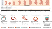
research on animals stem cell

A roadmap for the Human Developmental Cell Atlas
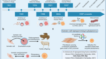
research on animals stem cell

Induced pluripotent stem cells (iPSCs): molecular mechanisms of induction and applications

Introduction.

The 1995 Nobel Prize in Physiology or Medicine, awarded to Edward B. Lewis, Christiane Nüsslein-Volhard, and Eric F. Wieschaus, marked a turning point in the field of modern developmental biology. By introducing mutations in Drosophila embryos, these researchers found key genes associated with regulating several significant early developmental processes, such as segmentation and polarization in Drosophila . Their groundbreaking discoveries in the genetic control of embryonic development laid the foundation for a deeper understanding of the molecular mechanisms governing the formation of multicellular organisms 1 , 2 .

Over the past three decades, substantial progress has been made in developmental biology ever since the foundation of several techniques, such as genetic engineering techniques, wherein various animal model systems were actively utilized to further enhance the understanding of genetic functions within the processes of embryonic development. However, studying human development has remained a significant challenge due to the inherent differences between conventional model organisms and humans 3 , ethical concerns about using human embryos 4 , and physical inaccessibility due to the intrauterine development of in vivo human embryos.

Recently, the rapid advancement of stem cell research has enabled the generation of stem cell-derived human organoids 5 , which are three-dimensional, self-organizing structures that mimic various human organs. This remarkable progress has expanded the boundaries of human biology research, allowing the investigation of human biological processes in a more physiologically relevant context 6 . In addition to organoids, recent advances in stem cell research have even facilitated the development of early embryonic models that mimic human embryos. Early embryonic models provide an opportunity to investigate molecular mechanisms that regulate human embryogenesis with better scalability and in a relatively ethical manner, thereby overcoming the major limitations of using natural human embryos 7 , 8 , 9 , 10 .

Early embryonic models were generated by using embryonic stem cells (ESCs) 11 , which possess the ability to self-renew and differentiate into a variety of specific cell types of the body. The potency of ESCs varies depending on their epigenetic traits and culture environment 12 . These shifts in potency allow the induction of various in vitro models, including synthetic whole embryo models that incorporate all significant cell types, such as those derived from both the embryo and extraembryonic tissues 8 . This review describes the process by which stem cells, including ESCs in relation to their potency, are utilized to create these in vitro models.

This review aims to explore developments in stem cell research, focusing on stem cell-based in vitro early embryonic developmental models. Overall, this review article aims to provide a comprehensive analysis of the new era of stem cell research, examining the potential uses of developmental-stage-specific embryo models for disease modeling and possible clinical applications. By providing a thorough discussion of these technologies, we hope to inspire further research and advancements in the field.

Embryonic stem cells (ESCs): the building blocks of synthetic embryos

Stem cells are self-renewing cells with the potential to differentiate into a wide variety of cell types 13 . Based on their origin, they are divided into two major categories: ESCs and adult stem cells (AdSCs). ESCs are derived from the inner cell mass (ICM) of blastocysts and can differentiate into any type of cell in the body 14 , whereas AdSCs are found in various tissues in our body and help maintain tissue homeostasis but can differentiate into only a limited number of cell types relative to ESCs 13 , 15 . Early embryonic in vitro models were all derived from ESCs or induced pluripotent stem cells (iPSCs) 16 of equivalent potency. This section focuses on ESCs, which serve as the foundation for creating in vitro models, and analyzes their diverse potencies (Fig. 1a ).
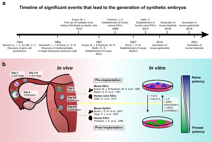
figure 1

a Timeline of significant events in stem cell research facilitating the generation of synthetic embryos. b Illustration of early mammalian development in vivo (left), as well as the in vitro counterparts—naïve and primed ES cells—represented as cell states (right). Each developmental stage of the mammalian embryo is depicted from the fertilization of the egg (Day 0) to the implantation of the blastocyst (Day 9). Pre- (top) and post-implantation (bottom) stem cells derived in vitro are listed in the middle, as well as the original research in which they were discovered. The ESCs that correspond to the pre- and post-implantation states are naïve (top) and primed (bottom) ESCs, respectively, and the degrees of the states are represented on the right (ascending, more naïve; descending, more primed). Morphogens that regulate their states are written in between.

Birth of ESCs

The concept of ESCs was first proposed in the 1950s by Stevens and Little, who discovered that spontaneous testicular teratomas in an inbred mouse strain originated from primordial germ cells and that these cells can differentiate into a variety of cell types 17 . This discovery illuminated the pluripotency of germ cells, opening the way for further investigation into the origin and differentiation potential of other stem cell types. Kleinsmith and Pierce expanded upon these findings by demonstrating the multipotentiality of single embryonal carcinoma cells 18 .

The development of in vitro culture methods also played a crucial role in the establishment of ESCs. Evans pioneered the use of irradiated chick embryo fibroblasts as feeder cells to culture mammalian stem cells in vitro, which enabled the maintenance and expansion of pluripotent stem cells (PSCs) 19 . Based on these findings, Evans, Kaufman, and Martin successfully derived mouse ESCs (mESCs) from the ICM of blastocysts, marking the beginning of modern stem cell research 20 , 21 . Building on the advancements in mESC research, the scientific focus shifted to human ESCs (hESCs), leading to a significant breakthrough in 1998 when Thomson et al. successfully derived and cultured hESCs in vitro by isolating them from the ICM of human blastocysts 14 . Both mouse and human ESCs possess the capacity for self-renewal and pluripotency, which allows them to maintain an undifferentiated state while proliferating in culture and to differentiate into any cell type of the three germ layers (endoderm, mesoderm, and ectoderm) 14 , 20 .

Although initially established mESCs and hESCs share certain traits, they also display distinct differences in morphological, transcriptional, and epigenetic features 22 . mESCs, for instance, have a tendency to form a dome-like structure, whereas hESCs typically exhibit a flattened shape 22 . Both mESCs and hESCs express the pluripotency-related transcription factor octamer-binding transcription factor 4 (OCT4), albeit with distinct regulatory mechanisms. In mESCs, OCT4 expression is regulated by its distal enhancer, while in hESCs, its expression is mainly regulated by its proximal enhancer 23 , 24 .

As subsequent research progressed, a method was developed to culture mESCs that exhibited characteristics similar to hESCs 25 , 26 . These cells, derived from the epiblast of post-implantation embryos, were termed mouse epiblast stem cells (mEpiSCs) and shared morphology and a variety of epigenetic properties similar to hESCs 26 . Continuous investigations have revealed that the differences between mESCs and hESCs originate from the fact that these ESCs represent different stages of development, thereby influencing their slightly different differentiation potentials.

Naïve and primed pluripotent states of ESCs

Mammalian ESCs are known to exist in a continuum of configurations, which at either end of the spectrum lie two different pluripotent states: naïve and primed 12 . These states have distinct molecular and functional characteristics, and understanding their differences is crucial for optimizing the use of ESCs in modeling early embryos in vitro.

Naïve ESCs, compared to primed ESCs, correspond to an earlier developmental stage of pluripotent cells. They are derived from the pre-implantation ICM during the early blastocyst stage 27 (Fig. 1b ). These cells exhibit characteristics such as having a relatively unrestricted differentiation potential, allowing them to differentiate into both embryonic (epiblast) and extraembryonic tissue (hypoblast). In addition, they have a more open chromatin structure on developmental regulatory gene promoters, accompanied by a global reduction in DNA methylation 27 , 28 .

In contrast, primed ESCs are derived from a developmentally more advanced stage, specifically from post-implantation epiblast cells 27 (Fig. 1b ). These cells exhibit a relatively restricted differentiation potential; they predominantly contribute to embryonic components, unlike their naïve counterparts 25 . This restriction is likely due to their relatively closed chromatin structure on lineage regulatory genes 29 .

The first mESCs established were in the naïve state 20 , 21 , while the initial hESCs and mEpiSCs that were established resembled the primed state of pluripotency 14 , 25 . Over several years, efforts to establish human naïve ESCs have led researchers to gain insights into the molecular mechanisms and signaling pathways regulating pluripotency. These key insights included the identification of factors essential for maintaining naïve pluripotency in mESCs, such as leukemia inhibitory factor (LIF), which activates the Janus kinase-signal transducer and activator of transcription 3 (JAK-STAT3) signaling pathway 30 , 31 . The development of specific culture conditions to derive and maintain mEpiSCs facilitated further investigation into the signaling pathways that regulate primed pluripotency, including the fibroblast growth factor/extracellular signal-regulated kinase (FGF/ERK) and Activin/Nodal pathways 25 , 26 .

The establishment of naïve hESCs required the identification of culture conditions that could support the naïve state while suppressing the primed state. This requirement led researchers to focus on modulating signaling pathways, including the inhibition of the FGF/ERK 32 and transforming growth factor-beta (TGF-β)/Activin/Nodal pathways 33 , which are known for maintaining primed pluripotency. The application of small molecules to target these pathways enabled the conversion of primed hESCs into a state resembling naïve pluripotency 23 , 34 .

After years of extensive efforts, the landmark achievement of establishing naïve hESCs was made by Gafni et al. in 2013. Gafni and colleagues successfully established naïve hESCs using a combination of small molecules and growth factors 35 . They showed that inhibiting the FGF/ERK, TGF-β, and WNT/β-catenin pathways while simultaneously activating the LIF/STAT3 signaling pathway could prompt the conversion of primed hESCs to a naïve state 36 . These newly established naïve hESCs exhibited key features of naïve pluripotency, including the acquisition of epigenetic states similar to those of naïve mESCs, the expression of naïve-specific markers, and the attainment of an enhanced differentiation potential 23 , 35 .

Blastoids modeling early embryo development

The early stages of mammalian embryogenesis, leading up to implantation, encompass a series of conserved, systematic steps across various species, including ovulation, fertilization, cleavage, and implantation. Upon ovulation, the oocyte travels through the oviduct, where fertilization takes place in the ampulla. As the embryo travels toward the uterus, it undergoes asynchronous cleavages. At this juncture, the mammalian genome is activated, facilitating embryonic development through the expression of early transcribed proteins.

In humans, major waves of zygotic gene activation occur at the 8-cell stage, whereas in mice, activation arises at the 2-cell stage (minor waves transpire at the 4-cell stage and late zygotic stage for human and mouse embryos, respectively) 37 , 38 , 39 , 40 . Researchers have endeavored to model this process by generating human eight-cell-like cells (8CLCs) to probe the mechanism of totipotency 41 . In 2022, Mazid et al. developed a method to produce 8CLCs from human PSCs (hPSCs), revealing key roles of developmental pluripotency associated 3 (DPPA3), a master regulator of DNA methylation in oocytes, and tetrapeptide repeat homeobox 1 (TPRX1), a eutherian totipotent cell homeobox (ETCHbox) family transcription factor, in this process. Using this embryo model, the researchers demonstrated that 8CLCs can contribute to embryonic and extraembryonic lineages, providing a valuable resource for the study of the earliest stages of human embryogenesis.

Compaction is one of the most significant events of mammalian cleavage. In mice, after the embryo reaches the 8-cell stage, compaction occurs as blastomeres express cell adhesion proteins and coalesce into a sphere of cells 42 . This compact embryo matures into a 16-cell morula, where its inner cells later become constituents of the ICM, and its outer cells give rise to the trophectoderm (TE) 43 . The ICM is situated on one side within the TE, culminating in the formation of the blastocyst. In essence, the blastocyst comprises two primary cell populations: the ICM and the TE.

The peri-implantation period designates the phase during which the blastocyst is unattached in the uterus, preceding its implantation into the uterine wall, and differentiates into several fundamental structures. The nascent cells of the ICM segregate to form the primitive endoderm (PrE) and the epiblast. These intricate processes, which are fundamental to early embryonic development, have historically been difficult to study in depth. However, recent advances in stem cell research have now made it possible to create in vitro models that closely mimic these early stages of development (Fig. 2 ).
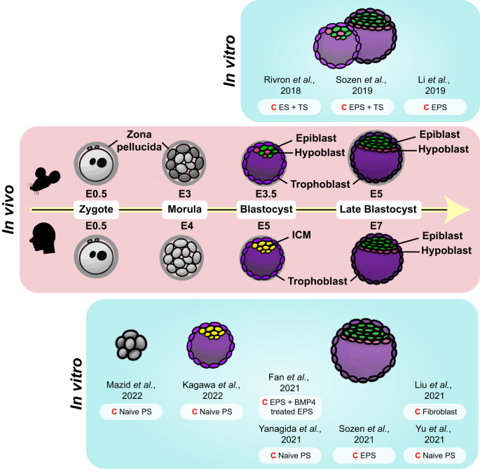
figure 2

A depiction of the early developmental stages, spanning from the zygote phase (E0.5 of both mouse and human embryo) to the blastocyst stage (E5 and E7 of the mouse and human embryo, respectively), of mouse and human embryos are portrayed in the middle. In vitro models corresponding to the in vivo embryo of each developmental stage are illustrated on either side (top, mouse; bottom, human). Specific cells are color-coded throughout the figure (epiblast, green; hypoblast, pink; trophoblast, purple; ICM, yellow); the same color represents similar states at which they are in. Starting cells (C) of each model are noted adjacent to each illustration.

Since the isolation of mESCs in the 1980s 20 , 21 , stem cell research has significantly advanced. With the subsequent discovery of trophoblast stem cells (TSCs) 44 and extraembryonic endoderm stem cells (XENs) 45 , researchers have been equipped with essential cellular tools for the creation of in vitro mouse embryo-like structures known as mouse blastoids. This marked a pivotal breakthrough in embryonic research, signifying the first successful attempt to replicate an entire embryo in vitro.

Rivron et al. set a precedent by successfully generating the first mouse blastoids 46 . Following this breakthrough, numerous research groups have also developed similar in vitro structures that mimic in vivo blastocysts by employing a variety of methods 46 , 47 , 48 . Mouse blastoids are generated by aggregating various types of stem cells, including pluripotent and extraembryonic stem cells, in non-adherent hydrogel microwells, utilizing the self-organizing property of the mammalian embryo 43 . These cells are cultured with a blend of morphogens that influence the specification of the epiblast and TE lineages. Through this self-assembly process, in vitro structures that strikingly resemble in vivo blastocysts are created.

A pivotal event in successful blastoid formation is cavitation, underpinned by TE formation. TE formation is fostered by inhibiting the Hippo and TGF-β pathways. Concurrently, the addition of LIF and inhibitors of mitogen-activated protein kinase kinase (MEK) and glycogen synthase kinase 3 (GSK3) (known as 2i) helps maintain the pluripotency of ESCs by activating the STAT3, ERK, and WNT signaling pathways 49 . Moreover, Y-27632, a Rho-associated protein kinase (ROCK) inhibitor, is utilized to prevent cell apoptosis during this complex process.

In just 3 to 4 days, blastoids successfully take shape, exhibiting features such as the blastocoel cavity, ICM, and TE layer, the organization of which closely replicates the spatial organization seen in in vivo blastocysts. As cells start the process of self-assembly into blastoids, additional transcription factors, including GATA binding protein 6 (GATA6) and caudal type homeobox 2 (CDX2), are activated. These transcription factors govern lineage specification and cell differentiation, with GATA6 promoting the formation of the PrE lineage and CDX2 playing a crucial role in inducing the proper differentiation of the TE lineage 43 , 50 , 51 . The precise activation of transcription factors within blastoids is essential for directing the self-organization and lineage segregation of ESCs and TSCs into a fully functional blastoid structure. These blastoids share several characteristics with natural blastocysts, such as the capacity to acquire apical‒basal polarity and tight junctions.

Expanding upon strategies used in the creation of mouse blastoids, researchers have successfully generated human blastoids by harnessing the unique properties of hPSCs. Human naïve ESCs, notable for their increased plasticity and lower lineage barrier compared to naïve mESCs, have the capacity to differentiate into both embryonic (epiblast) and all extraembryonic (TE and hypoblast) lineages 52 , 53 , 54 . This significant capability allows the formation of human blastoids without necessitating the mixture of ESCs and TSCs 55 , 56 , 57 . In a parallel manner, human extended pluripotent stem cells (hEPSCs) 58 , 59 , cultured to embody developmental potency for embryonic and extraembryonic cell lineages, have shown the potential to generate blastoids 60 .

Several techniques have been developed to generate human blastoids. For instance, human blastoids can be generated by aggregating naïve hPSCs or hEPSCs in rounded microwells using centrifugation 55 , and similar results can be achieved with standard microwells or pyramid wells 56 , 57 , 60 . It should be noted that the use of a single cell type, such as naïve hPSCs or EPSCs, is not the only option to induce blastoid formation in the medium. Indeed, blastoids can also be generated through a combination of hEPSCs and TE-like cells derived from EPSCs 61 . Similarly, 8CLCs have been used to derive blastoids that exhibit both morphological and transcriptomic similarities to human blastocysts 41 .

Another breakthrough was the direct reprogramming of human somatic cells, which was achieved by infecting human dermal fibroblasts with a virus capable of inducing the expression of OCT4, SRY-box 2 (SOX2), Kruppel-like factor 4 (KLF4), and c-MYC, which in turn generated induced blastoids, or iBlastoids 62 . These human blastoids bear transcriptomic and morphological resemblances to human blastocysts, and remarkably, some have even shown the potential to initiate events akin to implantation.

Blastoids have the potential to be implanted into a hormone-stimulated endometrial layer 55 , allowing the observation of post-implantation processes. In compliance with the International Society for Stem Cell Research (ISSCR) guidelines 63 , which allow human experimental culture up to 14 days, these human blastoids could potentially be implanted into a synthetic endometrial lining for the purpose of investigating post-implantation events. The implantation of blastoids revealed that this process took place in the polar TE region, which likely became polarized due to its nascent relation to epiblast-like cells 55 . After implantation, sustained proliferation of epiblasts, TEs, and PrE-like cells was observed 55 .

Although several blastoids have cellular compositions similar to those of human in vivo blastocysts, it is necessary to investigate them more closely to determine whether they precisely replicate the in vivo state. The similarity of most of the blastoids created thus far to in vivo blastocysts has been validated using single-cell transcriptomics. Occasionally, however, the cells formed in blastoids do not match those of human early blastocysts but rather those of later developmental phases (such as gastrulation after embryonic day (E) 14) 64 . Therefore, further analysis is required to determine how accurately these in vitro models represent the in vivo context, and ongoing research should focus on increasing their similarity.

Early gastruloids recapitulating the formation of three germ layers

Gastrulation is one of the most important processes of the developing mammalian embryo, where it incorporates several combinations of cell movements in newly positioning the embryonic tissue to form the three germ layers—ectoderm, mesoderm, and endoderm—which set a platform that further enables the navigation of cellular lineage specification by placing the tissues accordingly throughout the anterior-posterior (AP), dorsal-ventral, and lateral (left and right) axes. By appropriately positioning the embryonic tissue layers, organizing axes, and facilitating progressive specialization, the embryo is able to undergo processes such as organogenesis and limb morphogenesis that lead to the formation of more complex structures.

Going back to mark the initiation of the creation of synthetic embryos, as mentioned earlier, developmental biologists Martin and Evans found that ES-cell-like 2D clonal pluripotent teratocarcinoma cells suspended into aggregates self-organize themselves to acquire a cellular organization pattern similar to that of the differentiation of the early mouse embryo 65 . Following their discovery, other studies have likewise continued to generate 3D aggregates that were able to recapitulate early embryonic differentiation and thus were collectively termed “embryoid bodies (EBs)”. When EBs derived from ES cells of mouse blastocyst ICM origin were cultured under in vivo-like conditions with appropriate developmental cues, they acquired the ability to differentiate into all three germ layers 66 , 67 .

Taking the potency of pluripotent stem cells to advantage, the addition of various signaling factors parallel to those that work at the corresponding developmental stage to EBs coaxed the formation of models that recapitulate the early development of the embryo. Due to the established role of signaling factors around this stage of embryonic development, small molecules added to induce the growth of in vitro models are short-range and similar among different protocols. Agonists of the FGF, BMP, and WNT signaling pathways (such as FGF2, BMP4, and CHIR99021, respectively)—key signaling pathways that govern development—as well as inhibitors (such as PD173074, LDN193189, and XAV939, respectively) are used to induce or inhibit characteristics of the developing embryo (Fig. 3 ).
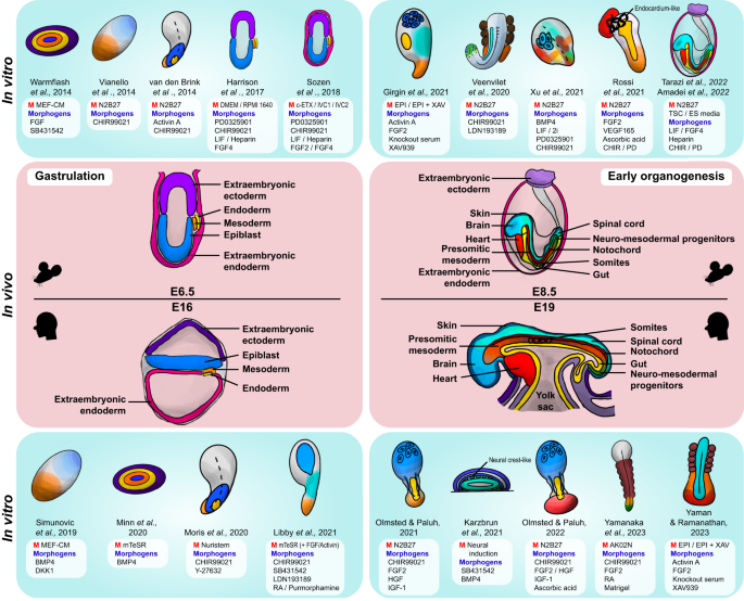
figure 3

In vivo developmental stages of the mouse and human embryos are depicted in the middle, from the process of gastrulation (E6.5 and E16 of the mouse and human embryo, respectively) to early organogenesis (E8.5 and E19 of the mouse and human embryo, respectively). In vitro models corresponding to the in vivo embryo of each developmental stage are illustrated on either side (top, mouse; bottom, human), as well as to note the original paper they were formed in. Specific tissues/organs are color-coded throughout the figure for both the embryos in gastrulation (mouse extraembryonic ectoderm, light purple; human extraembryonic ectoderm, dark purple; extraembryonic endoderm, pink; mesoderm, orange; endoderm, yellow; epiblast, blue) and those in early organogenesis (mouse extraembryonic ectoderm, light purple; extraembryonic endoderm, pink; brain, sky blue; spinal cord, turquoise; skin, dark navy; heart, red; notochord, dark red; somites, brown; presomitic mesoderm, dark orange; mesoderm, orange; gut and/or endoderm, yellow; neuromesodermal progenitors, green). Specific medium (M) or morphogens, including growth factors, cytokines, and inhibitors, used in the protocols for generating each in vitro model are listed adjacent to each gastruloid illustration.

As such, incorporating signaling pathways into in vitro cell aggregates yields models that recapitulate important hallmarks of gastrulation. For example, symmetry breaking is achievable in EBs in vitro, where the local activation of the WNT signaling pathway induces AP polarity and the formation of a primitive streak-like region, resulting in differentiation into a mesendodermal compartment (in contrast, neurectodermal differentiation is achieved by WNT signaling inhibition) 68 . Furthermore, the activation of WNT signaling at an appropriate time frame (at the initial stages of culture, which is usually within 74 h post-aggregation) induces the formation of a progressive elongating domain similar to that of the in vivo tail bud of the embryo, as well as the specification of the endoderm, which all lead to simulating aspects of the gastrulation period of the embryo 69 . Additionally, the precise timing regulated by WNT/β-catenin and Nodal signaling induces symmetry breaking in gastruloids even without the presence of extraembryonic tissue or localized signaling, allowing further polarization to take place, which is evidenced by distinct T/Brachyury expression 70 . Reducing FGF signaling yielded a diminished tail phenotype in the embryo models, indicating that the inducement of this signaling pathway is important for elongation 71 . With the knowledge of biological mechanisms underlying embryonic development and the use of key signaling factors, several models that mimic aspects of gastrulation were generated, starting from micropatterned 2D models (“2D gastruloids”), which encompass the endoderm, mesoderm, and ectoderm 72 , 73 , 74 , 75 , 76 , to more advanced free-floating 3D gastrulation models (“3D gastruloids”).

Early works to recapitulate aspects of gastrulation were conducted using mouse stem cells. The first absolute 3D gastruloid was created by van den Brink and colleagues in 2014 69 . Consisting of only 300 mouse ES cells, the EB was cultured in vitro to create a 3D gastruloid, where hallmarks of early mouse embryonic development, such as symmetry breaking and axial organization, were observable under the activation of WNT/β-catenin signaling, which further caused germ layer specification and minor axial elongation upon the addition of specific morphogens such as CHIR99021 and Activin A 77 . The usual 3D gastruloids are cultured through an initial few-hour-to-day EB suspension, whereafter they are then further matured under shaking culture conditions while adding certain morphogens to induce multi-axial organization and a temporal pattern similar to the gene expression of the embryo. Some gastruloids also acquire in vivo patterning integrity by expressing the collinear Hox gene pattern throughout the AP axis 77 .

Through the advancement of the creation of mESC embryonic models, adjustments in the culture conditions of these models enabled more developed structures as well as new discoveries. Although mouse gastruloids effectively mimic embryo-like axial morphogenesis and patterning, they lack regions such as the anterior embryonic region (which includes the brain). To improve this inaccurate depiction of the in vivo embryo, studies have inhibited WNT signaling during the early stages of development to induce gastruloids with anterior neural tissue 68 , 78 , since elevated expression of genes of the posterior mesoderm was observable with the inducement of WNT signaling 78 . However, WNT signaling is key in breaking axial symmetry in embryonic development, where its set dose induces primitive streak markers and spontaneously breaks AP symmetry. The aforementioned AP axis establishment enables the organization of embryonic tissues. Despite genetic studies that state that this process relies on the embryo’s exposure to extraembryonic spatiotemporally located signals—WNT/β-catenin and Nodal signaling—studies performed with mouse gastruloid models proved that this might not necessarily be the case. Mouse ESC-based gastruloids induced without surrounding extraembryonic tissues showed localized T/Brachyury expression with polarity and extension to one side, suggesting the notion of AP axis development. Thus, this discovery suggested that extraembryonic tissues are not necessarily required for the embryo to undergo self-patterning 69 .

Gastrulation occurs at approximately 16 days post-fertilization (dpf) in human embryos, and in vitro biomimetic models that recapitulate this stage are also achievable by utilizing primed hESCs and hiPSCs, enabling the formation of human gastruloids without the violation of major ethical issues. As in the case of initial murine gastruloid models, human gastruloids were also developed under 2D conditions that portrayed aspects of gastrulation, such as the development of primitive streak-like structures and the three germ layer domains 72 , 79 .

The development of 3D human gastruloids facilitated the study of human development, as they more accurately depict the in vivo gastrulation process than earlier 2D models. Human PSCs embedded in the matrix, such as by utilizing the commercial extracellular matrix (ECM) formula Matrigel, and cultured under conditions that are supplemented with the right morphogens construct aggregates that undergo symmetry breaking and gene expression patterning 80 . For example, the addition of BMP and WNT agonists was found to be crucial in the establishment of AP polarity and the elongation of the tail bud 81 . Adding extraembryonic compartments resulted in models that more closely resembled the in vivo gastrulating embryo 82 by mimicking attachment and AP symmetry breaking and further exhibiting human gastrula-specific cells. 3D aggregates that broke symmetry formed extensions along the AP axis with organized germ layers 83 , 84 . Through the development of the technology of generating human gastruloids, specific gastruloids that addressed parts of the early stages of post-implantation human embryonic development were created, such as the processes of primitive streak formation 80 , neurulation 84 , 85 , 86 , and somitogenesis 87 .

Late gastruloids modeling somitogenesis and organogenesis

At E7.5 in mice and approximately 20 dpf in humans, the presomitic mesoderm (PSM) condenses and gives rise to somites along the lateral sides of the central neural tube in an anterior-to-posterior fashion. These somites later form structures that act as anchors that hold parts of the embryo, such as the vertebrae, skeletal muscles, cartilage, and dermis. Shortly after somitogenesis has begun, organogenesis is launched, with the heart being the first organ to be generated. Models that mimic somitogenesis, as well as organogenesis, have been created over recent years (specific models are further discussed in the section “Modeling congenital diseases using synthetic embryos”). Segmentation clock waves are observable in mesoderm-based hPSC models (termed “axioloids” or “somatoids”) that mimic segmentation and somitogenesis in the human embryo 88 . Simpler models that recreate oscillatory movements were also generated 89 . Inducing AP symmetry breaking in hPSC models allowed scientists to study the mechanisms underlying human somitogenesis.

Gastruloids have also been able to acquire regions that resemble the gut tube. The formation of the AP and dorsal-ventral axes enables the patterning of the primitive gut tube in mouse gastruloids 90 . More specifically, the anterior foregut, midgut, and hindgut were induced in the gastruloid through the creation of a primordium that covers the overall region of the structure 91 . Furthermore, several gastruloid models, such as trunk-like structure (TLS) gastruloids (using mESCs) 92 and elongating multi-lineage organized (EMLO) gastruloids (using hiPSCs) 84 , portray gut tube-like structures.

Development of synthetic whole embryos

Instead of models that recapitulate only a small aspect of the early developing embryo, attempts were made to model its overall characteristics. In mice, the cylindrical shape of the post-implantation embryo is due to the polar TE invagination and actomyosin contractility that give rise to the extraembryonic ectoderm 39 . Stem cell models that resemble these early “egg-cylinders” were generated by combining mESCs with TSCs and incorporating tissues such as those that resemble the extraembryonic ectoderm without recapitulating the blastocyst stage 93 . Adding XEN cells that resemble extraembryonic endoderm cells to these models (ETX or iETX embryos) enabled the production of a visceral endoderm-like epithelium that lines the in vitro conceptus 94 , 95 . When synthetic embryos were constituents of extraembryonic compartments, they could initiate implantation 95 and further induce gastrulation, neurulation, and organogenesis 96 .

Several culture techniques that assist in establishing proper embryonic development ex vivo were created, such as circulator systems 97 and roller culture systems 98 . By taking advantage of these systems, the process of generating whole embryos in vitro was conducted. Due to the aforementioned ISSCR regulation restricting the culture of human embryos to a maximum of 14 days in vitro for research purposes (the “14-day rule”), attempts to create synthetic whole embryos in long-term culture conditions were generally proceeded using mESCs.

To create synthetic whole embryo models, an environment similar to in vivo conditions is needed. With the adaptation of the electronically controlled ex utero roller device that previously cultured a mouse embryo until E11 by the incorporation of the ex utero culture medium (EUCM), which is a mixture of 50% rat serum, 25% Dulbecco’s modified Eagle’s medium (DMEM), and 25% human umbilical cord blood serum 99 , mouse naïve ESCs were cultured until E8.5, which yielded a mouse synthetic whole embryo model that was able to undergo gastrulation and to form organ precursors 100 .

Attempts to create human synthetic whole embryos were also made. For human synthetic whole embryo models, blastoids are currently the most advanced tool, mimicking the entire pre-implantation stage of the human embryo. Although researchers have made attempts to push these blastoids toward the post-implantation stages in vitro, they have not been able to match them with the development of in vivo blastocysts 55 , 56 , 60 , 61 , 62 . Recently, there have been strides toward creating a human synthetic whole embryo that can model more advanced developmental processes mimicking post-implantation stages while maintaining the morphological integrity of a natural embryo.

Two groups of scientists have recently reported their attempts at creating human post-implantation embryo models 101 , 102 . They drew on strategies previously used to create a mouse synthetic whole embryo, which involved differentiating embryonic and extraembryonic cells separately before aggregating them to produce a post-implantation embryo. Weatherbee et al. generated two types of extraembryonic-like cells from hESCs through the overexpression of transcription factors: GATA6 and SRY-box transcription factor 17 (SOX17) for the hypoblast and GATA3 and transcription factor AP-2 gamma (TFAP2C) for TSCs 101 . On the other hand, Oldak et al. utilized ectopic expression of lineage-promoting transgenes to induce the formation of the hypoblast and trophoblast 102 .

Both groups reported human synthetic whole embryo models that replicated the hallmarks of 13-14 dpf human embryos. Weatherbee et al.’s model consisted of structures such as the amnion, extraembryonic mesenchyme, and primordial germ cell-like cells 101 . Oldak et al. reported the formation of the bilaminar disc, epiblast lumenogenesis, amniogenesis, primordial germ cell specification, yolk sac formation, and the expansion of extraembryonic mesoderm in their model 102 . While the creation of human synthetic whole embryo models is expected to continue to evolve, it is essential to closely monitor these developments within the framework of ethical considerations and international agreements.

Modeling congenital diseases using synthetic embryos

One of the main advantages of synthetic embryos is their feasibility for modeling diseases that arise during the early stages of embryonic development. Thus, congenital diseases that occur due to initial abnormal development are of interest. Furthermore, because synthetic embryo models such as gastruloids can be easily manipulated to imitate specific stages of development, diseases that occur at a particular event could be observed. Although only a few- considering the subject’s relatively recent advancement into the field, attempts have been made to incorporate synthetic embryoids to model human diseases. This section includes an overview of models that recapitulate key processes of embryonic development with respect to disease modeling (Fig. 4 ).
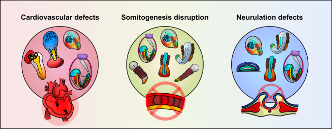
figure 4

Cardiovascular defects (left), diseases associated with somitogenesis disruption (middle), and neurulation defects (right) may be modeled by current gastruloids. Appropriate gastrulation models are depicted in each category.

Congenital heart disease (CHD) is one of the leading causes of death in newborns 103 . Several models that mimic the heart have been made to recapitulate aspects of cardiogenesis 104 , 105 , 106 , 107 , 108 ; however, to fully understand the dynamics of heart development, observing the organ alone is insufficient. Taking advantage of the gastruloid characteristic of encapsulating multiple cell types of all germ layers, cardiogenesis development is more accurately portrayed by gastruloid models since the formation of the heart necessitates complex interactions such as those among the organ, cardiac progenitor cells, and endothelial cells. Recent studies have mimicked cardiogenesis by manipulating embryo models by incorporating well-known cardiogenic growth factors, such as vascular endothelial growth factor 165 (VEGF-165), FGF2, and ascorbic acid. Rossi et al. captured the process of cardiogenesis via mESC gastruloids, in which the spatiotemporal accuracy was highly conserved compared to its in vivo counterpart 109 . The model incorporates both cardiac progenitors of the first heart field (FHF) and the second heart field (SHF), which anteriorly organize into a cardiac crescent-like region and form tissues that imitate the beating of the heart.

Expanding upon synthetic embryo models that recapitulate only cardiogenesis, new models that include both cardiac muscle and neurons to portray neuro-cardiac lineages have been made. Olmsted and Paluh have added the aspect of cardiogenesis 110 to the “elongating multi-lineage organized” (EMLO) gastruloids that they had previously generated 111 , creating EMLOC gastruloids that enable the remodeling of both cardiogenesis and neurogenesis to explain the complex interconnected lineages between the processes. Various developmental features that resemble those of the in vivo heart were observed, including the formation of the heart tube, putative outflow tract, ventricular wall, and epicardium, as well as the differentiation of cardiomyocytes. This neuro-cardiac model may offer a platform to provide further understanding of cardiac diseases such as neural-based heart arrhythmia diseases and congenital heart diseases.

Vertebrates acquire segmented structures through a process called somitogenesis. This process occurs in early development when somites, blocks of paraxial mesoderm, form on either side of the neural tube and the notochord in a bilateral position along the AP axis. Presenting phenotypes similar to those of mouse embryos, mouse gastruloids have been offered as appropriate models to study vertebrate segmentation. For instance, genetic alteration of gastruloids yielded phenotypes that matched those of previous knockout mouse embryo models. Veenvliet et al. used mESCs deleted with the T-box transcription factor 6 ( Tbx6 ) gene to generate gastruloids (TLSs) 92 . The resulting phenotype was the loss of somites and ectopic neural tubes, which correlates with the in vivo phenomenon of PSM transdifferentiation and ectopic neural tube formation at the expense of somites and PSM. Van den Brink et al. performed a screening test that showed that decreased levels of FGF signaling resulted in the generation of fewer somites, which was similar to the defects observed in Fgf mutant mice, and that incorporating 10% Matrigel may induce somitogenesis in gastruloids 71 .

Aberrations in somitogenesis can lead to several diseases, such as vertebral malformations and spinal defects. Specifically, Yamanaka et al. created axioloids that recapitulated human somitogenesis in vitro and applied them to investigate congenital diseases associated with the human spine 88 . They introduced loss-of-function mutations in the segmentation clock gene hes family bHLH transcription factor 7 (HES7) and the marker for the anterior PSM mesoderm posterior bHLH transcription factor 2 (MESP2), which are associated with segmentation defects of the vertebrae (SDV), and generated axioloids with them. Two different HES7 knockout hiPSC lines were used to create axioloids, and their phenotypes were assessed. There were losses of segments and rostro-caudal patterning, as well as the formation of the epithelial somites. Regarding the oscillatory activity of the segmentation clock, Yamanaka and colleagues found a definite loss of HES7 oscillation. In axioloids derived from iPS cell lines introduced with a point mutation in HES7, there were also losses of rostro-caudal patterning, yet these models managed to express normal HES7 oscillation in the tail bud as well as a few somitic mesoderm markers. MESP2 is known to be mutated in patients with SDV. Yamanaka and colleagues also assessed the effects of axioloids with MESP2 knockout. MESP2-knockout axioloids also lacked segments and epithelial somites and exhibited abnormal rostro-caudal patterning, despite their normal elongation. However, MESP2 knockout did not result in the loss of oscillation of HES7. As such, these axioloids serve as models that offer insights into the segmentation development of the human body and a platform to study the pathogenesis of the spine.

Neurulation is a highly orchestrated and complex morphogenetic process in which the neural plate fuses to form the neural tube, the precursor of the brain and spinal cord of the central nervous system. When the process of neurulation is disrupted, severe congenital abnormalities such as neural tube defects (NTDs) arise. Several studies have shown that gastruloids are capable of recapitulating the formation of the neural tube. Libby et al. generated a human neural tube gastruloid that demonstrated aspects of early spinal cord development, such as axial elongation, neuromesodermal progenitor (NMP) cell population maintenance, and neuroepithelial cell generation 81 . With exposure to doses of the WNT agonist CHIR99021, the elongation of hPSC aggregates and the formation of NMP cells could be observed, as well as the presence of the mesoderm and neuroectoderm. Neural tubes were also induced in mouse TLSs created by Veenvliet et al. when cultured in a 5% growth factor-reduced Matrigel condition, as well as by the addition of the WNT agonist CHIR99021 and the BMP signaling inhibitor LDN193189 92 .

Furthermore, neurulation was depicted in partially 3D gastruloids induced by 2D micropatterning by Karzbrun et al., where a neural-tube-like structure was detectable 85 . In this model, Karzbrun and colleagues incorporated three small-molecule inhibitors—the ROCK inhibitor Y-27632, the HSP-90 inhibitor novobiocin, and the NTD-associated drug valproic acid—to test the suitability of these gastruloids for modeling NTDs. The application of the small molecules resulted in morphological abnormalities, including less curvature of the tube and overall folding defects, which indicated that their model may indeed be utilized to represent the neural tube to study defects to some extent.

Perspectives

Historically, investigating human development has been an ongoing endeavor with significant limitations, including restricted understanding due to the complexity of the process, the lack of appropriate models (not to mention the inaccessibility of human embryos), and ethical concerns. Herein, we have illustrated how the generation of synthetic models of early mammalian development has assisted in overcoming these challenges.

Through the discovery of stem cells and research progress made due to their manipulation, the scope of human embryo experimentation, once available only through direct practice on authentic embryos, became more diverse. By inducing human stem cells to become structures characterized by organized systems, the need for human embryos has diminished, thereby compensating for the inaccessibility of human embryos due to the shortage of samples and ethical issues. Ethical concerns are inevitable; accordingly, regulations have been made by officials such as the ISSCR with recurring discussions among scientists 112 .

Synthetic models do not act merely as models that recapitulate early development; they also provide a platform for which numerous diseases, and their underlying mechanisms, could be further studied. Since these early embryo models may reliably replicate the embryo’s state at certain developmental stages, specific diseases that arise at specific time points may be investigated by recreating a developmentally similar embryonic structure with temporal accuracy. For example, blastoids allow the further study of implantation mechanisms, which then may suggest possible factors associated with implantation failures, and gastruloids may model congenital diseases, including but not limited to those stated above. Current mammalian embryo models are able to recapitulate in vivo embryos of Carnegie stages 1 to 10, in which diseases that arise may be modeled.

As a shared advantage of in vitro structures, synthetic embryo models may be produced in bulk, offering compatibility with systems that require large amounts of samples, such as drug screening and toxicological assays. By using stem cell-based models, researchers have studied the effects of small molecules on mammalian development 113 , 114 . Furthermore, teratogenicity could be tested through quantifiable gastruloids, which yield statistical robustness, and further applications may be tested, such as species-specific reactions to the molecules 115 .

Nonetheless, a limitation that remains is the total inducement of the synthetic whole embryo in vitro. Several attempts have been made to create synthetic whole embryos, yet results show that there is still a need for further understanding of developmental mechanisms, given the morphological difference of the cultured in vitro embryo with little resemblance to the in vivo embryo and the fact that these synthetic embryos cannot further develop after a set time—not to mention their incompetence to be born.

Furthermore, it should be noted that many in vitro early embryogenesis models representing various developmental stages and dimensions often lack fidelity to in vivo conditions in their cellular composition. Recent work 64 has revealed that several in vitro models are composed of cells that differ significantly from the transcriptome of their in vivo counterparts. For modes of fidelity validation, the current tools we have to validate the integrity of cell lineages in in vitro models are largely limited to single-cell analyses. At present, single-cell transcriptomics represents the most advanced tool to assess the fidelity of these models. However, even if parts of the in vitro model display gene expression patterns similar to those of its in vivo counterpart, reliance on single-cell transcriptomics alone is insufficient. The number of genes considered per cell in a single-cell analysis ranges from as few as 200 genes to an average of 2000–6000 genes, suggesting that comparisons of similarities between in vitro models and their in vivo counterparts cannot be completely accurate considering the approximate 30,000 genes in the human genome. Much like how the invention of telescopes unveiled previously unseen galaxies and microscopes provided insights into cellular structures, the advancement of tools that govern degrees of accuracy for analyzing differences between in vitro and in vivo embryos will enable us to see new perspectives that lie within the two objects. Thus, there is a clear need for a more sophisticated tool capable of accurately discerning even the subtlest differences. Such innovation would enable the creation of in vitro models that more closely—if not exactly—mirror the in vivo embryo. Further technological improvements would enhance investigations in modeling diseases and pragmatic applications of these models to clinical areas, such as cell-type-specific transplantation in regenerative medicine.

Given that previous works were conducted on less relevant structures via 2D culture and that the field of stem cell and developmental biology has made substantial progress since these advancements in pursuit of generating proper mammalian in vitro embryos that accurately recapitulate developmental stages elicit anticipations of further research improvements and discoveries. The availability of once-seemingly impossible technical applications of lineage-specific stem cell differentiation via these advances will enable further research into discovering new mechanisms that underlie the development of the embryo.

Lewis, E. B. A gene complex controlling segmentation in Drosophila . Nature 276 , 565–570 (1978).

Article   CAS   PubMed   Google Scholar  

Nusslein-Volhard, C. & Wieschaus, E. Mutations affecting segment number and polarity in Drosophila . Nature 287 , 795–801 (1980).

Perlman, R. L. Mouse models of human disease: an evolutionary perspective. Evol. Med. Public Health 2016 , 170–176 (2016).

PubMed   PubMed Central   Google Scholar  

Steinbock, B. Life Before Birth: The Moral and Legal Status of Embryos and Fetuses 2nd edn (Oxford University Press, 2011).

Clevers, H. Modeling development and disease with organoids. Cell 165 , 1586–1597 (2016).

Kim, J., Koo, B. K. & Knoblich, J. A. Human organoids: model systems for human biology and medicine. Nat. Rev. Mol. Cell Biol. 21 , 571–584 (2020).

Article   CAS   PubMed   PubMed Central   Google Scholar  

van den Brink, S. C. & van Oudenaarden, A. 3D gastruloids: a novel frontier in stem cell-based in vitro modeling of mammalian gastrulation. Trends Cell Biol. 31 , 747–759 (2021).

Article   PubMed   Google Scholar  

Shahbazi, M. N., Siggia, E. D. & Zernicka-Goetz, M. Self-organization of stem cells into embryos: a window on early mammalian development. Science 364 , 948–951 (2019).

Terhune, A. H., Bok, J., Sun, S. & Fu, J. Stem cell-based models of early mammalian development. Development 149 , dev201015 (2022).

Bredenoord, A. L., Clevers, H. & Knoblich, J. A. Human tissues in a dish: the research and ethical implications of organoid technology. Science 355 , eaaf9414 (2017).

Wobus, A. M. & Boheler, K. R. Embryonic stem cells: prospects for developmental biology and cell therapy. Physiol. Rev. 85 , 635–678 (2005).

Weinberger, L., Ayyash, M., Novershtern, N. & Hanna, J. H. Dynamic stem cell states: naive to primed pluripotency in rodents and humans. Nat. Rev. Mol. Cell Biol. 17 , 155–169 (2016).

Fuchs, E. & Segre, J. A. Stem cells: a new lease on life. Cell 100 , 143–155 (2000).

Thomson, J. A. et al. Embryonic stem cell lines derived from human blastocysts. Science 282 , 1145–1147 (1998).

Weissman, I. L. Stem cells: units of development, units of regeneration, and units in evolution. Cell 100 , 157–168 (2000).

Okita, K., Ichisaka, T. & Yamanaka, S. Generation of germline-competent induced pluripotent stem cells. Nature 448 , 313–317 (2007).

Stevens, L. C. & Little, C. C. Spontaneous testicular teratomas in an inbred strain of mice. Proc. Natl Acad. Sci. USA 40 , 1080–1087 (1954).

Kleinsmith, L. J. & Pierce, G. B. Jr Multipotentiality of single embryonal carcinoma cells. Cancer Res. 24 , 1544–1551 (1964).

CAS   PubMed   Google Scholar  

Evans, M. J. The isolation and properties of a clonal tissue culture strain of pluripotent mouse teratoma cells. J. Embryol. Exp. Morphol. 28 , 163–176 (1972).

Evans, M. J. & Kaufman, M. H. Establishment in culture of pluripotential cells from mouse embryos. Nature 292 , 154–156 (1981).

Martin, G. R. Isolation of a pluripotent cell line from early mouse embryos cultured in medium conditioned by teratocarcinoma stem cells. Proc. Natl Acad. Sci. USA 78 , 7634–7638 (1981).

Ginis, I. et al. Differences between human and mouse embryonic stem cells. Dev. Biol. 269 , 360–380 (2004).

Hanna, J. et al. Human embryonic stem cells with biological and epigenetic characteristics similar to those of mouse ESCs. Proc. Natl Acad. Sci. USA 107 , 9222–9227 (2010).

Loh, Y. H. et al. The Oct4 and Nanog transcription network regulates pluripotency in mouse embryonic stem cells. Nat. Genet. 38 , 431–440 (2006).

Brons, I. G. et al. Derivation of pluripotent epiblast stem cells from mammalian embryos. Nature 448 , 191–195 (2007).

Tesar, P. J. et al. New cell lines from mouse epiblast share defining features with human embryonic stem cells. Nature 448 , 196–199 (2007).

Nichols, J. & Smith, A. Naive and primed pluripotent states. Cell Stem Cell 4 , 487–492 (2009).

Theunissen, T. W. & Jaenisch, R. Mechanisms of gene regulation in human embryos and pluripotent stem cells. Development 144 , 4496–4509 (2017).

Marks, H. et al. The transcriptional and epigenomic foundations of ground state pluripotency. Cell 149 , 590–604 (2012).

Smith, A. G. et al. Inhibition of pluripotential embryonic stem cell differentiation by purified polypeptides. Nature 336 , 688–690 (1988).

Niwa, H., Burdon, T., Chambers, I. & Smith, A. Self-renewal of pluripotent embryonic stem cells is mediated via activation of STAT3. Genes Dev. 12 , 2048–2060 (1998).

Lanner, F. & Rossant, J. The role of FGF/Erk signaling in pluripotent cells. Development 137 , 3351–3360 (2010).

James, D., Levine, A. J., Besser, D. & Hemmati-Brivanlou, A. TGFbeta/activin/nodal signaling is necessary for the maintenance of pluripotency in human embryonic stem cells. Development 132 , 1273–1282 (2005).

Ware, C. B. et al. Derivation of naive human embryonic stem cells. Proc. Natl Acad. Sci. USA 111 , 4484–4489 (2014).

Gafni, O. et al. Derivation of novel human ground state naive pluripotent stem cells. Nature 504 , 282–286 (2013).

Bayerl, J. et al. Principles of signaling pathway modulation for enhancing human naive pluripotency induction. Cell Stem Cell 28 , 1549–1565.e1512 (2021).

Braude, P., Bolton, V. & Moore, S. Human gene expression first occurs between the four- and eight-cell stages of preimplantation development. Nature 332 , 459–461 (1988).

Schulz, K. N. & Harrison, M. M. Mechanisms regulating zygotic genome activation. Nat. Rev. Genet. 20 , 221–234 (2019).

Mole, M. A., Weberling, A. & Zernicka-Goetz, M. Comparative analysis of human and mouse development: from zygote to pre-gastrulation. Curr. Top. Dev. Biol. 136 , 113–138 (2020).

Schultz, R. M. Regulation of zygotic gene activation in the mouse. Bioessays 15 , 531–538 (1993).

Mazid, M. A. et al. Rolling back human pluripotent stem cells to an eight-cell embryo-like stage. Nature 605 , 315–324 (2022).

Fleming, T. P., Sheth, B. & Fesenko, I. Cell adhesion in the preimplantation mammalian embryo and its role in trophectoderm differentiation and blastocyst morphogenesis. Front. Biosci. 6 , D1000–D1007 (2001).

Zhu, M. & Zernicka-Goetz, M. Principles of self-organization of the mammalian embryo. Cell 183 , 1467–1478 (2020).

Tanaka, S., Kunath, T., Hadjantonakis, A. K., Nagy, A. & Rossant, J. Promotion of trophoblast stem cell proliferation by FGF4. Science 282 , 2072–2075 (1998).

Kunath, T. et al. Imprinted X-inactivation in extra-embryonic endoderm cell lines from mouse blastocysts. Development 132 , 1649–1661 (2005).

Rivron, N. C. et al. Blastocyst-like structures generated solely from stem cells. Nature 557 , 106–111 (2018).

Li, R. et al. Generation of blastocyst-like structures from mouse embryonic and adult cell cultures. Cell 179 , 687–702.e618 (2019).

Sozen, B. et al. Self-organization of mouse stem cells into an extended potential blastoid. Dev. Cell 51 , 698–712.e698 (2019).

Ying, Q. L. et al. The ground state of embryonic stem cell self-renewal. Nature 453 , 519–523 (2008).

Arnold, S. J. & Robertson, E. J. Making a commitment: cell lineage allocation and axis patterning in the early mouse embryo. Nat. Rev. Mol. Cell Biol. 10 , 91–103 (2009).

Yamanaka, Y., Lanner, F. & Rossant, J. FGF signal-dependent segregation of primitive endoderm and epiblast in the mouse blastocyst. Development 137 , 715–724 (2010).

Guo, G. et al. Human naive epiblast cells possess unrestricted lineage potential. Cell Stem Cell 28 , 1040–1056.e1046 (2021).

Yang, Y. et al. Derivation of pluripotent stem cells with in vivo embryonic and extraembryonic potency. Cell 169 , 243–257.e225 (2017).

Io, S. et al. Capturing human trophoblast development with naive pluripotent stem cells in vitro. Cell Stem Cell 28 , 1023–1039.e1013 (2021).

Kagawa, H. et al. Human blastoids model blastocyst development and implantation. Nature 601 , 600–605 (2022).

Yu, L. et al. Blastocyst-like structures generated from human pluripotent stem cells. Nature 591 , 620–626 (2021).

Yanagida, A. et al. Naive stem cell blastocyst model captures human embryo lineage segregation. Cell Stem Cell 28 , 1016–1022.e1014 (2021).

Yang, J. et al. Establishment of mouse expanded potential stem cells. Nature 550 , 393–397 (2017).

Gao, X. et al. Establishment of porcine and human expanded potential stem cells. Nat. Cell Biol. 21 , 687–699 (2019).

Sozen, B. et al. Reconstructing aspects of human embryogenesis with pluripotent stem cells. Nat. Commun. 12 , 5550 (2021).

Article   PubMed   PubMed Central   Google Scholar  

Fan, Y. et al. Generation of human blastocyst-like structures from pluripotent stem cells. Cell Discov. 7 , 81 (2021).

Liu, X. et al. Modelling human blastocysts by reprogramming fibroblasts into iBlastoids. Nature 591 , 627–632 (2021).

Lovell-Badge, R. et al. ISSCR Guidelines for Stem Cell Research and Clinical Translation: the 2021 update. Stem Cell Rep. 16 , 1398–1408 (2021).

Article   Google Scholar  

Zhao, C. et al. Reprogrammed blastoids contain amnion-like cells but not trophectoderm. Preprint at bioRxiv https://doi.org/10.1101/2021.05.07.442980 (2021).

Martin, G. R. & Evans, M. J. Differentiation of clonal lines of teratocarcinoma cells: formation of embryoid bodies in vitro. Proc. Natl Acad. Sci. USA 72 , 1441–1445 (1975).

Ying, Q. L., Stavridis, M., Griffiths, D., Li, M. & Smith, A. Conversion of embryonic stem cells into neuroectodermal precursors in adherent monoculture. Nat. Biotechnol. 21 , 183–186 (2003).

D'Amour, K. A. et al. Efficient differentiation of human embryonic stem cells to definitive endoderm. Nat. Biotechnol. 23 , 1534–1541 (2005).

ten Berge, D. et al. Wnt signaling mediates self-organization and axis formation in embryoid bodies. Cell Stem Cell 3 , 508–518 (2008).

van den Brink, S. C. et al. Symmetry breaking, germ layer specification and axial organisation in aggregates of mouse embryonic stem cells. Development 141 , 4231–4242 (2014).

Turner, D. A. et al. Anteroposterior polarity and elongation in the absence of extra-embryonic tissues and of spatially localised signalling in gastruloids: mammalian embryonic organoids. Development 144 , 3894–3906 (2017).

CAS   PubMed   PubMed Central   Google Scholar  

van den Brink, S. C. et al. Single-cell and spatial transcriptomics reveal somitogenesis in gastruloids. Nature 582 , 405–409 (2020).

Warmflash, A., Sorre, B., Etoc, F., Siggia, E. D. & Brivanlou, A. H. A method to recapitulate early embryonic spatial patterning in human embryonic stem cells. Nat. Methods 11 , 847–854 (2014).

Etoc, F. et al. A balance between secreted inhibitors and edge sensing controls gastruloid self-organization. Dev. Cell 39 , 302–315 (2016).

Martyn, I., Siggia, E. D. & Brivanlou, A. H. Mapping cell migrations and fates in a gastruloid model to the human primitive streak. Development 146 , dev179564 (2019).

Blin, G. et al. Geometrical confinement controls the asymmetric patterning of brachyury in cultures of pluripotent cells. Development 145 , dev166025 (2018).

Muncie, J. M. et al. Mechanical tension promotes formation of gastrulation-like nodes and patterns mesoderm specification in human embryonic stem cells. Dev. Cell 55 , 679–694.e611 (2020).

Beccari, L. et al. Multi-axial self-organization properties of mouse embryonic stem cells into gastruloids. Nature 562 , 272–276 (2018).

Girgin, M. U., Broguiere, N., Mattolini, L. & Lutolf, M. P. Gastruloids generated without exogenous Wnt activation develop anterior neural tissues. Stem Cell Rep. 16 , 1143–1155 (2021).

Article   CAS   Google Scholar  

Minn, K. T. et al. High-resolution transcriptional and morphogenetic profiling of cells from micropatterned human ESC gastruloid cultures. Elife 9 , e59445 (2020).

Simunovic, M. et al. A 3D model of a human epiblast reveals BMP4-driven symmetry breaking. Nat. Cell Biol. 21 , 900–910 (2019).

Libby, A. R. G. et al. Axial elongation of caudalized human organoids mimics aspects of neural tube development. Development 148 , dev198275 (2021).

Simunovic, M., Siggia, E. D. & Brivanlou, A. H. In vitro attachment and symmetry breaking of a human embryo model assembled from primed embryonic stem cells. Cell Stem Cell 29 , 962–972.e964 (2022).

Moris, N. et al. An in vitro model of early anteroposterior organization during human development. Nature 582 , 410–415 (2020).

Olmsted, Z. T. & Paluh, J. L. Co-development of central and peripheral neurons with trunk mesendoderm in human elongating multi-lineage organized gastruloids. Nat. Commun. 12 , 3020 (2021).

Karzbrun, E. et al. Human neural tube morphogenesis in vitro by geometric constraints. Nature 599 , 268–272 (2021).

Whye, D. et al. Dynamic 3D combinatorial generation of hPSC-derived neuromesodermal organoids with diverse regional and cellular identities. Curr. Protoc. 2 , e568 (2022).

Yaman, Y. I. & Ramanathan, S. Controlling human organoid symmetry breaking reveals signaling gradients drive segmentation clock waves. Cell 186 , 513–527.e519 (2023).

Yamanaka, Y. et al. Reconstituting human somitogenesis in vitro. Nature 614 , 509–520 (2023).

Diaz-Cuadros, M. et al. In vitro characterization of the human segmentation clock. Nature 580 , 113–118 (2020).

Xu, P. F. et al. Construction of a mammalian embryo model from stem cells organized by a morphogen signalling centre. Nat. Commun. 12 , 3277 (2021).

Vianello, S. & Lutolf, M. P. In vitro endoderm emergence and self-organisation in the absence of extraembryonic tissues and embryonic architecture. PhD thesis, EPFL, Preprint at bioRxiv https://www.biorxiv.org/content/10.1101/2020.06.07.138883v3 (2021).

Veenvliet, J. V. et al. Mouse embryonic stem cells self-organize into trunk-like structures with neural tube and somites. Science 370 , eaba4937 (2020).

Harrison, S. E., Sozen, B., Christodoulou, N., Kyprianou, C. & Zernicka-Goetz, M. Assembly of embryonic and extraembryonic stem cells to mimic embryogenesis in vitro. Science 356 , eaal1810 (2017).

Sozen, B. et al. Self-assembly of embryonic and two extra-embryonic stem cell types into gastrulating embryo-like structures. Nat. Cell Biol. 20 , 979–989 (2018).

Zhang, S. et al. Implantation initiation of self-assembled embryo-like structures generated using three types of mouse blastocyst-derived stem cells. Nat. Commun. 10 , 496 (2019).

Amadei, G. et al. Embryo model completes gastrulation to neurulation and organogenesis. Nature 610 , 143–153 (2022).

New, D. A. Development of explanted rat embryos in circulating medium. J. Embryol. Exp. Morphol. 17 , 513–525 (1967).

New, D. A., Coppola, P. T. & Terry, S. Culture of explanted rat embryos in rotating tubes. J. Reprod. Fertil. 35 , 135–138 (1973).

Aguilera-Castrejon, A. et al. Ex utero mouse embryogenesis from pre-gastrulation to late organogenesis. Nature 593 , 119–124 (2021).

Tarazi, S. et al. Post-gastrulation synthetic embryos generated ex utero from mouse naive ESCs. Cell 185 , 3290–3306.e3225 (2022).

Weatherbee, B. A. T. et al. Pluripotent stem cell-derived model of the post-implantation human embryo. Nature https://doi.org/10.1038/s41586-023-06368-y (2023).

Oldak B. et al. Complete human day 14 post-implantation embryo models from naïve ES cells. Nature https://doi.org/10.1038/s41586-023-06604-5 (2023).

Williams, J. L. et al. Causes of death in infants and children with congenital heart disease. Pediatr. Cardiol. 42 , 1308–1315 (2021).

Nguyen, D. C. et al. Microscale generation of cardiospheres promotes robust enrichment of cardiomyocytes derived from human pluripotent stem cells. Stem Cell Rep. 3 , 260–268 (2014).

Giacomelli, E. et al. Three-dimensional cardiac microtissues composed of cardiomyocytes and endothelial cells co-differentiated from human pluripotent stem cells. Development 144 , 1008–1017 (2017).

Polonchuk, L. et al. Cardiac spheroids as promising in vitro models to study the human heart microenvironment. Sci. Rep. 7 , 7005 (2017).

Andersen, P. et al. Precardiac organoids form two heart fields via Bmp/Wnt signaling. Nat. Commun. 9 , 3140 (2018).

Hofbauer, P. et al. Cardioids reveal self-organizing principles of human cardiogenesis. Cell 184 , 3299–3317.e3222 (2021).

Rossi, G. et al. Capturing cardiogenesis in gastruloids. Cell Stem Cell 28 , 230–240.e236 (2021).

Olmsted, Z. T. & Paluh, J. L. A combined human gastruloid model of cardiogenesis and neurogenesis. iScience 25 , 104486 (2022).

Olmsted, Z. T. & Paluh, J. L. Generation of human elongating multi-lineage organized (EMLO) gastruloids. Res. Square https://doi.org/10.21203/rs.3.pex-1441/v1 (2021).

Rivron, N. et al. Debate ethics of embryo models from stem cells. Nature 564 , 183–185 (2018).

Yuan, C. J. & Marikawa, Y. Developmental toxicity assessment of common excipients using a stem cell-based in vitro morphogenesis model. Food Chem. Toxicol. 109 , 376–385 (2017).

Li, A. S. W. & Marikawa, Y. Methoxyacetic acid inhibits histone deacetylase and impairs axial elongation morphogenesis of mouse gastruloids in a retinoic acid signaling-dependent manner. Birth Defects Res. 112 , 1043–1056 (2020).

Mantziou, V. et al. In vitro teratogenicity testing using a 3D, embryo-like gastruloid system. Reprod. Toxicol. 105 , 72–90 (2021).

Download references

Acknowledgements

This research was supported by grants (NRF-2022R1A2C300270212, Bio&Medical Technology Development Program: RS-2023-00223277) from the National Research Foundation of Korea funded by the Korean government (MSIT), Samsung Science and Technology Foundation (SSTF-BA2101-12), New Faculty Startup Fund from Seoul National University, the BK21FOUR Research Fellowship, and Samsung Advanced Institute of Technology (IO220818-02090-01).

Author information

These authors contributed equally: Yunhee Kim, Inha Kim.

Authors and Affiliations

School of Biological Sciences, College of Natural Sciences, Seoul National University, Seoul, 08826, Republic of Korea

Yunhee Kim, Inha Kim & Kunyoo Shin

Institute of Molecular Biology and Genetics, Seoul National University, Seoul, 08826, Republic of Korea

You can also search for this author in PubMed   Google Scholar

Contributions

Y.K., I.K., and K.S. wrote the manuscript.

Corresponding author

Correspondence to Kunyoo Shin .

Ethics declarations

Competing interests.

The authors declare no competing interests.

Additional information

Publisher’s note Springer Nature remains neutral with regard to jurisdictional claims in published maps and institutional affiliations.

Rights and permissions

Open Access This article is licensed under a Creative Commons Attribution 4.0 International License, which permits use, sharing, adaptation, distribution and reproduction in any medium or format, as long as you give appropriate credit to the original author(s) and the source, provide a link to the Creative Commons license, and indicate if changes were made. The images or other third party material in this article are included in the article’s Creative Commons license, unless indicated otherwise in a credit line to the material. If material is not included in the article’s Creative Commons license and your intended use is not permitted by statutory regulation or exceeds the permitted use, you will need to obtain permission directly from the copyright holder. To view a copy of this license, visit http://creativecommons.org/licenses/by/4.0/ .

Reprints and permissions

About this article

Cite this article.

Kim, Y., Kim, I. & Shin, K. A new era of stem cell and developmental biology: from blastoids to synthetic embryos and beyond. Exp Mol Med 55 , 2127–2137 (2023). https://doi.org/10.1038/s12276-023-01097-8

Download citation

Received : 26 May 2023

Revised : 17 July 2023

Accepted : 17 July 2023

Published : 02 October 2023

Issue Date : October 2023

DOI : https://doi.org/10.1038/s12276-023-01097-8

Share this article

Anyone you share the following link with will be able to read this content:

Sorry, a shareable link is not currently available for this article.

Provided by the Springer Nature SharedIt content-sharing initiative

This article is cited by

Harnessing the potential of hydrogels for advanced therapeutic applications: current achievements and future directions.

  • Dongxue Ruan
  • Zecong Xiao

Signal Transduction and Targeted Therapy (2024)

Quick links

  • Explore articles by subject
  • Guide to authors
  • Editorial policies

research on animals stem cell

IMAGES

  1. Induced Pluripotent Stem Cells from Animal Models: Applications on

    research on animals stem cell

  2. Figure 2 from Advances in the Use of Stem Cells in Veterinary Medicine

    research on animals stem cell

  3. Perspectives of pluripotent stem cells in livestock

    research on animals stem cell

  4. Bovine stem cells: methodology and applications

    research on animals stem cell

  5. Stem Cell Therapy

    research on animals stem cell

  6. Stem cell experiments on animals. These experiments are one of the many

    research on animals stem cell

VIDEO

  1. STEM CELLS: Long Term Live Imaging of Mouse Embryonic Stem Cells for 48 hours

  2. Unique Animals #science #animals

  3. Top 5 Mind-Blowing Animal Facts

  4. Animal research aims to improve the prospects for future organ transplant patients

  5. Life-long all-rounders: Stem cells of plants

  6. Pluripotent Stem Cells: The future of Animal Cells (2024)

COMMENTS

  1. Animal Stem Cells—A Perspective on Their Use in Human Health

    Stem cell research in farm animals such as buffaloes, goats, and cattle is of interest for similar reasons as shown for mice and humans. Clinicians use adult stem cells to develop therapies for cancer, stroke, SCI, autoimmune diseases, and regeneration of bone, cartilage, and other tissues.

  2. Stem Cells in Veterinary Medicine—Current State and Treatment ...

    Stem cells are undifferentiated cells with the capability to self—renew and differentiate into tissue cells with specialized functions. Stem cell therapies are therefore used to overcome the body's inability to regenerate damaged tissues and metabolic processes after acute or chronic insult.

  3. Stem Cells in Domestic Animals: Applications in Health and ...

    The contributors published in this first book collection, “Stem Cells in Domestic Animals: Applications in Health and Production,” are excellent examples of recent advances made in the field of stem/stromal cell research in veterinary medicine.

  4. Advances in the Use of Stem Cells in Veterinary Medicine ...

    This review shows the most commonly used stem cell types, with emphasis on mesenchymal stem cells and their mechanistic potential to repair, as well as the experimental protocols, studied diseases, and species with the highest amount of studies and applications.

  5. Progress and challenges in stem cell biology - Nature

    Since stem cells were first discovered, researchers have identified distinct stem cell populations in different organs and with various functions, converging on the unique abilities of...

  6. A new era of stem cell and developmental biology: from ...

    Here, we present an overview of current techniques that model early mammalian embryogenesis, specifically noting models created from stem cells derived from two significant species: Homo...