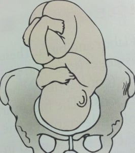
Fetal Position And Presentation Quiz Questions And Answers

Settings

Hey, are you studying maternal and child health nursing concepts? Try these fetal position and presentation quiz questions and answers that we have prepared below and test your knowledge on this topic? This test consists of well-researched and intensive questions and answers that will keep you engaged till the end. You will also get to know your final score by the end of the quiz. So, if you're someone who is studying this subject or have studied it earlier, you should play this quiz and freshen up your memory!
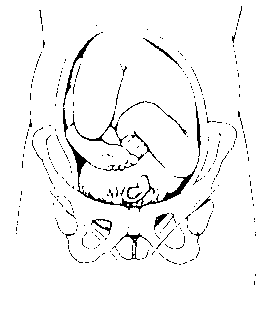
fetal presentation quiz

Determine the fetal position presented in the picture above.

Right occipitotransverse

Left occipitotransverse

Right occipitoposterior

Left occipitoposterior

Rate this question:
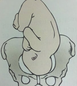
fetal presentation quiz

Identify the fetal position presented above.

Left occipitoanterior

Right occipitoanterior
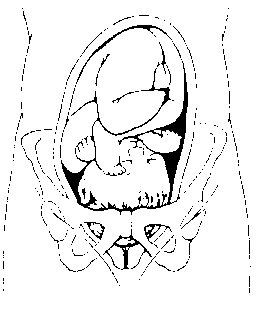
fetal presentation quiz

Determine the fetal position above.

fetal presentation quiz

Determine the fetal position provided in the picture above.
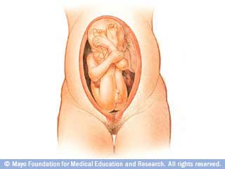
fetal presentation quiz

Determine the fetal presentation presented in this picture.

Shoulder presentation

Frank breech

Single footling breech

Compete breech

Determine the fetal presentation shown in this image.

Complete breech

Shoulder breech
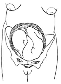
fetal presentation quiz

Determine the fetal presentation presented above by using the Leopold maneuvers.
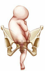
fetal presentation quiz

Determine thefetal position that is presented above.

Single or (double) footling breech

A student nurse is differentiating TRUE labor from FALSE labor to a 39th-week pregnant woman admitted to the labor and delivery department last night. The student nurse needs further teaching if she mentioned which of the following?

"True labor usually begins in your lower back and it may extend to your abdomen"

"There will be thinning or disappearance of the cervix during true labor"

"Ambulation will relieve your abdominal pain when you're in true labor"

"There are no cervical changes in false labor"

What are the prodromal labor signs? Select all that apply.

Braxton Hicks contraction

Left occipital anterior position of the fetus for delivery

Cervical softening

Slight effacement

Bloody show or expulsion of the mucus plug

The nurse practitioner described to the patient that she is 3 cm effacement. The mother was shy to ask questions to the nurse practitioner. When you went to the room and gave her towels, she asked you what effacement means. As an LPN you know that effacement means:

"It is the degree of cervical dilation which is usually 0-10 centimeters "

"it is the thinning or disappearance of the cervix during cervical dilation or labor"

"The nurse practitioner is pertaining to the fetal location in relation to the ischial spine located in your hip bone"

"The nurse practitioner is just hungry at that time, she doesn't know what she's talking about, don't worry about what she said."

What anatomical part of the fetus is the BEST part to hear the fetal heart rate (FHR)?

The back of the fetus

The apical pulse located in the heart

Brachial artery

The popliteal

The patient in labor is hyperventilating with a respiratory rate of 29. You are expecting that the client will have an acid-base imbalance and is in a respiratory alkalosis condition because she is releasing too much carbon dioxide outside her body. As an LPN, you would notice the patient to have which of the following presenting manifestations? Select all that apply.

Productive and persistent coughing

Tingling in the fingers

When should a laboring mother begin to voluntarily push the fetus?

When the cervix is at 10 cm dilation

When the cervix is at 3 cm dilation

When the fetal station is at -2

When the fetal station is engaged

What are the FIVE symptoms of respiratory distress in the newborn? Select all that apply.

Tachycardia

Pallor or dusky color

Flaring of the nares

Chest retraction

Decreased blood pressure

Smiling and crying

What medication should be given to the newly born child to prevent ophthalmia neonatorum from gonorrheal exposure through the birth canal in a vaginal delivery?

Silver nitrate

Butorphanol

What are the CARDINAL MOVEMENTS of the fetus during delivery? Select all that apply.

Internal rotation

Restitution

External rotation

When should we administer a pudendal block?

First stage of labor

Second stage of labor

Third stage of labor

Fourth stage of labor

What are the FIRST signs of regional block effectiveness?

Warmth and tingling sensation of the big toe

Cool and tingling sensation of the big toe

Tingling sensation in the lateral portion of the mouth and slight dizziness

Tingling sensation in the arms and chest pain

The nurse is applying Erythromycin on the newborn to prevent ophthalmia neonatorum. She is aware that to apply this medication correctly to the newborn, she must consider which nursing implication?

Instruct the parents that can wipe the excess medications around the baby's eye after 1 minute

Place a thin line of ointment along the entire lower lid in conjunctival sac

Irrigate the neonate's eye first with normal saline and instill Erythromycin from inner to outer portion of the lower eyelid

Inject the medication in the newborn's eye until it bleeds

When is the appropriate time to give oxytocin to the patient?

After the placenta is delivered

Before the placenta is delivered

Before laboring

If pain does not go away during laboring

What are the normal Puerperium changes relating to the body of the postpartal woman? Select all that apply.

Rugae in the vagina reappears within 3 weeks

Pulse may decrease to 50

The fundus is at the level of the umbilicus after delivery

25,000 WBC count

Palmar erythema declines quickly

Pelvic muscle regain tone in 6 weeks unless diastasis recti

Abdominal muscle regain tone in 3-6 weeks

Bowel movement is expected for 2 to 3 days

Colostrum is expressed first, and then milk

30000 mL/day of urine is normal after delivery because of 40% gain during pregnancy

Chloasma and hyperpigmentation decreases

Coagulation factors postpartally increases

ESR value is elevated for the first 10 days postpartum

Hematocrit decreases because of hemodilution

They are risk for UTI

What is the most common cause of uterine atony in the first 24 hours postpartum?

Decreased lochial flow

Full bladder

Retained placenta

Too late administration of morphine sulfate

What are the objective manifestations of positive maternal-infant bonding? Select all that apply. 

Eye contact between mother and infant

Exploration of infant from head to toe

Stroking, kissing, and fondling the neonate

Smiling, talking,singing and kicking the neonate

Use of exclaiming expressions

Naming the newborn as alien's child

Taking out the clothes of the infant and letting the child cry

A postpartum mother feels unexplained tearfulness, feeling down and "not feeling to eat well". As a nurse, your correct response would be:

"I understand how you feel, this is normal especially if you have a bad looking child"

This is called the postpartum blues, they are normal behaviors of the postpartum mother especially 5 days following delivery"

"You don't feel well?, take a walk with your husband in the hallway with the newborn child."

"I completely understand how you feel, let me refer you to a psychiatrist, this signifies that you have dementia."

A woman who just delivered twenty four hours ago with a full term infant sleeps the whole day and needs assistance in doing activities of daily living. The nurse identified this as normal especially during the first 24 to 48 hours postpartum. According to Riva Rubin, what stage of maternal psychological adaptation  is the postpartum mother in?

Taking-in stage

Taking-hold stage

Letting-go stage

Let-down stage

What are the interventions necessary for episiotomy wounds? Select all that apply.

Provide good perineal care

Lavage the perineum with several squirts of warm water

Blot dry the perineal area without touching the anal area

Carefully wiping the perineal area from back to front to avoid contamination of the vaginal area

Drink at least 3 liters of fluids (at least 4-6 glasses of water) daily

Quiz Review Timeline +

Our quizzes are rigorously reviewed, monitored and continuously updated by our expert board to maintain accuracy, relevance, and timeliness.

  • Current Version
  • Feb 27, 2024 Quiz Edited by ProProfs Editorial Team Expert Reviewed by Farah Naz
  • May 24, 2012 Quiz Created by Nursejbv21

Related Topics

  • Breastfeeding

Recent Quizzes

Featured Quizzes

Popular Topics

  • ACLS Quizzes
  • AHS Quizzes
  • CEN Quizzes
  • CMA Quizzes
  • CPR Quizzes
  • EMT Quizzes
  • GPAT Quizzes
  • MLT Quizzes
  • NBME Quizzes
  • NCCT Quizzes
  • NCLEX Quizzes
  • RMT Quizzes
  • USMLE Quizzes

Back to Top

Related Quizzes

Wait! Here's an interesting quiz for you.

Fetal positions

There is a printable worksheet available for download here so you can take the quiz with pen and paper.

From the quiz author

Cephalic position Position is designated by three-letter abbreviation: First letter designates the location of presenting part to the left (L) right (R) of the womans pelvis Second - specific fetal part presenting: occiput (O),sacrum (S), metum (M) and shoulder (A) Third - desingate relationship of the presenting fetal part to the womans pelvis such as anterior (A), posterior (P), or transverse (T)

The scorecard of a champion

100% needed

Midwest States and Capitals

Other Games of Interest

inferior and lateral view of skull

Latest Quiz Activities

  • An unregistered player played the game 4 hours ago
  • An unregistered player played the game 5 hours ago
  • An unregistered player played the game 11 hours ago
  • An unregistered player played the game 3 days ago

Fetal positions — Quiz Information

This is an online quiz called Fetal positions

You can use it as Fetal positions practice, completely free to play.

Cloud Types

Appointments at Mayo Clinic

  • Pregnancy week by week
  • Fetal presentation before birth

The way a baby is positioned in the uterus just before birth can have a big effect on labor and delivery. This positioning is called fetal presentation.

Babies twist, stretch and tumble quite a bit during pregnancy. Before labor starts, however, they usually come to rest in a way that allows them to be delivered through the birth canal headfirst. This position is called cephalic presentation. But there are other ways a baby may settle just before labor begins.

Following are some of the possible ways a baby may be positioned at the end of pregnancy.

Head down, face down

When a baby is head down, face down, the medical term for it is the cephalic occiput anterior position. This the most common position for a baby to be born in. With the face down and turned slightly to the side, the smallest part of the baby's head leads the way through the birth canal. It is the easiest way for a baby to be born.
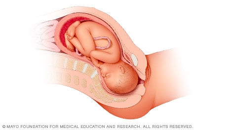
Illustration of the head-down, face-down position

Head down, face up

When a baby is head down, face up, the medical term for it is the cephalic occiput posterior position. In this position, it might be harder for a baby's head to go under the pubic bone during delivery. That can make labor take longer.

Most babies who begin labor in this position eventually turn to be face down. If that doesn't happen, and the second stage of labor is taking a long time, a member of the health care team may reach through the vagina to help the baby turn. This is called manual rotation.

In some cases, a baby can be born in the head-down, face-up position. Use of forceps or a vacuum device to help with delivery is more common when a baby is in this position than in the head-down, face-down position. In some cases, a C-section delivery may be needed.
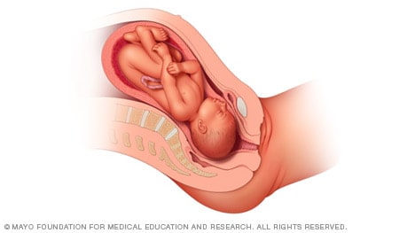
Illustration of the head-down, face-up position

Frank breech

When a baby's feet or buttocks are in place to come out first during birth, it's called a breech presentation. This happens in about 3% to 4% of babies close to the time of birth. The baby shown below is in a frank breech presentation. That's when the knees aren't bent, and the feet are close to the baby's head. This is the most common type of breech presentation.

If you are more than 36 weeks into your pregnancy and your baby is in a frank breech presentation, your health care professional may try to move the baby into a head-down position. This is done using a procedure called external cephalic version. It involves one or two members of the health care team putting pressure on your belly with their hands to get the baby to roll into a head-down position.

If the procedure isn't successful, or if the baby moves back into a breech position, talk with a member of your health care team about the choices you have for delivery. Most babies in a frank breech position are born by planned C-section.

Illustration of the frank breech position

Complete and incomplete breech

A complete breech presentation, as shown below, is when the baby has both knees bent and both legs pulled close to the body. In an incomplete breech, one or both of the legs are not pulled close to the body, and one or both of the feet or knees are below the baby's buttocks. If a baby is in either of these positions, you might feel kicking in the lower part of your belly.

If you are more than 36 weeks into your pregnancy and your baby is in a complete or incomplete breech presentation, your health care professional may try to move the baby into a head-down position. This is done using a procedure called external cephalic version. It involves one or two members of the health care team putting pressure on your belly with their hands to get the baby to roll into a head-down position.

If the procedure isn't successful, or if the baby moves back into a breech position, talk with a member of your health care team about the choices you have for delivery. Many babies in a complete or incomplete breech position are born by planned C-section.

Illustration of a complete breech presentation

When a baby is sideways — lying horizontal across the uterus, rather than vertical — it's called a transverse lie. In this position, the baby's back might be:

  • Down, with the back facing the birth canal.
  • Sideways, with one shoulder pointing toward the birth canal.
  • Up, with the hands and feet facing the birth canal.

Although many babies are sideways early in pregnancy, few stay this way when labor begins.

If your baby is in a transverse lie during week 37 of your pregnancy, your health care professional may try to move the baby into a head-down position. This is done using a procedure called external cephalic version. External cephalic version involves one or two members of your health care team putting pressure on your belly with their hands to get the baby to roll into a head-down position.

If the procedure isn't successful, or if the baby moves back into a transverse lie, talk with a member of your health care team about the choices you have for delivery. Many babies who are in a transverse lie are born by C-section.
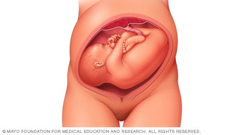
Illustration of baby lying sideways

If you're pregnant with twins and only the twin that's lower in the uterus is head down, as shown below, your health care provider may first deliver that baby vaginally.

Then, in some cases, your health care team may suggest delivering the second twin in the breech position. Or they may try to move the second twin into a head-down position. This is done using a procedure called external cephalic version. External cephalic version involves one or two members of the health care team putting pressure on your belly with their hands to get the baby to roll into a head-down position.

Your health care team may suggest delivery by C-section for the second twin if:

  • An attempt to deliver the baby in the breech position is not successful.
  • You do not want to try to have the baby delivered vaginally in the breech position.
  • An attempt to move the baby into a head-down position is not successful.
  • You do not want to try to move the baby to a head-down position.

In some cases, your health care team may advise that you have both twins delivered by C-section. That might happen if the lower twin is not head down, the second twin has low or high birth weight as compared to the first twin, or if preterm labor starts.
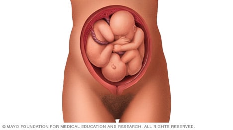
Illustration of twins before birth

  • Landon MB, et al., eds. Normal labor and delivery. In: Gabbe's Obstetrics: Normal and Problem Pregnancies. 8th ed. Elsevier; 2021. https://www.clinicalkey.com. Accessed May 19, 2023.
  • Holcroft Argani C, et al. Occiput posterior position. https://www.updtodate.com/contents/search. Accessed May 19, 2023.
  • Frequently asked questions: If your baby is breech. American College of Obstetricians and Gynecologists https://www.acog.org/womens-health/faqs/if-your-baby-is-breech. Accessed May 22, 2023.
  • Hofmeyr GJ. Overview of breech presentation. https://www.updtodate.com/contents/search. Accessed May 22, 2023.
  • Strauss RA, et al. Transverse fetal lie. https://www.updtodate.com/contents/search. Accessed May 22, 2023.
  • Chasen ST, et al. Twin pregnancy: Labor and delivery. https://www.updtodate.com/contents/search. Accessed May 22, 2023.
  • Cohen R, et al. Is vaginal delivery of a breech second twin safe? A comparison between delivery of vertex and non-vertex second twins. The Journal of Maternal-Fetal & Neonatal Medicine. 2021; doi:10.1080/14767058.2021.2005569.
  • Marnach ML (expert opinion). Mayo Clinic. May 31, 2023.

Products and Services

  • A Book: Obstetricks
  • A Book: Mayo Clinic Guide to a Healthy Pregnancy
  • 3rd trimester pregnancy
  • Fetal development: The 3rd trimester
  • Overdue pregnancy
  • Pregnancy due date calculator
  • Prenatal care: 3rd trimester

Mayo Clinic does not endorse companies or products. Advertising revenue supports our not-for-profit mission.

  • Opportunities

Mayo Clinic Press

Check out these best-sellers and special offers on books and newsletters from Mayo Clinic Press .

  • Mayo Clinic on Incontinence - Mayo Clinic Press Mayo Clinic on Incontinence
  • The Essential Diabetes Book - Mayo Clinic Press The Essential Diabetes Book
  • Mayo Clinic on Hearing and Balance - Mayo Clinic Press Mayo Clinic on Hearing and Balance
  • FREE Mayo Clinic Diet Assessment - Mayo Clinic Press FREE Mayo Clinic Diet Assessment
  • Mayo Clinic Health Letter - FREE book - Mayo Clinic Press Mayo Clinic Health Letter - FREE book
  • Healthy Lifestyle

Your gift holds great power – donate today!

Make your tax-deductible gift and be a part of the cutting-edge research and care that's changing medicine.

  • Getting Pregnant
  • Registry Builder
  • Baby Products
  • Birth Clubs
  • See all in Community
  • Ovulation Calculator
  • How To Get Pregnant
  • How To Get Pregnant Fast
  • Ovulation Discharge
  • Implantation Bleeding
  • Ovulation Symptoms
  • Pregnancy Symptoms
  • Am I Pregnant?
  • Pregnancy Tests
  • See all in Getting Pregnant
  • Due Date Calculator
  • Pregnancy Week by Week
  • Pregnant Sex
  • Weight Gain Tracker
  • Signs of Labor
  • Morning Sickness
  • COVID Vaccine and Pregnancy
  • Fetal Weight Chart
  • Fetal Development
  • Pregnancy Discharge
  • Find Out Baby Gender
  • Chinese Gender Predictor
  • See all in Pregnancy
  • Baby Name Generator
  • Top Baby Names 2023
  • Top Baby Names 2024
  • How to Pick a Baby Name
  • Most Popular Baby Names
  • Baby Names by Letter
  • Gender Neutral Names
  • Unique Boy Names
  • Unique Girl Names
  • Top baby names by year
  • See all in Baby Names
  • Baby Development
  • Baby Feeding Guide
  • Newborn Sleep
  • When Babies Roll Over
  • First-Year Baby Costs Calculator
  • Postpartum Health
  • Baby Poop Chart
  • See all in Baby
  • Average Weight & Height
  • Autism Signs
  • Child Growth Chart
  • Night Terrors
  • Moving from Crib to Bed
  • Toddler Feeding Guide
  • Potty Training
  • Bathing and Grooming
  • See all in Toddler
  • Height Predictor
  • Potty Training: Boys
  • Potty training: Girls
  • How Much Sleep? (Ages 3+)
  • Ready for Preschool?
  • Thumb-Sucking
  • Gross Motor Skills
  • Napping (Ages 2 to 3)
  • See all in Child
  • Photos: Rashes & Skin Conditions
  • Symptom Checker
  • Vaccine Scheduler
  • Reducing a Fever
  • Acetaminophen Dosage Chart
  • Constipation in Babies
  • Ear Infection Symptoms
  • Head Lice 101
  • See all in Health
  • Second Pregnancy
  • Daycare Costs
  • Family Finance
  • Stay-At-Home Parents
  • Breastfeeding Positions
  • See all in Family
  • Baby Sleep Training
  • Preparing For Baby
  • My Custom Checklist
  • My Registries
  • Take the Quiz
  • Best Baby Products
  • Best Breast Pump
  • Best Convertible Car Seat
  • Best Infant Car Seat
  • Best Baby Bottle
  • Best Baby Monitor
  • Best Stroller
  • Best Diapers
  • Best Baby Carrier
  • Best Diaper Bag
  • Best Highchair
  • See all in Baby Products
  • Why Pregnant Belly Feels Tight
  • Early Signs of Twins
  • Teas During Pregnancy
  • Baby Head Circumference Chart
  • How Many Months Pregnant Am I
  • What is a Rainbow Baby
  • Braxton Hicks Contractions
  • HCG Levels By Week
  • When to Take a Pregnancy Test
  • Am I Pregnant
  • Why is Poop Green
  • Can Pregnant Women Eat Shrimp
  • Insemination
  • UTI During Pregnancy
  • Vitamin D Drops
  • Best Baby Forumla
  • Postpartum Depression
  • Low Progesterone During Pregnancy
  • Baby Shower
  • Baby Shower Games

Breech, posterior, transverse lie: What position is my baby in?

Layan Alrahmani, M.D.

Fetal presentation, or how your baby is situated in your womb at birth, is determined by the body part that's positioned to come out first, and it can affect the way you deliver. At the time of delivery, 97 percent of babies are head-down (cephalic presentation). But there are several other possibilities, including feet or bottom first (breech) as well as sideways (transverse lie) and diagonal (oblique lie).

Fetal presentation and position

During the last trimester of your pregnancy, your provider will check your baby's presentation by feeling your belly to locate the head, bottom, and back. If it's unclear, your provider may do an ultrasound or an internal exam to feel what part of the baby is in your pelvis.

Fetal position refers to whether the baby is facing your spine (anterior position) or facing your belly (posterior position). Fetal position can change often: Your baby may be face up at the beginning of labor and face down at delivery.

Here are the many possibilities for fetal presentation and position in the womb.

Medical illustrations by Jonathan Dimes

Head down, facing down (anterior position)

A baby who is head down and facing your spine is in the anterior position. This is the most common fetal presentation and the easiest position for a vaginal delivery.

This position is also known as "occiput anterior" because the back of your baby's skull (occipital bone) is in the front (anterior) of your pelvis.

Head down, facing up (posterior position)

In the posterior position , your baby is head down and facing your belly. You may also hear it called "sunny-side up" because babies who stay in this position are born facing up. But many babies who are facing up during labor rotate to the easier face down (anterior) position before birth.

Posterior position is formally known as "occiput posterior" because the back of your baby's skull (occipital bone) is in the back (posterior) of your pelvis.

Frank breech

In the frank breech presentation, both the baby's legs are extended so that the feet are up near the face. This is the most common type of breech presentation. Breech babies are difficult to deliver vaginally, so most arrive by c-section .

Some providers will attempt to turn your baby manually to the head down position by applying pressure to your belly. This is called an external cephalic version , and it has a 58 percent success rate for turning breech babies. For more information, see our article on breech birth .

Complete breech

A complete breech is when your baby is bottom down with hips and knees bent in a tuck or cross-legged position. If your baby is in a complete breech, you may feel kicking in your lower abdomen.

Incomplete breech

In an incomplete breech, one of the baby's knees is bent so that the foot is tucked next to the bottom with the other leg extended, positioning that foot closer to the face.

Single footling breech

In the single footling breech presentation, one of the baby's feet is pointed toward your cervix.

Double footling breech

In the double footling breech presentation, both of the baby's feet are pointed toward your cervix.

Transverse lie

In a transverse lie, the baby is lying horizontally in your uterus and may be facing up toward your head or down toward your feet. Babies settle this way less than 1 percent of the time, but it happens more commonly if you're carrying multiples or deliver before your due date.

If your baby stays in a transverse lie until the end of your pregnancy, it can be dangerous for delivery. Your provider will likely schedule a c-section or attempt an external cephalic version , which is highly successful for turning babies in this position.

Oblique lie

In rare cases, your baby may lie diagonally in your uterus, with his rump facing the side of your body at an angle.

Like the transverse lie, this position is more common earlier in pregnancy, and it's likely your provider will intervene if your baby is still in the oblique lie at the end of your third trimester.

Was this article helpful?

What to know if your baby is breech

diagram of breech baby, facing head-up in uterus

What's a sunny-side up baby?

pregnant woman resting on birth ball

What happens to your baby right after birth

A newborn baby wrapped in a receiving blanket in the hospital.

How your twins’ fetal positions affect labor and delivery

illustration of twin babies head down in utero

BabyCenter's editorial team is committed to providing the most helpful and trustworthy pregnancy and parenting information in the world. When creating and updating content, we rely on credible sources: respected health organizations, professional groups of doctors and other experts, and published studies in peer-reviewed journals. We believe you should always know the source of the information you're seeing. Learn more about our editorial and medical review policies .

Ahmad A et al. 2014. Association of fetal position at onset of labor and mode of delivery: A prospective cohort study. Ultrasound in obstetrics & gynecology 43(2):176-182. https://www.ncbi.nlm.nih.gov/pubmed/23929533 Opens a new window [Accessed September 2021]

Gray CJ and Shanahan MM. 2019. Breech presentation. StatPearls.  https://www.ncbi.nlm.nih.gov/books/NBK448063/ Opens a new window [Accessed September 2021]

Hankins GD. 1990. Transverse lie. American Journal of Perinatology 7(1):66-70.  https://www.ncbi.nlm.nih.gov/pubmed/2131781 Opens a new window [Accessed September 2021]

Medline Plus. 2020. Your baby in the birth canal. U.S. National Library of Medicine. https://medlineplus.gov/ency/article/002060.htm Opens a new window [Accessed September 2021]

Kate Marple

Where to go next

doctor holding ultrasound probe

fetal presentation quiz

Fetal Presentation, Position, and Lie (Including Breech Presentation)

  • Variations in Fetal Position and Presentation |

During pregnancy, the fetus can be positioned in many different ways inside the mother's uterus. The fetus may be head up or down or facing the mother's back or front. At first, the fetus can move around easily or shift position as the mother moves. Toward the end of the pregnancy the fetus is larger, has less room to move, and stays in one position. How the fetus is positioned has an important effect on delivery and, for certain positions, a cesarean delivery is necessary. There are medical terms that describe precisely how the fetus is positioned, and identifying the fetal position helps doctors to anticipate potential difficulties during labor and delivery.

Presentation refers to the part of the fetus’s body that leads the way out through the birth canal (called the presenting part). Usually, the head leads the way, but sometimes the buttocks (breech presentation), shoulder, or face leads the way.

Position refers to whether the fetus is facing backward (occiput anterior) or forward (occiput posterior). The occiput is a bone at the back of the baby's head. Therefore, facing backward is called occiput anterior (facing the mother’s back and facing down when the mother lies on her back). Facing forward is called occiput posterior (facing toward the mother's pubic bone and facing up when the mother lies on her back).

Lie refers to the angle of the fetus in relation to the mother and the uterus. Up-and-down (with the baby's spine parallel to mother's spine, called longitudinal) is normal, but sometimes the lie is sideways (transverse) or at an angle (oblique).

For these aspects of fetal positioning, the combination that is the most common, safest, and easiest for the mother to deliver is the following:

Head first (called vertex or cephalic presentation)

Facing backward (occiput anterior position)

Spine parallel to mother's spine (longitudinal lie)

Neck bent forward with chin tucked

Arms folded across the chest

If the fetus is in a different position, lie, or presentation, labor may be more difficult, and a normal vaginal delivery may not be possible.

Variations in fetal presentation, position, or lie may occur when

The fetus is too large for the mother's pelvis (fetopelvic disproportion).

The uterus is abnormally shaped or contains growths such as fibroids .

The fetus has a birth defect .

There is more than one fetus (multiple gestation).

fetal presentation quiz

Position and Presentation of the Fetus

Variations in fetal position and presentation.

Some variations in position and presentation that make delivery difficult occur frequently.

Occiput posterior position

In occiput posterior position (sometimes called sunny-side up), the fetus is head first (vertex presentation) but is facing forward (toward the mother's pubic bone—that is, facing up when the mother lies on her back). This is a very common position that is not abnormal, but it makes delivery more difficult than when the fetus is in the occiput anterior position (facing toward the mother's spine—that is facing down when the mother lies on her back).

When a fetus faces up, the neck is often straightened rather than bent,which requires more room for the head to pass through the birth canal. Delivery assisted by a vacuum device or forceps or cesarean delivery may be necessary.

Breech presentation

In breech presentation, the baby's buttocks or sometimes the feet are positioned to deliver first (before the head).

When delivered vaginally, babies that present buttocks first are more at risk of injury or even death than those that present head first.

The reason for the risks to babies in breech presentation is that the baby's hips and buttocks are not as wide as the head. Therefore, when the hips and buttocks pass through the cervix first, the passageway may not be wide enough for the head to pass through. In addition, when the head follows the buttocks, the neck may be bent slightly backwards. The neck being bent backward increases the width required for delivery as compared to when the head is angled forward with the chin tucked, which is the position that is easiest for delivery. Thus, the baby’s body may be delivered and then the head may get caught and not be able to pass through the birth canal. When the baby’s head is caught, this puts pressure on the umbilical cord in the birth canal, so that very little oxygen can reach the baby. Brain damage due to lack of oxygen is more common among breech babies than among those presenting head first.

In a first delivery, these problems may occur more frequently because a woman’s tissues have not been stretched by previous deliveries. Because of risk of injury or even death to the baby, cesarean delivery is preferred when the fetus is in breech presentation, unless the doctor is very experienced with and skilled at delivering breech babies or there is not an adequate facility or equipment to safely perform a cesarean delivery.

Breech presentation is more likely to occur in the following circumstances:

Labor starts too soon (preterm labor).

The uterus is abnormally shaped or contains abnormal growths such as fibroids .

Other presentations

In face presentation, the baby's neck arches back so that the face presents first rather than the top of the head.

In brow presentation, the neck is moderately arched so that the brow presents first.

Usually, fetuses do not stay in a face or brow presentation. These presentations often change to a vertex (top of the head) presentation before or during labor. If they do not, a cesarean delivery is usually recommended.

In transverse lie, the fetus lies horizontally across the birth canal and presents shoulder first. A cesarean delivery is done, unless the fetus is the second in a set of twins. In such a case, the fetus may be turned to be delivered through the vagina.

quizzes_lightbulb_red

  • Cookie Preferences

This icon serves as a link to download the eSSENTIAL Accessibility assistive technology app for individuals with physical disabilities. It is featured as part of our commitment to diversity and inclusion.

Copyright © 2024 Merck & Co., Inc., Rahway, NJ, USA and its affiliates. All rights reserved.

fetal presentation quiz

Fetal Presentation, Position, and Lie (Including Breech Presentation)

  • Key Points |

Abnormal fetal lie or presentation may occur due to fetal size, fetal anomalies, uterine structural abnormalities, multiple gestation, or other factors. Diagnosis is by examination or ultrasonography. Management is with physical maneuvers to reposition the fetus, operative vaginal delivery , or cesarean delivery .

Terms that describe the fetus in relation to the uterus, cervix, and maternal pelvis are

Fetal presentation: Fetal part that overlies the maternal pelvic inlet; vertex (cephalic), face, brow, breech, shoulder, funic (umbilical cord), or compound (more than one part, eg, shoulder and hand)

Fetal position: Relation of the presenting part to an anatomic axis; for transverse presentation, occiput anterior, occiput posterior, occiput transverse

Fetal lie: Relation of the fetus to the long axis of the uterus; longitudinal, oblique, or transverse

Normal fetal lie is longitudinal, normal presentation is vertex, and occiput anterior is the most common position.

Abnormal fetal lie, presentation, or position may occur with

Fetopelvic disproportion (fetus too large for the pelvic inlet)

Fetal congenital anomalies

Uterine structural abnormalities (eg, fibroids, synechiae)

Multiple gestation

Several common types of abnormal lie or presentation are discussed here.

fetal presentation quiz

Transverse lie

Fetal position is transverse, with the fetal long axis oblique or perpendicular rather than parallel to the maternal long axis. Transverse lie is often accompanied by shoulder presentation, which requires cesarean delivery.

Breech presentation

There are several types of breech presentation.

Frank breech: The fetal hips are flexed, and the knees extended (pike position).

Complete breech: The fetus seems to be sitting with hips and knees flexed.

Single or double footling presentation: One or both legs are completely extended and present before the buttocks.

Types of breech presentations

Breech presentation makes delivery difficult ,primarily because the presenting part is a poor dilating wedge. Having a poor dilating wedge can lead to incomplete cervical dilation, because the presenting part is narrower than the head that follows. The head, which is the part with the largest diameter, can then be trapped during delivery.

Additionally, the trapped fetal head can compress the umbilical cord if the fetal umbilicus is visible at the introitus, particularly in primiparas whose pelvic tissues have not been dilated by previous deliveries. Umbilical cord compression may cause fetal hypoxemia.

fetal presentation quiz

Predisposing factors for breech presentation include

Preterm labor

Uterine abnormalities

Fetal anomalies

If delivery is vaginal, breech presentation may increase risk of

Umbilical cord prolapse

Birth trauma

Perinatal death

fetal presentation quiz

Face or brow presentation

In face presentation, the head is hyperextended, and position is designated by the position of the chin (mentum). When the chin is posterior, the head is less likely to rotate and less likely to deliver vaginally, necessitating cesarean delivery.

Brow presentation usually converts spontaneously to vertex or face presentation.

Occiput posterior position

The most common abnormal position is occiput posterior.

The fetal neck is usually somewhat deflexed; thus, a larger diameter of the head must pass through the pelvis.

Progress may arrest in the second phase of labor. Operative vaginal delivery or cesarean delivery is often required.

Position and Presentation of the Fetus

If a fetus is in the occiput posterior position, operative vaginal delivery or cesarean delivery is often required.

In breech presentation, the presenting part is a poor dilating wedge, which can cause the head to be trapped during delivery, often compressing the umbilical cord.

For breech presentation, usually do cesarean delivery at 39 weeks or during labor, but external cephalic version is sometimes successful before labor, usually at 37 or 38 weeks.

quizzes_lightbulb_red

  • Cookie Preferences

This icon serves as a link to download the eSSENTIAL Accessibility assistive technology app for individuals with physical disabilities. It is featured as part of our commitment to diversity and inclusion. M

Copyright © 2024 Merck & Co., Inc., Rahway, NJ, USA and its affiliates. All rights reserved.

Legal stuff

  • Self Guided Tutorial
  • EFM In-Depth
  • Assessments
  • Fetal Tracing Index

Electronic Fetal Monitoring

Basic and Advanced Study

Practice Quizzes 1-5

Try your hand at the following quizzes. Scroll down for another when you’re done. When you’ve finished these first five, here are five more .

fetal presentation quiz

  • Mammary Glands
  • Fallopian Tubes
  • Supporting Ligaments
  • Reproductive System
  • Gametogenesis
  • Placental Development
  • Maternal Adaptations
  • Menstrual Cycle
  • Antenatal Care
  • Small for Gestational Age
  • Large for Gestational Age
  • RBC Isoimmunisation
  • Prematurity
  • Prolonged Pregnancy
  • Multiple Pregnancy
  • Miscarriage
  • Recurrent Miscarriage
  • Ectopic Pregnancy
  • Hyperemesis Gravidarum
  • Gestational Trophoblastic Disease
  • Breech Presentation
  • Abnormal lie, Malpresentation and Malposition
  • Oligohydramnios
  • Polyhydramnios
  • Placenta Praevia
  • Placental Abruption
  • Pre-Eclampsia
  • Gestational Diabetes
  • Headaches in Pregnancy
  • Haematological
  • Obstetric Cholestasis
  • Thyroid Disease in Pregnancy
  • Epilepsy in Pregnancy
  • Induction of Labour
  • Operative Vaginal Delivery
  • Prelabour Rupture of Membranes
  • Caesarean Section
  • Shoulder Dystocia
  • Cord Prolapse
  • Uterine Rupture
  • Amniotic Fluid Embolism
  • Primary PPH
  • Secondary PPH
  • Psychiatric Disease
  • Postpartum Contraception
  • Breastfeeding Problems
  • Primary Dysmenorrhoea
  • Amenorrhoea and Oligomenorrhoea
  • Heavy Menstrual Bleeding
  • Endometriosis
  • Endometrial Cancer
  • Adenomyosis
  • Cervical Polyps
  • Cervical Ectropion
  • Cervical Intraepithelial Neoplasia + Cervical Screening
  • Cervical Cancer
  • Polycystic Ovary Syndrome (PCOS)
  • Ovarian Cysts & Tumours
  • Urinary Incontinence
  • Genitourinary Prolapses
  • Bartholin's Cyst
  • Lichen Sclerosus
  • Vulval Carcinoma
  • Introduction to Infertility
  • Female Factor Infertility
  • Male Factor Infertility
  • Female Genital Mutilation
  • Barrier Contraception
  • Combined Hormonal
  • Progesterone Only Hormonal
  • Intrauterine System & Device
  • Emergency Contraception
  • Pelvic Inflammatory Disease
  • Genital Warts
  • Genital Herpes
  • Trichomonas Vaginalis
  • Bacterial Vaginosis
  • Vulvovaginal Candidiasis
  • Obstetric History
  • Gynaecological History
  • Sexual History
  • Obstetric Examination
  • Speculum Examination
  • Bimanual Examination
  • Amniocentesis
  • Chorionic Villus Sampling
  • Hysterectomy
  • Endometrial Ablation
  • Tension-Free Vaginal Tape
  • Contraceptive Implant
  • Fitting an IUS or IUD

Abnormal Fetal lie, Malpresentation and Malposition

Original Author(s): Anna Mcclune Last updated: 1st December 2018 Revisions: 12

  • 1 Definitions
  • 2 Risk Factors
  • 3.2 Presentation
  • 3.3 Position
  • 4 Investigations
  • 5.1 Abnormal Fetal Lie
  • 5.2 Malpresentation
  • 5.3 Malposition

The lie, presentation and position of a fetus are important during labour and delivery.

In this article, we will look at the risk factors, examination and management of abnormal fetal lie, malpresentation and malposition.

Definitions

  • Longitudinal, transverse or oblique
  • Cephalic vertex presentation is the most common and is considered the safest
  • Other presentations include breech, shoulder, face and brow
  • Usually the fetal head engages in the occipito-anterior position (the fetal occiput facing anteriorly) – this is ideal for birth
  • Other positions include occipito-posterior and occipito-transverse.

Note: Breech presentation is the most common malpresentation, and is covered in detail here .

fetal presentation quiz

Fig 1 – The two most common fetal presentations: cephalic and breech.

Risk Factors

The risk factors for abnormal fetal lie, malpresentation and malposition include:

  • Multiple pregnancy
  • Uterine abnormalities (e.g fibroids, partial septate uterus)
  • Fetal abnormalities
  • Placenta praevia
  • Primiparity

Identifying Fetal Lie, Presentation and Position

The fetal lie and presentation can usually be identified via abdominal examination. The fetal position is ascertained by vaginal examination.

For more information on the obstetric examination, see here .

  • Face the patient’s head
  • Place your hands on either side of the uterus and gently apply pressure; one side will feel fuller and firmer – this is the back, and fetal limbs may feel ‘knobbly’ on the opposite side

Presentation

  • Palpate the lower uterus (above the symphysis pubis) with the fingers of both hands; the head feels hard and round (cephalic) and the bottom feels soft and triangular (breech)
  • You may be able to gently push the fetal head from side to side

The fetal lie and presentation may not be possible to identify if the mother has a high BMI, if she has not emptied her bladder, if the fetus is small or if there is polyhydramnios .

During labour, vaginal examination is used to assess the position of the fetal head (in a cephalic vertex presentation). The landmarks of the fetal head, including the anterior and posterior fontanelles, indicate the position.

fetal presentation quiz

Fig 2 – Assessing fetal lie and presentation.

Investigations

Any suspected abnormal fetal lie or malpresentation should be confirmed by an ultrasound scan . This could also demonstrate predisposing uterine or fetal abnormalities.

Abnormal Fetal Lie

If the fetal lie is abnormal, an external cephalic version (ECV) can be attempted – ideally between 36 and 38 weeks gestation.

ECV is the manipulation of the fetus to a cephalic presentation through the maternal abdomen.

It has an approximate success rate of 50% in primiparous women and 60% in multiparous women. Only 8% of breech presentations will spontaneously revert to cephalic in primiparous women over 36 weeks gestation.

Complications of ECV are rare but include fetal distress , premature rupture of membranes, antepartum haemorrhage (APH) and placental abruption. The risk of an emergency caesarean section (C-section) within 24 hours is around 1 in 200.

ECV is contraindicated in women with a recent APH, ruptured membranes, uterine abnormalities or a previous C-section .

fetal presentation quiz

Fig 3 – External cephalic version.

Malpresentation

The management of malpresentation is dependent on the presentation.

  • Breech – attempt ECV before labour, vaginal breech delivery or C-section
  • Brow – a C-section is necessary
  • If the chin is anterior (mento-anterior) a normal labour is possible; however, it is likely to be prolonged and there is an increased risk of a C-section being required
  • If the chin is posterior (mento-posterior) then a C-section is necessary
  • Shoulder – a C-section is necessary

Malposition

90% of malpositions spontaneously rotate to occipito-anterior as labour progresses. If the fetal head does not rotate, rotation and operative vaginal delivery can be attempted. Alternatively a C-section can be performed.

  • Usually the fetal head engages in the occipito-anterior position (the fetal occiput facing anteriorly) - this is ideal for birth

If the fetal lie is abnormal, an external cephalic version (ECV) can be attempted - ideally between 36 and 38 weeks gestation.

  • Breech - attempt ECV before labour, vaginal breech delivery or C-section

Found an error? Is our article missing some key information? Make the changes yourself here!

Once you've finished editing, click 'Submit for Review', and your changes will be reviewed by our team before publishing on the site.

We use cookies to improve your experience on our site and to show you relevant advertising. To find out more, read our privacy policy .

Privacy Overview

Fetal Development & Fetal Growth Assessment

fetal presentation quiz

In the realm of maternal care, understanding and monitoring fetal development and fetal growth are paramount for ensuring the well-being of both the mother and the developing baby. The intricate processes of fetal development, from conception to birth, lay the foundation for a healthy and successful transition into the world outside the womb. Likewise, monitoring fetal growth throughout pregnancy aids in identifying potential complications early on, enabling healthcare professionals to provide timely interventions and personalized care.

This article aims to provide nursing professionals with an essential guide to fetal development and fetal growth assessment . By delving into the latest research and evidence-based practices, we aim to equip nurses with the knowledge and skills necessary to promote optimal outcomes for expectant mothers and their unborn children.

Table of Contents

Fertilization, implantation, chorionic villi, amniotic membranes, amniotic fluid, umbilical cord.

  • 4th Week of Gestation
  • 8th Week of Gestation
  • 12th Week of Gestation
  • 16th Week of Gestation
  • 20th Week of Gestation
  • 24th Week of Gestation
  • 28th Week of Gestation
  • 32nd Week of Gestation
  • 36th Week of Gestation
  • 40th Week of Gestation

McDonald’s Rule

Fetal movement, rhythm strip testing, nonstress test, contraction stress testing, ultrasonography, electrocardiography, magnetic resonance imaging, maternal serum alpha fetoprotein, amniocentesis, biophysical profile, fetal development.

Fertilization is the process wherein the ovum and the spermatozoa unite at the ampullary portion of the fallopian tube, the usual site of fertilization.

  • A mature ovum can only be fertilized within 24 to 48 hours after being released.
  • The functional life of the spermatozoa is only 48 to 72 hours.
  • The best time that fertilization would occur is 72 hours after sexual intercourse.
  • The fertilized ovum is propelled along the tube through the help of the peristaltic movements of the fallopian tube and the tube’s cilia.
  • The average time that the sperm can reach the cervix is within 90 seconds, and it can reach the outer fallopian tube within 5 minutes.
  • If the ovum has already been penetrated by a spermatozoon, it changes its composition so that it becomes impermeable to other spermatozoa.
  • After penetration, the chromosomal materials of both the ovum and the spermatozoon combine to form a zygote.
  • the maturation of both the sperm and the ovum;
  • the ability of the sperm to reach the ovum; and
  • the ability of the sperm to penetrate the cell membrane and achieve fertilization.
  • The migration of the zygote towards the uterus reaches 3 to 4 days, and it is propelled by the muscular contractions in the fallopian tube.
  • Mitotic cell division or cleaving starts after 24 hours. The rate of cleaving is an average of one every 22 hours.
  • As the zygote reaches the uterus, it already has 16 to 50 cell divisions, and it is now called a morula because of its bumpy appearance.
  • The morula floats freely along the body of the uterus within 3 to 4 days, and it becomes a blastocyst.
  • The blastocyst attaches into the endometrium, and this process is called implantation, which occurs 8 to 10 days after fertilization.
  • Apposition, or the brushing of the blastocyst against the endometrium, is the first part of implantation.
  • Adhesion occurs afterwards as the blastocyst attaches to the surface of the endometrium, then invasion, as it settles into the folds of the endometrium.
  • On the day of implantation, the woman may experience a small amount of vaginal spotting as the capillaries by the implanting blastocyst.
  • As implantation occurs, the zygote now becomes an embryo.

Fetal Structures

Decidua

  • The uterine endometrium continues to thicken because of the corpus luteum that is influenced by hCG, and instead of sloughing off in a usual menstrual cycle , it becomes the deciduas.
  • The deciduas are divided into three parts: basalis, capsularis, and vera.
  • The decidua basalis is the innermost portion of the layer which rests directly under the embryo.
  • The decidua capsularis encapsulates the trophoblast’s surface.
  • The decidua vera becomes the remaining portion of the uterine lining,and sheds as the lochias.
  • Eventually, the deciduas vera and capsularis fuse because of the enlarging embryo.

Chorionic Villi

  • On the 11 th or 12 th day, the chorionic villi start to form from the miniature villi that protrude from a single layer of cells to start the formation of placenta.
  • The chorionic villi have a central core and fetal capillaries, and a double layer of trophoblast cells.
  • The syncytial layer or the outer portion of the two layers produces placental hormones such as hPL, hCG, estrogen , and progesterone.
  • The Langhans’ layer is the middle layer and it protects the embryo and fetus from infectious diseases. This layer appears to function as early as 12 days’ gestation.
  • The layer disappears on the 20 th to 24 th week of gestation, however, leaving the fetus more susceptible to infections.

The placenta, which is a Latin term for “pancake” for its appearance came from the trophoblast tissues and has a lot of functions that benefit the fetus.

Placenta

  • Nutrients such as glucose , amino acids, vitamins, minerals, fatty acids, and water as well as oxygen are transported through the placenta from the maternal blood supply to the fetus.
  • Placental osmosis also plays an essential part in maintaining the health of the fetus. it is impermeable to a few harmful substances, thereby it does not allow the crossing of these substances towards the fetal blood circulation .
  • The syncytial layer produces various hormones that benefit both the mother and the fetus.
  • The human chorionic gonadotropin is the first placental hormone to be produced, and it ensures that the corpus luteum would continue to produce estrogen and progesterone to support the pregnancy.
  • Estrogen is also one of the hormones produced by the syncytial cells and it aids in the uterine growth and the development of the mammary glands in preparation for lactation.
  • Progesterone is responsible for maintaining the lining of the uterus during pregnancy. It also reduces the contractility of the uterus to prevent preterm labor .
  • Human placental lactogen promotes lactogenic properties and mammary growth in preparation for the lactation of the mother.

amniotic membrane

  • The smooth portion of the chorionic villi eventually becomes the chorionic membrane which forms the sac that contains the amniotic fluid.
  • The amniotic membrane forms under the chorion, giving an appearance that seem like they are only one membrane.
  • The amniotic membrane is also responsible for producing the amniotic fluid and the phospholipids that triggers the formation of prostaglandins, the hormone that initiates uterine contractions.
  • The normal amount of amniotic fluid is 800 to 1000 mL.
  • The role of the amniotic fluid in the safety of the fetus is it protects the fetus from trauma or pressure to the mother’s abdomen. It also regulates the temperature for the fetus and aids in muscular development allowing the fetus to move freely
  • The amniotic fluid also protects the umbilical cord from trauma and pressure, thereby protecting the fetal oxygen supply.
  • The amnion and chorion compose the umbilical cord which connects the embryo to the chorionic villi of the placenta.
  • The main function of the umbilical cord is the transport of oxygen and nutrients from the placenta to the fetus and the return of waste products from the fetus to the placenta.
  • The cord is made up of a gelatinous mucopolysaccharide called Wharton’s jelly that protects the vein and arteries from trauma.
  • The umbilical cord contains only one vein, which carries blood from the placenta to the fetus, and two arteries, which carries blood from the fetus to the placenta.

Fetal Milestones

4 th week of gestation.

  • Spinal cord is formed and fused at the midpoint.
  • Head folds forward and is prominent.
  • The back is bent, which makes the head almost touch the tail.
  • A prominent bulge appears which would later form as the heart.
  • Lateral wings, the body, folds forward and fuse at midline.
  • Arms and legs are budlike structures.
  • Eyes, ears, and nose are barely recognizable.

8 th Week of Gestation

  • Organogenesis is achieved and complete.
  • The heart already developed a septum and valves and is beating rhythmically.
  • Arms and legs have developed.
  • Facial features are noticeable.
  • The genital starts to form but is not yet recognizable.
  • Fetal intestine is rapidly growing.
  • Results of an ultrasound would show a gestational sac which confirms pregnancy.

12 th Week of Gestation

  • The toes and fingers already have nail beds.
  • Faint fetal movements are starting.
  • Early reflexes are present.
  • Tooth buds are forming.
  • Formation of bone ossification centers initiate.
  • The genital is already recognizable through its appearance.
  • Urine secretion begins but is not yet evident.
  • Heartbeat could be detected by Doppler.

16 th Week of Gestation

  • An ordinary stethoscope could detect the fetus’ heart beat.
  • Lanugo has started to form.
  • The pancreas and liver are forming.
  • Urine is present in the amniotic fluid.
  • Fetus starts to swallow the amniotic fluid.
  • Ultrasound could determine the sex of the fetus.

20 th Week of Gestation

  • Mother could sense spontaneous fetal movements.
  • There is hair formation on the head until the eyebrows.
  • The upper intestine contains meconium .
  • Brown fat starts to form behind the kidneys, sternum , and posterior neck.
  • Vernix caseosa also starts to form and covers the skin.
  • Passive antibody transfer begins.
  • The sleep and activity patterns of the fetus are evident.

24 th Week of Gestation

  • Lung surfactant begins to develop.
  • Meconium is present at the rectum.
  • Eyebrows and eyelashes are distinguishable.
  • Eyelids can now open.
  • Pupils react to light.
  • The fetus has reached the age of viability, wherein they could survive externally if cared for in a modern intensive facility.
  • Responds to sudden sounds.

28 th Week of Gestation

  • Surfactant is demonstrated in the amniotic fluid.
  • Alveoli are starting to mature.
  • Testes descend into the scrotal sac.
  • Retinal blood vessels start to form but are highly susceptible to damage.

32 nd Week of Gestation

  • Subcutaneous fat is deposited.
  • Fetus responds to sounds outside the mother’s body through movements.
  • Active Moro reflex is present.
  • Iron stores are starting to develop.
  • Fingernails are starting to grow.

36 th Week of Gestation

  • Depositions of iron, carbohydrate, calcium , and glycogen stores are in the body.
  • Additional subcutaneous fats are deposited.
  • One or two creases are present at the sole of the foot.
  • Lanugo starts to diminish.
  • Some babies turn and assume a vertex presentation.

40 th Week of Gestation

  • Fetus now kicks very actively and hard enough to cause discomfort.
  • The fetal hemoglobin is being converted to adult hemoglobin.
  • Vernix caseosa is fully formed.
  • Fingernails extend to the fingertips.
  • The soles of the feet have creases that cover at least two-thirds of the surface.

The slow but sure development of the fetus inside a woman’s body should be monitored to ensure the delivery of a healthy and safe baby. Fetal development is a critical stage in a mother’s responsibility over her children. The role of a mother starts not only during the time that the baby is born, but most especially when she decides that she wants to conceive an offspring.

Fetal Growth Assessment

Estimating fetal growth.

  • McDonald’s rule is the measurement of the fundal height from the symphysis pubis.
  • To measure, instruct the woman to lie supine and start measuring from the symphysis pubis to the uterine fundus.
  • The distance between in centimeters depicts the week of gestation between the 20 th to the 31 st weeks of pregnancy.
  • At 12 weeks, the uterine fundus should be at the level of the symphysis pubis.
  • At 20 weeks, the uterine fundus should be at the level of the umbilicus.
  • At 36 weeks, the uterine fundus should be at the level of the xiphoid process.
  • Quickening or the first fetal movement that is felt by the mother usually starts at 18 to 20 weeks of pregnancy.
  • A healthy fetus moves at an average of at least 10 times a day.
  • In the Sandovsky method , to assess the fetal movement, ask the woman to lie in a recumbent position after a meal and record the number of fetal movements within an hour.
  • In every 10 minutes, the fetus normally moves at least twice or 10 to 12 times in an hour.
  • If there is less than 10 movements in an hour, the woman should repeat the procedure for the next hour.
  • The Cardiff method or the “Count-to-Ten” method , the woman records the time interval between every 10 fetal movements she feels within 60 minutes.

Fetal Heart Rate

  • The normal fetal heart rate is 120 to 160 beats per minute.
  • In rhythm strip testing, the fetal heart rate is assessed if a good baseline heart rate or a degree of variability is present.
  • The results are categorized as absent (none apparent), minimal (extremely small fluctuations), moderate (a range of 6-25 beats per minute), and marked (range over 25 beats per minute).
  • The rhythm strip testing is done as the woman is asked to remain in a fixed position for 20 minutes.
  • In a nonstress testing, the response of the fetal heart rate is measured in response to the fetal movement.
  • The woman is attached to a fetal heart rate and uterine contraction monitor.
  • The woman should push the button of the monitor whenever she feels the fetus move.
  • Normally, when the fetus moves, the fetal heart should increase for about 15 beats per minute and remain elevated for 15 seconds.
  • The nonstress test is done for 10 to 20 minutes.
  • The result is reactive if there are two accelerations of fetal heart rate lasting for 15 seconds that occurs after movement.
  • The result is non reactive if there are no fetal accelerations after a fetal movement, or there is no fetal movement.
  • If the nonstress test is nonreactive, a contraction stress test or biophysical profile will be scheduled.
  • In contraction stress testing, the fetal heart rate is assessed in conjunction with uterine contractions.
  • The woman is attached to an external uterine contraction and fetal heart rate monitor.
  • The woman is instructed to roll a nipple between her fingers and thumb to produce uterine contractions.
  • Within a 10-minute window, three contractions with a duration of 40 seconds or longer must be present.
  • The test is negative or normal if there are no decelerations in the fetal heart rate during contractions.
  • It is positive or abnormal if there is a late deceleration at the end of a contraction and even after the contraction.
  • Ultrasonography measures the response of sound waves against solid objects.
  • It can diagnose a pregnancy of 6 weeks’ gestation, confirm the presence, size, and location of the placenta, establish that the fetus is growing, detect any gross anomalies, establish the fetal sex, and determine the presentation and position of the fetus.
  • The woman has to have a full bladder at the time of the procedure.
  • Have the woman drink a full glass of water every 15 minutes 90 minutes before the procedure until the start of the procedure.
  • Ultrasonography is also used to predict fetal maturity by the measurement of the biparietal diameter of the fetal head.
  • Placental grading can also be done through ultrasound as 0 (12 to 24 weeks), 1 (30 to 32 weeks), 2 (36 weeks), and 3 (38 weeks).
  • The amount of amniotic fluid present can also be detected through ultrasonography and is also a way to estimate fetal health.
  • As early as the 11 th week of pregnancy, fetal ECG can be recorded.
  • However, fetal ECG is inaccurate before the 20 th week as the fetal electrical conduction is still weak.
  • MRI does not have any harmful effects to both the mother and the fetus, and is now largely considered as one of the preferred fetal assessment techniques.
  • MRI can diagnose complications like ectopic pregnancy and trophoblastic disease or H-mole because fetal movements could hide the findings later in pregnancy.

AFP is found in the amniotic fluid and the maternal serum and is produced by the fetal liver.

  • MSAFP levels start to increase at 11 weeks’ gestation and increases steadily until term.
  • The MSAFP level is abnormally high if there is a spina bifida defect or abdominal defect.
  • The MSAFP level is low if the fetus has a chromosomal defect such as Down syndrome .
  • The MSAFP is assessed at the 15 th week of pregnancy and can detect 85% to 90% of neural tube defects and 80% of Down syndrome.

Amniocentesis is the aspiration of amniotic fluid from the pregnant uterus for examination.

  • The test is typically done between the 14 th and 16 th weeks of pregnancy so that there is a generous amount of amniotic fluid present.
  • Before the procedure, instruct the woman to void, and then place her on a supine position.
  • Fetal heart rate and uterine contraction monitors are attached to the woman, and blood pressure and fetal heart rate are taken.
  • An ultrasound is performed first to determine the position of the fetus and the location of a pocket of amniotic fluid and the placenta.
  • Antiseptic solution is applied to the abdomen and local anesthetic is injected.
  • Inform the woman that she might feel pressure as the needle is introduced, but do not advise her to take a deep breath and hold it in.
  • About 15 mL of amniotic fluid is aspirated.
  • Amniotic fluid is analyzed for AFP, bilirubin determination, chromosome analysis, color, fetal fibronectin, inborn errors of metabolism, lecithin-sphingomyelin ratio, and phosphatidylglycerol and desaturated phosphatidylcholine.
  • The biophysical profile combines five parameters into one assessment.
  • Fetal heart rate and breathing measure short-term central nervous system function.
  • The amniotic fluid volume measures long-term adequacy of placental function.
  • The biophysical profile is more accurate than any other single assessment method.
  • The score ranges from 2-10, with 10 as the highest.
  • If the fetus has a score of 8 to 10, it is doing well.
  • If the score is 6, this is considered suspicious.
  • A score of 4 denotes that fetus might be in jeopardy.
  • The assessment is similar to that of an Apgar scoring , and it is commonly called as fetal Apgar.

Fetal assessment is just one of the many assessments that a pregnant woman must undergo to ensure the health of the fetus and even her own health. Undergoing these tests can give comfort to the mother regarding the status of her baby’s health, and compliance of her health care provider’s orders is the key to a healthy and safe pregnancy.

Leave a Comment Cancel reply

logo

Have an account?

pencil-icon

Fetal position

8 questions

Player avatar

Introducing new   Paper mode

No student devices needed.   Know more

Right occiput transverse (ROT)

Right occiput posterior (ROP)

Left occiput anterior (LOA)

Left occiput posterior (LOP)

Right occiput anterior

Left occiput posterior

Right occiput posterior

Left occiput anterior

Occiput posterior (OP)

Occiput anterior (OA)

Right occiput anterior (ROA)

Left occiput transverse (LOT)

Explore all questions with a free account

Google Logo

Continue with email

Continue with phone

Registered Nurse RN

Registered Nurse, Free Care Plans, Free NCLEX Review, Nurse Salary, and much more. Join the nursing revolution.

Fetal Circulation Quiz Maternity Nursing NCLEX

Fetal circulation quiz  for nursing students taking maternity nursing !

It is important to be familiar with fetal circulation as you study maternity nursing. The student should be familiar with the basic structures of the fetal circulatory system. For example, what structures within the circulatory system shunt blood away from the liver and lungs. In addition, to how the fetal circulation changes after birth.

Don’t forget to watch the lecture on fetal circulation before taking the quiz. Also, this quiz is part of a NCLEX review series over maternity nursing , so be sure to check out those other reviews as well.

fetal circulation, baby, nclex, quiz, nursing, maternity

Fetal Circulation Quiz NCLEX

This quiz will test your knowledge on fetal circulation in preparation for maternity nursing for the NCLEX exam.

  • A. Ductus arteriosus
  • B. Ductus venosus
  • C. Umbilical artery
  • D. Foramen ovale
  • E. Umbilical vein
  • A. Umbilical vein
  • C. Foramen ovale
  • D. Umbilical artery
  • E. Ductus arteriosus
  • A. “The ductus arteriosus helps connect the umbilical artery to the inferior vena cava.”
  • B. “The ductus arteriosus is found between the right and left atrium.”
  • C. “In fetal circulation the pulmonary artery and aorta are connected via the ductus arteriosus.”
  • D. “The ductus arteriosus only carries oxygenated blood from the left side of the heart to the right side.”
  • A. umbilical artery, lungs, aorta, ductus arteriosus
  • B. umbilical vein, liver, inferior vena cava, ductus venosus
  • C. umbilical vein, liver, superior vena cava, ductus arteriosus
  • D. umbilical artery, liver, inferior vena cava, ductus venosus
  • A. high, pulmonary artery, aorta, ductus arteriosus
  • B. high, pulmonary vein, aorta, ductus venosus
  • C. low, aorta, pulmonary artery, pulmonary vein, ductus arteriosus
  • D. low, right atrium, left atrium, foramen ovale
  • A. lower, right, left, foramen ovale
  • B. higher, left, right, ductus arteriosus
  • C. higher, right, left, foramen ovale
  • D. lower, left, right, ductus venosus
  • A. The pressure in the right side of the heart decreases compared to the left side.
  • B. The resistance in the lungs decreases.
  • C. Prostaglandin production increases.
  • D. Oxygen levels in the baby’s body increase.
  • A. deoxygenated blood, oxygenated blood.
  • B. deoxygenated/oxygenated blood, oxygen blood.
  • C. oxygenated blood, deoxygenated blood.
  • A. Oxygenated
  • B. Deoxygenated

(NOTE: When you hit submit, it will refresh this same page. Scroll down to see your results.)

Fetal Circulation Quiz

1.      What structures in fetal circulation play a role in shunting blood away from the LUNGS? Select all that apply:

A.     Ductus arteriosus

B.     Ductus venosus

C.     Umbilical artery

D.     Foramen ovale

E.      Umbilical vein

The answers are A and D. The ductus arteriosus and foramen ovale are the structures that help blood flow bypass (or shunt) away from the lungs. These structures seal off and become nonfunctional after birth. The ductus venosus plays a role with shunting blood from the LIVER (not lungs).

2.      Select the structures in fetal circulation that play a role with shunting blood away from the lungs and liver? Select all that apply:

A.     Umbilical vein

C.     Foramen ovale

D.     Umbilical artery

E.      Ductus arteriosus

The answers are B, C, and E. These structures play a role with shunting blood from the lungs and liver. The ductus venosus shunts some blood from the LIVER, and the foramen ovale and ductus arteriosus shunt blood from the LUNGS.

3.      Which statement below accurately describes the role of the ductus arteriosus?

A.     “The ductus arteriosus helps connect the umbilical artery to the inferior vena cava.”

B.     “The ductus arteriosus is found between the right and left atrium.”

C.     “In fetal circulation the pulmonary artery and aorta are connected via the ductus arteriosus.”

D.     “The ductus arteriosus only carries oxygenated blood from the left side of the heart to the right side.”

The answer is C. This is the only correct statement about the ductus arteriosus. This structure connects the pulmonary artery and aorta, which helps carry mixed blood (oxygenated and deoxygenated blood) to the lower body and back to the placenta via the umbilical arteries (which branch off the descending aorta). This structure helps shunt blood away from the lungs.

4.      Fill in the blank: The ______________ carries oxygenated blood from the placenta to the fetus. Some of the blood flow from this structure is shunted from the __________ to the ___________ via the _______________.

A.     umbilical artery, lungs, aorta, ductus arteriosus

B.     umbilical vein, liver, inferior vena cava, ductus venosus

C.     umbilical vein, liver, superior vena cava, ductus arteriosus

D.     umbilical artery, liver, inferior vena cava, ductus venosus

The answer is B. The UMBILICAL VEIN carries oxygenated blood from the placenta to the fetus. Some of the blood flow from this structure is shunted from the LIVER to the INFERIOR VENA CAVA via the DUCTUS VENOSUS.

5.      Fill in the blank: The pressure in the fetal lungs before birth is __________, which allows blood from the _____________ to shunt into the ______________ via the __________________.

A.     high, pulmonary artery, aorta, ductus arteriosus

B.     high, pulmonary vein, aorta, ductus venosus

C.     low, aorta, pulmonary artery, pulmonary vein, ductus arteriosus

D.     low, right atrium, left atrium, foramen ovale

The answer is A. The pressure in the fetal lungs is HIGH, which allows blood from the PULMONARY ARTERY to shunt into the AORTA via the DUCTUS ARTERIOSUS.

6.      Fill in the blank: In the fetus’ circulation before birth the pressure is ____________ on the right side of the heart compared to the left side. This causes some of the blood from the _________ atrium to flow into the __________ atrium via the ______________.

A.     lower, right, left, foramen ovale

B.     higher, left, right, ductus arteriosus

C.     higher, right, left, foramen ovale

D.     lower, left, right, ductus venosus

The answer is C. In the fetus before birth, the pressure in HIGHER on the right side of the heart compared to the left side. This causes the blood from the RIGHT atrium to flow into the LEFT atrium via the FORAMEN OVALE.

7.      True or False: The umbilical cord is made up of two umbilical veins and one umbilical artery.

The answer is FALSE: The statement should say: The umbilical cord is made up of ONE (not two) umbilical vein and TWO (not one) umbilical arteries.

8.      After the birth of the baby, heart circulation changes due to the closure of the shunting structures in the baby’s circulatory system. Select below all the reasons for the closure of these shunting structures:

A.     The pressure in the right side of the heart decreases compared to the left side.

B.     The resistance in the lungs decreases.

C.     Prostaglandin production increases.

D.     Oxygen levels in the baby’s body increase.

The answers are: A, B, and D. The only incorrect statement is Option C. The placenta produces prostaglandins. When it is removed the production of prostaglandin production decreases, which causes the ductus arteriosus to close.

9.      Fill in the blank: In fetal circulation the umbilical artery carries _____________, while the umbilical vein carries ________________.

A.     deoxygenated blood, oxygenated blood.

B.     deoxygenated/oxygenated blood, oxygen blood.

C.     oxygenated blood, deoxygenated blood.

The answer is A. The umbilical artery in fetal circulation carries DEOXYGENATED, while the umbilical vein carries OXYGENATED.

10.   The right ventricle pumps what type of blood up through the pulmonary artery?

A.     Oxygenated

B.     Deoxygenated

C.     Mixed

The answer is C. The blood pumped from the right ventricle contains both oxygenated and deoxygenated blood. Therefore, it is mixed.

Don’t forget to tell your friends about this quiz by sharing it your Facebook, Twitter, and other social media. You can also take more fun nursing quizzes .

*Disclaimer: While we do our best to provide students with accurate and in-depth study quizzes, this quiz/test is for educational and entertainment purposes only. Please refer to the latest NCLEX review books for the latest updates in nursing. This quiz is copyright RegisteredNurseRn.com. Please do not copy this quiz directly; however, please feel free to share a link to this page with students, friends, and others.

Please Share:

  • Click to print (Opens in new window)
  • Click to share on Facebook (Opens in new window)
  • Click to share on Twitter (Opens in new window)
  • Click to share on Pinterest (Opens in new window)
  • Click to share on Reddit (Opens in new window)
  • Click to share on LinkedIn (Opens in new window)
  • Click to share on WhatsApp (Opens in new window)
  • Click to share on Pocket (Opens in new window)
  • Click to share on Telegram (Opens in new window)

Disclosure and Privacy Policy

Important links, follow us on social media.

  • Facebook Nursing
  • Instagram Nursing
  • TikTok Nurse
  • Twitter Nursing
  • YouTube Nursing

Copyright Notice

  • Normal Normal and Abnormal First Trimester Exam Normal Fetal Ultrasound Biometry Fetal Heart Ultrasound Determining Fetal Situs Fetal Urinary System Umbilical Cord CNS Critical Anatomy CNS Embryology and Early Development CNS Later Development CNS Ultrasound Brain Anatomy
  • Fetal Anomalies Fetal Heart Fetal Chest Fetal Colon Fetal Central Nervous System Facial Clefts Skeletal Dysplasia Genetic Markers
  • Doppler Editorial Board Fetal Doppler Middle Cerebral Artery Doppler
  • Fetal/Maternal Conditions Transvaginal Ultrasound Fetal Anemia Interventions Preterm Labor Placenta and Umbilical Cord
  • Twins Key Views Twins: Videos Diamniotic Dichorionic and Diamniotic Monochorionic Twins Monochorionic Monoamniotic Twins Twin to Twin Transfusion Syndrome Fetal Growth Restriction Twin Reversed Arterial Perfusion Conjoined Twins
  • Video Topics
  • Access CMEs
  • Other Links Topics A-Z Links CME Videos Upload Your Case Editorial Board
  • Take A Free Tour
  • Free Videos
  • Editorial Board

fetal presentation quiz

Determining Normal Fetal Situs (Situs Solitus)

One of the first steps in obtaining cardiac views is to determine how the fetus is oriented within the uterus and to determine the right and left side of the abdominal contents versus the right and left side of the heart and thoracic contents.  This is not as easy as it might seem since the fetal left side can be on the maternal right and the opposite can be true.

In summary, situs refers to the right and left orientation of fetal organs.  For example situs solitus is the normal left to right axis arrangement in the fetus with the stomach and spleen on the left side of the body, and the liver and gallbladder on the right side.

FN.Situssolitus

Visual Summary of Normal Fetal Situs

Below are steps required to determine situs related to cephalic or breech presentation, and whether the spine or back is up (anterior) or down (posterior).

1.  Determine the lie of the fetus:

A.  Is the fetus head first with the head in front of the ultrasound screen?  This could also be termed cephalic or vertex presentation. B.  Is the fetal feet or bottom first with the head behind the screen?  This could also be termed footling or breech presentation. C.  Determine whether the spine or back is anterior (back up) or posterior (back down).

2.  Obtain a transverse cut of the thorax to demonstrate a 4-chamber view.  The left atrium is nearest the spine and the cardiac axis points to the left.

Detailed Method to Determine Fetal Situs

1.  Define within the uterus the presentation of the fetus (generally vertex or breech; less often the presentation is oblique or transverse.).

2.  Determine whether the fetal spine is parallel or transverse to the maternal spine.  In sagittal view, if the fetal and maternal spine are parallel, the fetus is in longitudinal lie.  When the fetal spine is perpendicular to the maternal spine, the fetus is in a transverse lie.

3.  Determine the position of the fetal left side.  The fetal left side will be as follows: A.  With respect to the maternal abdomen, the fetal left side is anterior and near to the ultrasound transducer. B.  With respect to the posterior uterine wall, the fetal left side is posterior and farthest from the transducer. C.  With respect to the right uterine wall, the fetal left side will be on the maternal right. D.  With respect to the left uterine wall, the fetal left side will be on the maternal left.

4.  Obtain a transverse view of the abdomen and define the fetal stomach which is positioned in the left side of the fetal abdomen.

5.  Obtain a 4-chamber view of the heart by obtaining a transverse view of the thorax.  The left atrium and descending aorta are nearest to the spine and the cardiac axis points to the left.

6.  Finally, ascertain if the stomach and heart are in their correct respective locations, i.e., the stomach is on the left side and the cardiac axis points to the left.

7.  Place a transverse image of the fetal abdomen and heart side by side and validate that the left side of the fetal abdomen (stomach near to the spine) is concordant with the left side of the fetal heart (left atrium and descending aorta near to the spine).  This is done by displaying a side by side comparison of a transverse view through the fetal stomach and a 4-chamber cardiac view.

FN.Chartfinal

Above.  Normal ultrasound orientation for situs solitus.

Right Hand Rule of Thumb:  Introduction

In their article “Sonographic definition of the fetal situs,” Bronshtein, Gover, and Zimmer [ 1 ]  describe a “right hand rule of thumb” to define fetal situs during transabdominal scanning, and a “left hand rule of thumb” for transvaginal scanning.

FN.hand2

Right Hand Rule of Thumb:  Cephalic, supine, back down

FN.Cep.BD

Left.   The sonographer’s right hand represents the fetus with the thumb pointing to the fetal left.  The palm of the hand is anterior, or represents the ventral or face surface of the fetus.  The fetus is therefore face up, back down, and the thumb points to the fetal left.

Right.   Again, the imaginary fetus is back down with the stomach and cardiac axis pointing to the left.  (Ignore color scheme of fetal heart and vessels.)

FN.CepUS.BD

Right Hand Rule of Thumb:  Cephalic, prone, back up

FN.Cep.BU

Left.   The sonographer’s right hand represents the fetus with the thumb pointing to the fetal left.  The top of the hand (dorsal surface or prone position) represents back up.

Right.  The imaginary fetus is back up with the stomach and cardiac axis pointing to the fetal left.

Fn.2Cep.BU

Right Hand Rule of Thumb:  Breech, supine, back down

FN.hndbabybrbkdw.us

Left.   The sonographer’s right hand represents the fetus with the thumb pointing to the fetal left.  The palm of the sonographer’s hand is anterior, or represents the ventral surface of the fetus.  The fetus is therefore face up, back down, and the thumb points to the fetal left.

Right .  The imaginary fetus is back down with the stomach and cardiac axis pointing to the left.  (Ignore color scheme of fetal heart and vessels).

FN.Br.BD

Right Hand Rule of Thumb:  Breech, prone, back up

FNBrBU

Right .  The imaginary fetus is back up with the stomach and cardiac axis pointing to the fetal left side.

FN.BRB.Uu.us

  • Normal CNS Anatomy
  • Fetal Situs
  • Normal Fetal Ultrasound Biometry

All are web-based courses. Subscribe Today!

  • Literature Reviews
  • Terms of Use

COMMENTS

  1. Fetal Position And Presentation Quiz Questions And Answers

    Determine the fetal position presented in the picture above. The correct answer is Left occipitotransverse. This means that the fetus is in a position where the back of its head (occiput) is towards the left side of the mother's pelvis, and the baby is facing towards the mother's side (transverse position). 2.

  2. Fetal Station Quiz Maternity Nursing

    A. Assist the mother onto the left side. B. Check the baby's heart rate. C. Prepare for delivery of the baby. D. Continue to monitor the labor. The answer is C. This is fetal station +4. At fetal station +4 to +5, the baby's presenting part is 4 to 5 cm BELOW the ischial spines and the baby is about to be born.

  3. Fetal Position and Presentation Quiz

    Fetal Position and Presentation — Quiz Information This is an online quiz called Fetal Position and Presentation You can use it as Fetal Position and Presentation practice, completely free to play.

  4. Fetal Presentation Flashcards

    What are the various fetal lies. Longitudinal, transverse, oblique. Transverse fetal lie. The fetus is positioned perpendicular to the long axis of the mother. When the fetus is in a transverse lie, what does the sonographer usually report. The position of the head- maternal right or maternal left and.

  5. OB Exam 1: Fetal Station & Fetal Presentation Flashcards

    describe fetal presentation. Refers to the part of the fetus that first enters the pelvic inlet and leads through the birth canal during labor. Mentum Anterior. vaginal delivery possible. Mentum Posterior. labor halts, impaction follows and further advancement of the fetus is NOT possible. Vertex presentation (head first, in the occiput)

  6. Fetal Attitude, Fetal Lie, Fetal Presentation, & Fetal Position

    fetal position. refers to the relationship of a designated landmark on the presenting fetal part to the front, sides, or back of the maternal pelvis. fetal position. 1. R or L of maternal pelvis. 2. landmark of presenting part (O, M, S, A) 3. anterior, posterior, transverse. Study with Quizlet and memorize flashcards containing terms like fetal ...

  7. Fetal positions Quiz

    Fetal positions — Quiz Information. This is an online quiz called Fetal positions . You can use it as Fetal positions practice, ... Fetal Position and Presentation. by Kathryn Montgomery. 84 plays. 15p Image Quiz. Status: Not logged in. Today . 0 p . Get busy! Next Flair. Player on Fire.

  8. Fetal presentation before birth

    Frank breech. When a baby's feet or buttocks are in place to come out first during birth, it's called a breech presentation. This happens in about 3% to 4% of babies close to the time of birth. The baby shown below is in a frank breech presentation. That's when the knees aren't bent, and the feet are close to the baby's head.

  9. Fetal presentation: Breech, posterior, transverse lie, and more

    Fetal presentation, or how your baby is situated in your womb at birth, is determined by the body part that's positioned to come out first, and it can affect the way you deliver. At the time of delivery, 97 percent of babies are head-down (cephalic presentation). But there are several other possibilities, including feet or bottom first (breech ...

  10. Fetal Presentation, Position, and Lie (Including Breech Presentation

    In face presentation, the baby's neck arches back so that the face presents first rather than the top of the head.. In brow presentation, the neck is moderately arched so that the brow presents first.. Usually, fetuses do not stay in a face or brow presentation. These presentations often change to a vertex (top of the head) presentation before or during labor.

  11. Fetal Presentation, Position, and Lie (Including Breech Presentation)

    Fetal lie: Relation of the fetus to the long axis of the uterus; longitudinal, oblique, or transverse. Normal fetal lie is longitudinal, normal presentation is vertex, and occiput anterior is the most common position. Abnormal fetal lie, presentation, or position may occur with. Fetopelvic disproportion (fetus too large for the pelvic inlet)

  12. Nursing 2410

    Study with Quizlet and memorize flashcards containing terms like LOA, LOT, LOP and more.

  13. Fetal Positions, Presentation, & Station Explained in 5 ...

    👉I know OB-Maternity can be overwhelming. Let me help YOU!👉Fetal Positioning, Presentation, and Station can be complex concepts that I tried to break down ...

  14. Fetal Station Assessment and Engagement Nursing NCLEX ...

    Fetal station assessment review for nursing students preparing for maternity nursing. For maternity nursing exams, you want to be familiar with fetal station...

  15. Practice Quizzes 1-5

    Practice Quizzes 1-5. Try your hand at the following quizzes. Scroll down for another when you're done. When you've finished these first five, here are five more. Powered by. Powered by.

  16. Abnormal Fetal lie, Malpresentation and Malposition

    Abnormal Fetal Lie. If the fetal lie is abnormal, an external cephalic version (ECV) can be attempted - ideally between 36 and 38 weeks gestation. ECV is the manipulation of the fetus to a cephalic presentation through the maternal abdomen. It has an approximate success rate of 50% in primiparous women and 60% in multiparous women.

  17. Fetal Development and Fetal Growth Assessment for Nurses

    The score ranges from 2-10, with 10 as the highest. If the fetus has a score of 8 to 10, it is doing well. If the score is 6, this is considered suspicious. A score of 4 denotes that fetus might be in jeopardy. The assessment is similar to that of an Apgar scoring, and it is commonly called as fetal Apgar.

  18. Fetal Position Practice problems Flashcards

    What are the four main fetal presentations. cephalic/vertex. What is this presentation. Breech, footling breach. head on maternal right. For a longitudinal image (long on mom), if the stomach is at 3 o'clock on a transverse image of the fetus, how is the baby lying. head on maternal left. for a longitidinal image, if the stomach is at 9 o'clock ...

  19. Fetal position

    7. Multiple Choice. 8. Multiple Choice. Already have an account? Fetal position quiz for University students. Find other quizzes for Education and more on Quizizz for free!

  20. Fetal Circulation Quiz Maternity Nursing NCLEX

    Fetal circulation quiz for nursing students in maternity nursing! It is important to be familiar with fetal circulation as you study maternity nursing. The student should be familiar with the basic structures of the fetal circulatory system. For example, what structures within the circulatory system shunt blood away from the liver and lungs.

  21. fetal chest and presentation quiz

    Quiz yourself with questions and answers for fetal chest and presentation quiz , so you can be ready for test day. Explore quizzes and practice tests created by teachers and students or create one from your course material.

  22. Fetal Situs

    Detailed Method to Determine Fetal Situs. 1. Define within the uterus the presentation of the fetus (generally vertex or breech; less often the presentation is oblique or transverse.). 2. Determine whether the fetal spine is parallel or transverse to the maternal spine. In sagittal view, if the fetal and maternal spine are parallel, the fetus ...

  23. Test 2

    This letter refers to either LEFT or RIGHT position, indicating which side of the maternal pelvis the presenting part is toward. Fetal Position: 2nd Letter. This uses the letters O, M, S, A, which indicate the land mark of the fetal presentation. Fetal Position: 3rd Letter. This indicates the relationship of the fetal landmark of the presenting ...