
Want to create or adapt books like this? Learn more about how Pressbooks supports open publishing practices.

4 Writing the Materials and Methods (Methodology) Section
The Materials and Methods section briefly describes how you did your research. In other words, what did you do to answer your research question? If there were materials used for the research or materials experimented on you list them in this section. You also describe how you did the research or experiment. The key to a methodology is that another person must be able to replicate your research—follow the steps you take. For example if you used the internet to do a search it is not enough to say you “searched the internet.” A reader would need to know which search engine and what key words you used.
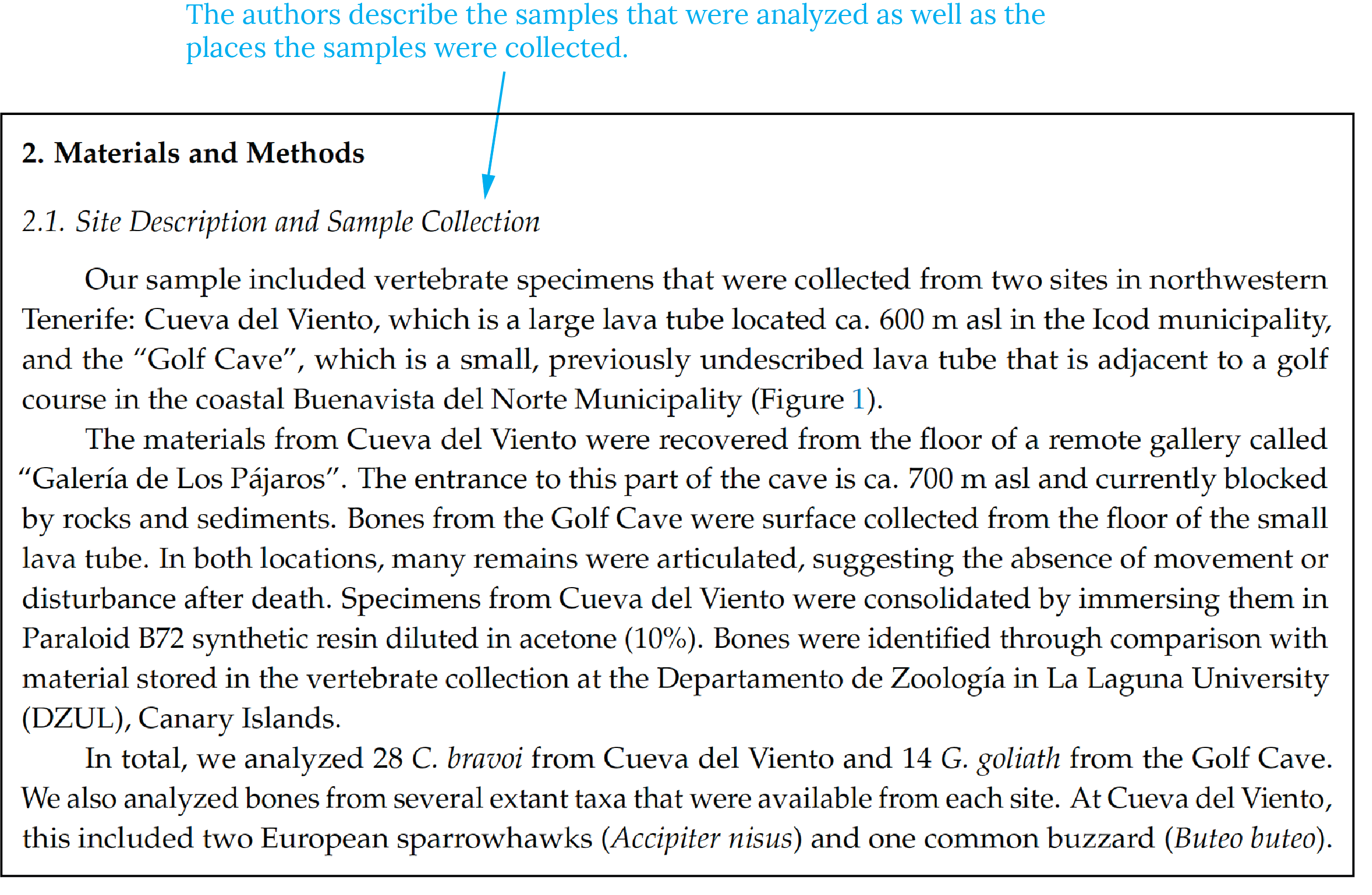
Open this section by describing the overall approach you took or the materials used. Then describe to the readers step-by-step the methods you used including any data analysis performed. See Fig. 2.5 below for an example of materials and methods section.
Writing tips:
- Explain procedures, materials, and equipment used
- Example: “We used an x-ray fluorescence spectrometer to analyze major and trace elements in the mystery mineral samples.”
- Order events chronologically, perhaps with subheadings (Field work, Lab Analysis, Statistical Models)
- Use past tense (you did X, Y, Z)
- Quantify measurements
- Include results in the methods! It’s easy to make this mistake!
- Example: “W e turned on the machine and loaded in our samples, then calibrated the instrument and pushed the start button and waited one hour. . . .”
Technical Writing @ SLCC Copyright © 2020 by Department of English, Linguistics, and Writing Studies at SLCC is licensed under a Creative Commons Attribution-NonCommercial 4.0 International License , except where otherwise noted.
Share This Book
Generate accurate APA citations for free
- Knowledge Base
- APA Style 7th edition
- How to write an APA methods section
How to Write an APA Methods Section | With Examples
Published on February 5, 2021 by Pritha Bhandari . Revised on June 22, 2023.
The methods section of an APA style paper is where you report in detail how you performed your study. Research papers in the social and natural sciences often follow APA style. This article focuses on reporting quantitative research methods .
In your APA methods section, you should report enough information to understand and replicate your study, including detailed information on the sample , measures, and procedures used.
Instantly correct all language mistakes in your text
Upload your document to correct all your mistakes in minutes

Table of contents
Structuring an apa methods section.
Participants
Example of an APA methods section
Other interesting articles, frequently asked questions about writing an apa methods section.
The main heading of “Methods” should be centered, boldfaced, and capitalized. Subheadings within this section are left-aligned, boldfaced, and in title case. You can also add lower level headings within these subsections, as long as they follow APA heading styles .
To structure your methods section, you can use the subheadings of “Participants,” “Materials,” and “Procedures.” These headings are not mandatory—aim to organize your methods section using subheadings that make sense for your specific study.
Note that not all of these topics will necessarily be relevant for your study. For example, if you didn’t need to consider outlier removal or ways of assigning participants to different conditions, you don’t have to report these steps.
The APA also provides specific reporting guidelines for different types of research design. These tell you exactly what you need to report for longitudinal designs , replication studies, experimental designs , and so on. If your study uses a combination design, consult APA guidelines for mixed methods studies.
Detailed descriptions of procedures that don’t fit into your main text can be placed in supplemental materials (for example, the exact instructions and tasks given to participants, the full analytical strategy including software code, or additional figures and tables).
Are your APA in-text citations flawless?
The AI-powered APA Citation Checker points out every error, tells you exactly what’s wrong, and explains how to fix it. Say goodbye to losing marks on your assignment!
Get started!

Begin the methods section by reporting sample characteristics, sampling procedures, and the sample size.
Participant or subject characteristics
When discussing people who participate in research, descriptive terms like “participants,” “subjects” and “respondents” can be used. For non-human animal research, “subjects” is more appropriate.
Specify all relevant demographic characteristics of your participants. This may include their age, sex, ethnic or racial group, gender identity, education level, and socioeconomic status. Depending on your study topic, other characteristics like educational or immigration status or language preference may also be relevant.
Be sure to report these characteristics as precisely as possible. This helps the reader understand how far your results may be generalized to other people.
The APA guidelines emphasize writing about participants using bias-free language , so it’s necessary to use inclusive and appropriate terms.
Sampling procedures
Outline how the participants were selected and all inclusion and exclusion criteria applied. Appropriately identify the sampling procedure used. For example, you should only label a sample as random if you had access to every member of the relevant population.
Of all the people invited to participate in your study, note the percentage that actually did (if you have this data). Additionally, report whether participants were self-selected, either by themselves or by their institutions (e.g., schools may submit student data for research purposes).
Identify any compensation (e.g., course credits or money) that was provided to participants, and mention any institutional review board approvals and ethical standards followed.
Sample size and power
Detail the sample size (per condition) and statistical power that you hoped to achieve, as well as any analyses you performed to determine these numbers.
It’s important to show that your study had enough statistical power to find effects if there were any to be found.
Additionally, state whether your final sample differed from the intended sample. Your interpretations of the study outcomes should be based only on your final sample rather than your intended sample.
Write up the tools and techniques that you used to measure relevant variables. Be as thorough as possible for a complete picture of your techniques.
Primary and secondary measures
Define the primary and secondary outcome measures that will help you answer your primary and secondary research questions.
Specify all instruments used in gathering these measurements and the construct that they measure. These instruments may include hardware, software, or tests, scales, and inventories.
- To cite hardware, indicate the model number and manufacturer.
- To cite common software (e.g., Qualtrics), state the full name along with the version number or the website URL .
- To cite tests, scales or inventories, reference its manual or the article it was published in. It’s also helpful to state the number of items and provide one or two example items.
Make sure to report the settings of (e.g., screen resolution) any specialized apparatus used.
For each instrument used, report measures of the following:
- Reliability : how consistently the method measures something, in terms of internal consistency or test-retest reliability.
- Validity : how precisely the method measures something, in terms of construct validity or criterion validity .
Giving an example item or two for tests, questionnaires , and interviews is also helpful.
Describe any covariates—these are any additional variables that may explain or predict the outcomes.
Quality of measurements
Review all methods you used to assure the quality of your measurements.
These may include:
- training researchers to collect data reliably,
- using multiple people to assess (e.g., observe or code) the data,
- translation and back-translation of research materials,
- using pilot studies to test your materials on unrelated samples.
For data that’s subjectively coded (for example, classifying open-ended responses), report interrater reliability scores. This tells the reader how similarly each response was rated by multiple raters.
Report all of the procedures applied for administering the study, processing the data, and for planned data analyses.
Data collection methods and research design
Data collection methods refers to the general mode of the instruments: surveys, interviews, observations, focus groups, neuroimaging, cognitive tests, and so on. Summarize exactly how you collected the necessary data.
Describe all procedures you applied in administering surveys, tests, physical recordings, or imaging devices, with enough detail so that someone else can replicate your techniques. If your procedures are very complicated and require long descriptions (e.g., in neuroimaging studies), place these details in supplementary materials.
To report research design, note your overall framework for data collection and analysis. State whether you used an experimental, quasi-experimental, descriptive (observational), correlational, and/or longitudinal design. Also note whether a between-subjects or a within-subjects design was used.
For multi-group studies, report the following design and procedural details as well:
- how participants were assigned to different conditions (e.g., randomization),
- instructions given to the participants in each group,
- interventions for each group,
- the setting and length of each session(s).
Describe whether any masking was used to hide the condition assignment (e.g., placebo or medication condition) from participants or research administrators. Using masking in a multi-group study ensures internal validity by reducing research bias . Explain how this masking was applied and whether its effectiveness was assessed.
Participants were randomly assigned to a control or experimental condition. The survey was administered using Qualtrics (https://www.qualtrics.com). To begin, all participants were given the AAI and a demographics questionnaire to complete, followed by an unrelated filler task. In the control condition , participants completed a short general knowledge test immediately after the filler task. In the experimental condition, participants were asked to visualize themselves taking the test for 3 minutes before they actually did. For more details on the exact instructions and tasks given, see supplementary materials.
Data diagnostics
Outline all steps taken to scrutinize or process the data after collection.
This includes the following:
- Procedures for identifying and removing outliers
- Data transformations to normalize distributions
- Compensation strategies for overcoming missing values
To ensure high validity, you should provide enough detail for your reader to understand how and why you processed or transformed your raw data in these specific ways.
Analytic strategies
The methods section is also where you describe your statistical analysis procedures, but not their outcomes. Their outcomes are reported in the results section.
These procedures should be stated for all primary, secondary, and exploratory hypotheses. While primary and secondary hypotheses are based on a theoretical framework or past studies, exploratory hypotheses are guided by the data you’ve just collected.
Scribbr Citation Checker New
The AI-powered Citation Checker helps you avoid common mistakes such as:
- Missing commas and periods
- Incorrect usage of “et al.”
- Ampersands (&) in narrative citations
- Missing reference entries
This annotated example reports methods for a descriptive correlational survey on the relationship between religiosity and trust in science in the US. Hover over each part for explanation of what is included.
The sample included 879 adults aged between 18 and 28. More than half of the participants were women (56%), and all participants had completed at least 12 years of education. Ethics approval was obtained from the university board before recruitment began. Participants were recruited online through Amazon Mechanical Turk (MTurk; www.mturk.com). We selected for a geographically diverse sample within the Midwest of the US through an initial screening survey. Participants were paid USD $5 upon completion of the study.
A sample size of at least 783 was deemed necessary for detecting a correlation coefficient of ±.1, with a power level of 80% and a significance level of .05, using a sample size calculator (www.sample-size.net/correlation-sample-size/).
The primary outcome measures were the levels of religiosity and trust in science. Religiosity refers to involvement and belief in religious traditions, while trust in science represents confidence in scientists and scientific research outcomes. The secondary outcome measures were gender and parental education levels of participants and whether these characteristics predicted religiosity levels.
Religiosity
Religiosity was measured using the Centrality of Religiosity scale (Huber, 2003). The Likert scale is made up of 15 questions with five subscales of ideology, experience, intellect, public practice, and private practice. An example item is “How often do you experience situations in which you have the feeling that God or something divine intervenes in your life?” Participants were asked to indicate frequency of occurrence by selecting a response ranging from 1 (very often) to 5 (never). The internal consistency of the instrument is .83 (Huber & Huber, 2012).
Trust in Science
Trust in science was assessed using the General Trust in Science index (McCright, Dentzman, Charters & Dietz, 2013). Four Likert scale items were assessed on a scale from 1 (completely distrust) to 5 (completely trust). An example question asks “How much do you distrust or trust scientists to create knowledge that is unbiased and accurate?” Internal consistency was .8.
Potential participants were invited to participate in the survey online using Qualtrics (www.qualtrics.com). The survey consisted of multiple choice questions regarding demographic characteristics, the Centrality of Religiosity scale, an unrelated filler anagram task, and finally the General Trust in Science index. The filler task was included to avoid priming or demand characteristics, and an attention check was embedded within the religiosity scale. For full instructions and details of tasks, see supplementary materials.
For this correlational study , we assessed our primary hypothesis of a relationship between religiosity and trust in science using Pearson moment correlation coefficient. The statistical significance of the correlation coefficient was assessed using a t test. To test our secondary hypothesis of parental education levels and gender as predictors of religiosity, multiple linear regression analysis was used.
If you want to know more about statistics , methodology , or research bias , make sure to check out some of our other articles with explanations and examples.
- Normal distribution
- Measures of central tendency
- Chi square tests
- Confidence interval
- Quartiles & Quantiles
Methodology
- Cluster sampling
- Stratified sampling
- Thematic analysis
- Cohort study
- Peer review
- Ethnography
Research bias
- Implicit bias
- Cognitive bias
- Conformity bias
- Hawthorne effect
- Availability heuristic
- Attrition bias
- Social desirability bias
In your APA methods section , you should report detailed information on the participants, materials, and procedures used.
- Describe all relevant participant or subject characteristics, the sampling procedures used and the sample size and power .
- Define all primary and secondary measures and discuss the quality of measurements.
- Specify the data collection methods, the research design and data analysis strategy, including any steps taken to transform the data and statistical analyses.
You should report methods using the past tense , even if you haven’t completed your study at the time of writing. That’s because the methods section is intended to describe completed actions or research.
In a scientific paper, the methodology always comes after the introduction and before the results , discussion and conclusion . The same basic structure also applies to a thesis, dissertation , or research proposal .
Depending on the length and type of document, you might also include a literature review or theoretical framework before the methodology.
Cite this Scribbr article
If you want to cite this source, you can copy and paste the citation or click the “Cite this Scribbr article” button to automatically add the citation to our free Citation Generator.
Bhandari, P. (2023, June 22). How to Write an APA Methods Section | With Examples. Scribbr. Retrieved April 16, 2024, from https://www.scribbr.com/apa-style/methods-section/
Is this article helpful?

Pritha Bhandari
Other students also liked, how to write an apa results section, apa format for academic papers and essays, apa headings and subheadings, unlimited academic ai-proofreading.
✔ Document error-free in 5minutes ✔ Unlimited document corrections ✔ Specialized in correcting academic texts
When you choose to publish with PLOS, your research makes an impact. Make your work accessible to all, without restrictions, and accelerate scientific discovery with options like preprints and published peer review that make your work more Open.
- PLOS Biology
- PLOS Climate
- PLOS Complex Systems
- PLOS Computational Biology
- PLOS Digital Health
- PLOS Genetics
- PLOS Global Public Health
- PLOS Medicine
- PLOS Mental Health
- PLOS Neglected Tropical Diseases
- PLOS Pathogens
- PLOS Sustainability and Transformation
- PLOS Collections
- How to Write Your Methods

Ensure understanding, reproducibility and replicability
What should you include in your methods section, and how much detail is appropriate?
Why Methods Matter
The methods section was once the most likely part of a paper to be unfairly abbreviated, overly summarized, or even relegated to hard-to-find sections of a publisher’s website. While some journals may responsibly include more detailed elements of methods in supplementary sections, the movement for increased reproducibility and rigor in science has reinstated the importance of the methods section. Methods are now viewed as a key element in establishing the credibility of the research being reported, alongside the open availability of data and results.
A clear methods section impacts editorial evaluation and readers’ understanding, and is also the backbone of transparency and replicability.
For example, the Reproducibility Project: Cancer Biology project set out in 2013 to replicate experiments from 50 high profile cancer papers, but revised their target to 18 papers once they understood how much methodological detail was not contained in the original papers.

What to include in your methods section
What you include in your methods sections depends on what field you are in and what experiments you are performing. However, the general principle in place at the majority of journals is summarized well by the guidelines at PLOS ONE : “The Materials and Methods section should provide enough detail to allow suitably skilled investigators to fully replicate your study. ” The emphases here are deliberate: the methods should enable readers to understand your paper, and replicate your study. However, there is no need to go into the level of detail that a lay-person would require—the focus is on the reader who is also trained in your field, with the suitable skills and knowledge to attempt a replication.
A constant principle of rigorous science
A methods section that enables other researchers to understand and replicate your results is a constant principle of rigorous, transparent, and Open Science. Aim to be thorough, even if a particular journal doesn’t require the same level of detail . Reproducibility is all of our responsibility. You cannot create any problems by exceeding a minimum standard of information. If a journal still has word-limits—either for the overall article or specific sections—and requires some methodological details to be in a supplemental section, that is OK as long as the extra details are searchable and findable .
Imagine replicating your own work, years in the future
As part of PLOS’ presentation on Reproducibility and Open Publishing (part of UCSF’s Reproducibility Series ) we recommend planning the level of detail in your methods section by imagining you are writing for your future self, replicating your own work. When you consider that you might be at a different institution, with different account logins, applications, resources, and access levels—you can help yourself imagine the level of specificity that you yourself would require to redo the exact experiment. Consider:
- Which details would you need to be reminded of?
- Which cell line, or antibody, or software, or reagent did you use, and does it have a Research Resource ID (RRID) that you can cite?
- Which version of a questionnaire did you use in your survey?
- Exactly which visual stimulus did you show participants, and is it publicly available?
- What participants did you decide to exclude?
- What process did you adjust, during your work?
Tip: Be sure to capture any changes to your protocols
You yourself would want to know about any adjustments, if you ever replicate the work, so you can surmise that anyone else would want to as well. Even if a necessary adjustment you made was not ideal, transparency is the key to ensuring this is not regarded as an issue in the future. It is far better to transparently convey any non-optimal methods, or methodological constraints, than to conceal them, which could result in reproducibility or ethical issues downstream.
Visual aids for methods help when reading the whole paper
Consider whether a visual representation of your methods could be appropriate or aid understanding your process. A visual reference readers can easily return to, like a flow-diagram, decision-tree, or checklist, can help readers to better understand the complete article, not just the methods section.
Ethical Considerations
In addition to describing what you did, it is just as important to assure readers that you also followed all relevant ethical guidelines when conducting your research. While ethical standards and reporting guidelines are often presented in a separate section of a paper, ensure that your methods and protocols actually follow these guidelines. Read more about ethics .
Existing standards, checklists, guidelines, partners
While the level of detail contained in a methods section should be guided by the universal principles of rigorous science outlined above, various disciplines, fields, and projects have worked hard to design and develop consistent standards, guidelines, and tools to help with reporting all types of experiment. Below, you’ll find some of the key initiatives. Ensure you read the submission guidelines for the specific journal you are submitting to, in order to discover any further journal- or field-specific policies to follow, or initiatives/tools to utilize.
Tip: Keep your paper moving forward by providing the proper paperwork up front
Be sure to check the journal guidelines and provide the necessary documents with your manuscript submission. Collecting the necessary documentation can greatly slow the first round of peer review, or cause delays when you submit your revision.
Randomized Controlled Trials – CONSORT The Consolidated Standards of Reporting Trials (CONSORT) project covers various initiatives intended to prevent the problems of inadequate reporting of randomized controlled trials. The primary initiative is an evidence-based minimum set of recommendations for reporting randomized trials known as the CONSORT Statement .
Systematic Reviews and Meta-Analyses – PRISMA The Preferred Reporting Items for Systematic Reviews and Meta-Analyses ( PRISMA ) is an evidence-based minimum set of items focusing on the reporting of reviews evaluating randomized trials and other types of research.
Research using Animals – ARRIVE The Animal Research: Reporting of In Vivo Experiments ( ARRIVE ) guidelines encourage maximizing the information reported in research using animals thereby minimizing unnecessary studies. (Original study and proposal , and updated guidelines , in PLOS Biology .)
Laboratory Protocols Protocols.io has developed a platform specifically for the sharing and updating of laboratory protocols , which are assigned their own DOI and can be linked from methods sections of papers to enhance reproducibility. Contextualize your protocol and improve discovery with an accompanying Lab Protocol article in PLOS ONE .
Consistent reporting of Materials, Design, and Analysis – the MDAR checklist A cross-publisher group of editors and experts have developed, tested, and rolled out a checklist to help establish and harmonize reporting standards in the Life Sciences . The checklist , which is available for use by authors to compile their methods, and editors/reviewers to check methods, establishes a minimum set of requirements in transparent reporting and is adaptable to any discipline within the Life Sciences, by covering a breadth of potentially relevant methodological items and considerations. If you are in the Life Sciences and writing up your methods section, try working through the MDAR checklist and see whether it helps you include all relevant details into your methods, and whether it reminded you of anything you might have missed otherwise.
Summary Writing tips
The main challenge you may find when writing your methods is keeping it readable AND covering all the details needed for reproducibility and replicability. While this is difficult, do not compromise on rigorous standards for credibility!

- Keep in mind future replicability, alongside understanding and readability.
- Follow checklists, and field- and journal-specific guidelines.
- Consider a commitment to rigorous and transparent science a personal responsibility, and not just adhering to journal guidelines.
- Establish whether there are persistent identifiers for any research resources you use that can be specifically cited in your methods section.
- Deposit your laboratory protocols in Protocols.io, establishing a permanent link to them. You can update your protocols later if you improve on them, as can future scientists who follow your protocols.
- Consider visual aids like flow-diagrams, lists, to help with reading other sections of the paper.
- Be specific about all decisions made during the experiments that someone reproducing your work would need to know.

Don’t
- Summarize or abbreviate methods without giving full details in a discoverable supplemental section.
- Presume you will always be able to remember how you performed the experiments, or have access to private or institutional notebooks and resources.
- Attempt to hide constraints or non-optimal decisions you had to make–transparency is the key to ensuring the credibility of your research.
- How to Write a Great Title
- How to Write an Abstract
- How to Report Statistics
- How to Write Discussions and Conclusions
- How to Edit Your Work
The contents of the Peer Review Center are also available as a live, interactive training session, complete with slides, talking points, and activities. …
The contents of the Writing Center are also available as a live, interactive training session, complete with slides, talking points, and activities. …
There’s a lot to consider when deciding where to submit your work. Learn how to choose a journal that will help your study reach its audience, while reflecting your values as a researcher…
We have a new app!
Take the Access library with you wherever you go—easy access to books, videos, images, podcasts, personalized features, and more.
Download the Access App here: iOS and Android . Learn more here!
- Remote Access
- Save figures into PowerPoint
- Download tables as PDFs

Chapter 5: Materials and Methods
- Download Chapter PDF
Disclaimer: These citations have been automatically generated based on the information we have and it may not be 100% accurate. Please consult the latest official manual style if you have any questions regarding the format accuracy.
Download citation file:
- Search Book
Jump to a Section
- ORGANIZATION
- SUMMARY OF GUIDELINES FOR THE MATERIALS AND METHODS SECTION
- EXERCISE 5.1: A CLEARLY WRITTEN METHODS SECTION
- EXERCISE 5.2: CONTENT AND ORGANIZATION IN THE METHODS SECTION
- Full Chapter
- Supplementary Content
For hypothesis-testing papers, the function of the Materials and Methods section (often referred to as the Methods section) is to tell the reader what experiments you did to answer the question posed in the Introduction. Similarly, for descriptive studies, the Methods section tells what experiments you did to obtain the message stated in the Introduction. For methods papers, the Methods section has two functions: it describes the new method in complete detail and also tells what experiments you did to test the new method. For all types of paper, the Methods section should include sufficient detail and references to permit a trained scientist to evaluate your work fully or to repeat the experiments exactly as you did them.
Hypothesis-Testing and Descriptive Papers
We saw that the first step in the story line of a hypothesis-testing or a descriptive paper is presented in the Introduction. This first step is either the question being asked or the structure being described. In either case, the second step in the story line is an overview of the experiments you did. This overview of the experiments gives the strategy of the experiments, the plan that connects the methods to each other and to the question or the message.
Where the overview of the experiments is presented depends on the type of research:
Methods Papers
For a Methods paper, the first step in the story line is a statement that you are presenting a new or improved material, method, or apparatus. The second step in the story line has two parts: a complete description of the new method, material, or apparatus; and a description of how this new method, material, or apparatus was tested. These two steps are described in the Methods section.
In this chapter, we will consider only Methods sections for hypothesis-testing papers.
Sign in or create a free Access profile below to access even more exclusive content.
With an Access profile, you can save and manage favorites from your personal dashboard, complete case quizzes, review Q&A, and take these feature on the go with our Access app.
Pop-up div Successfully Displayed
This div only appears when the trigger link is hovered over. Otherwise it is hidden from view.
Please Wait
- SpringerLink shop
Materials and methods
The study’s methods are one of the most important parts used to judge the overall quality of the paper. In addition the Methods section should give readers enough information so that they can repeat the experiments. Reviewers should look for potential sources of bias in the way the study was designed and carried out, and for places where more explanation is needed.
The specific types of information in a Methods section will vary from field to field and from study to study. However, some general rules for Methods sections are:
- It should be clear from the Methods section how all of the data in the Results section were obtained.
- The study system should be clearly described. In medicine, for example, researchers need to specify the number of study subjects; how, when, and where the subjects were recruited, and that the study obtained appropriate ‘informed consent’ documents; and what criteria subjects had to meet to be included in the study.
- In most cases, the experiments should include appropriate controls or comparators. The conditions of the controls should be specified.
- The outcomes of the study should be defined, and the outcome measures should be objectively validated.
- The methods used to analyze the data must be statistically sound.
- For qualitative studies, an established qualitative research method (e.g. grounded theory is often used in sociology) must be used as appropriate for the study question.
- If the authors used a technique from a published study, they should include a citation and a summary of the procedure in the text. The method also needs to be appropriate to the present experiment.
- All materials and instruments should be identified, including the supplier’s name and location. For example, “Tests were conducted with a Vulcanizer 2.0 (XYZ Instruments, Mumbai, India).”
- The Methods section should not have information that belongs in another section (such as the Introduction or Results).
You may suggest if additional experiments would greatly improve the quality of the manuscript. Your suggestions should be in line with the study’s aims. Remember that almost any study could be strengthened by further experiments, so only suggest further work if you believe that the manuscript is not publishable without it.
Back │ Next
How to write a materials and methods section of a scientific article?
Affiliation.
- 1 Department of Urology, Faculty of Medicine, Gaziosmanpaşa University, Tokat, Turkey.
- PMID: 26328129
- PMCID: PMC4548564
- DOI: 10.5152/tud.2013.047
In contrast to past centuries, scientific researchers have been currently conducted systematically in all countries as part of an education strategy. As a consequence, scientists have published thousands of reports. Writing an effective article is generally a significant problem for researchers. All parts of an article, specifically the abstract, material and methods, results, discussion and references sections should contain certain features that should always be considered before sending a manuscript to a journal for publication. It is generally known that the material and methods section is a relatively easy section of an article to write. Therefore, it is often a good idea to begin by writing the materials and methods section, which is also a crucial part of an article. Because "reproducible results" are very important in science, a detailed account of the study should be given in this section. If the authors provide sufficient detail, other scientists can repeat their experiments to verify their findings. It is generally recommended that the materials and methods should be written in the past tense, either in active or passive voice. In this section, ethical approval, study dates, number of subjects, groups, evaluation criteria, exclusion criteria and statistical methods should be described sequentially. It should be noted that a well-written materials and methods section markedly enhances the chances of an article being published.
Keywords: Article; material; methods; publication.
University of Lethbridge
Science Toolkit
Materials and Methods
The Methods and Materials section of a paper often seems the least interesting to read, or to write, but it serves several essential purposes. First, it demonstrates to readers that the research was designed appropriately and conducted competently. Scientists are skeptical readers. They won’t have any confidence in your results unless the Methods section convinces them those results come from the correct experiment, carried out correctly. Second, this section allows other researchers to repeat the research for themselves. The ability to replicate a study and get the same results is a central part of science. If possible, your materials and methods section should be written in enough detail to allow another researcher to repeat what you did. (As science develops ever more complex techniques for probing nature this becomes increasingly difficult to achieve.) Where established protocols or techniques are used, it is often acceptable to simply cite a previously published work which sets out the procedure in detail. However, the procedures should always be described in sufficient detail that readers have a clear sense of the basic approach being taken.
What to include: One of the trickiest parts of writing the Methods section is determining the correct level of detail. Always ask yourself: Does the reader need to know this to understand and repeat the experiment? (Pechenik 1996) Let’s say you performed an experiment to test the effects of vitamin E by injecting lab rats with different doses. It would be essential for the reader to know the number of rats injected, and the dosages used, but it would probably not be necessary to include the brand of syringe used, since any standard sterilized syringe should give equivalent results. If in doubt, include the information. Students tend to include too little detail in their Methods rather than too much.
Know your audience: Part of the challenge of knowing what details to include is knowing what you can assume your audience already knows. You can always assume your audience has a basic understanding of biology, but how much detailed knowledge of your subject area you can expect will depend on the paper’s destination. You should never assume your reader has prior knowledge of your research. (Even an instructor who has coached you every step of the way as you prepared a paper will be reading and marking it from the perspective of someone seeing the research for the first time.)
No list of materials: Always describe your materials in the context of how they were used. A list of materials is a waste of space and tells your reader little. Simply describe the methods used to collect your data, and note the materials used for each.
Use figures: Pictures are often helpful in explaining methodology. These could include diagrams of apparatus, maps of the study area showing sampling locations, flowcharts for complicated protocols, etc. A picture really is often worth a thousand words. All graphics included in your paper should be numbered and captioned as figures.
Interpret when necessary: Methods should be concise and factual, but take the space to explain any choices which will not make sense to your readers. If one of your treatment groups was much smaller than the other because a badger ate several of the ground squirrels you were studying, point that out.
Stat tests are methods: Statistical tests and other types of analysis performed on your data are part of your methods. Commonly used statistical tests need not be described, but if any explanation of your analysis is needed, the methods section is the appropriate place. If you used a test which is not widely known, a short description and a citation of the source is warranted.
An official website of the United States government
The .gov means it’s official. Federal government websites often end in .gov or .mil. Before sharing sensitive information, make sure you’re on a federal government site.
The site is secure. The https:// ensures that you are connecting to the official website and that any information you provide is encrypted and transmitted securely.
- Publications
- Account settings
Preview improvements coming to the PMC website in October 2024. Learn More or Try it out now .
- Advanced Search
- Journal List
- J Indian Soc Periodontol
- v.26(3); May-Jun 2022
Materials and method: The “Recipe” of a research
Ashish kumar.
Editor, Journal of Indian Society of Periodontology, Professor and Head, Department of Periodontics, Dental College, Regional Institute of Medical Sciences (RIMS), Lamphelpat, Imphal-795004, Manipur, India. E-mail: moc.liamffider@79ramukhsihsa

In any research article, the detailed description and process of an experiment is provided in the section termed as “Materials and Method.” The Materials and Method section is also called Method section in few journals. This section describes how the experiment was conducted to arrive at the results. The aim of this section in any research article is to describe the process in detail for “reproducibility” which means that procedure of the experiment and related materials should be adequately described so that the other researchers working on the similar topic/area, should be able to conduct a similar experiment and replicate the results to allow corroboration of the inferences of the research. The reproducibility of the results is crucial for their scientific merit.[ 1 ] This section has been equated to “recipe section” which describes what to use, how much to use and how to use to come to the final product.[ 2 ]
Vital details of the research need to be described in this section. At the beginning of the section, the study design needs a description in terms of well-defined commonly used nomenclature (longitudinal, cross-over study”, “randomised controlled trial”, etc). The mention of the study design in the initial part of materials and method section is important as it helps the readers understand the research based on the merits and limitations of study design. The inclusion of study designs also help in understanding the type of statistical tests that can be appropriately applied in evaluating the data.[ 3 ] Randomisation being a crucial aspect of many clinical studies, has to be defined clearly.
The information about sample size, inclusion and exclusion criteria (sample characteristics) also should find a description in this aspect of the material and method section. An adequate sample size of a study would be able to provide the precision of our estimates and thus have adequate power of study to draw conclusions and justify answers to query being explored in the research.[ 3 ] The information of the sample characteristics is important to accomplish the aims of the experiment (hypothesis). Apart from this, the details of the approval from ethical board and trial registration should be mentioned here.[ 4 ]
The next aspect of Materials and Method should incorporate the description of materials in terms of quantity, precise technical descriptions and the method of preparations, if any. The details of the manufacturers of chemical reagents and equipment should also find a mention here. Generic names should be preferred over trade names. If study has usage of microorganisms or experimental animals, a clear description of such entities in terms of species/strains or genus species is required.[ 5 ]
The description of the method of the experiment should be accurate, concise but complete. The process should be written as a explanation of a process, not as a laboratory manual procedure. If the methods, devices, or techniques which have been used by authors, are in routine usage, and are widely known and published, then such methods do not require detailed description. But the authors should compulsorily mention the original article or references from where the readers can get information about the method in detail to replicate the procedure. If any treatment is being investigated, then exact treatment protocol should be described. Techniques/method which are new or uncommon should be explained fully and any related references should also be mentioned.
The statistical aspects should mention the statistical tests and the statistical computer packages that were used for data analysis. Use of an uncommon statistical test needs an explanation of its usage in the context of the study and a reference to the method for readers to refer.[ 5 ]
The material and method section may or may not have subheadings, depending upon the journal guidelines. The subdivisions can be: Study design, setting, subjects, data collection and data analysis[ 2 ] or overall design of the study, inclusion and exclusion criteria, sample sizes and statistical power.[ 6 ]
It is of paramount importance that a consistency is maintained between the “Materials and Method” section and “Results” section of the article. Procedures described in Methods section should correlate with the results described in the Results section for readers to understand the association of the specific methodology to results.[ 4 ]
Often, few issues arise while writing Materials and Method like inclusion of unnecessary details or results. Limitations on number of references that can be cited in journals, many times, leads to this section being extremely concise and lacks details required for the “reproducibility”.[ 7 ] The details of the procedure are not completely mentioned by authors sometimes because of commercial reasons.[ 7 ] These situations result in compromise with the basic principle of “reproducibility” while writing this section.
In certain cases, the authors are apprehensive of results being reproduced and validity of their results being challenged. To avoid any questions being raised on the methodology and results, the authors provide insufficient details in this section to avoid reproducibility.[ 7 ]
The aim of any research is progression of knowledge in that particular field. One of the essential requirement for progression of scientific knowledge is “reproducibility” and the assessment of the validity of available results. This is achievable only if the authors provide sufficient details in the “Materials and Method section”.[ 7 ]
Writing this section should be simple and easy especially when this part is written after the completion of the study, as the authors would have performed the experiment themselves. This is one of the first sections written while writing a research article.
“History has repeatedly shown that when a new method or material becomes available, new uses for it arise.”
Wilson Greatbatch

Research Paper Writing: 5. Methods / Materials
- 1. Getting Started
- 2. Abstract
- 3. Introduction
- 4. Literature Review
- 5. Methods / Materials
- 6. Results / Analysis
- 7. Discussion
- 8. Conclusion
- 9. Reference
Methods / Materials Overview
These sections of the research paper should be concise. The audience reading the paper will always want to know what materials or methods that were used. The methods and materials may be under subheadings in the section or incorporated together. The main objective for these sections is to provide specialized materials, general procedures, and methods to judge the scientific value of the paper.
What to include in the sections
- Described separately
- Include the chemicals, biological, and any equipment
- Do not include common supplies, such as test tubes, pipette tips, beakers, etc. or standard lab equipment
- Single out sources like a specific type of equipment, enzyme, or a culture
- These should be mentioned in a separate paragraph with its own heading or highlighted in the procedure section if there is one
- Refer to solutions by name and describe
- Describes in detail how the analysis was conducted
- Be brief when presenting methods under the title devoted to a specific technique or groups of procedures
- Simplify and report what the procedure was
- Report the method by name
- Use third person passive voice, and avoid using first person
- Use normal text in these sections
- Avoid informal lists
- Use complete sentences
Example of a Methods Section
Publication Manual of the American Psychological Association Sixth Ed. 2010
- << Previous: 4. Literature Review
- Next: 6. Results / Analysis >>
- Last Updated: Nov 7, 2023 7:37 AM
- URL: https://wiu.libguides.com/researchpaperwriting
Setting the Scene: Best Practices for Writing Materials and Methods
- Peer Review
- Research Process
This free white paper tackles the best ways to write the Materials and Methods section of a scientific manuscript.
Updated on March 3, 2014

The Materials and Methods (or “Methods section”) is the section of a research paper that provides the reader
with all the information needed to understand your work and how the reported results were produced. Having read
the Introduction, the reader already knows why your work is important, so the next step is to connect that section to
the experimental design used to address your research questions.
Below is a preview of our free white paper tackling the best way to write the Materials and Methods section of a scientific manuscript. It covers the following topics:
Purpose and Structure
- Key Information
- Notation and Terminology
- Equipment and Materials Citations
- Acquisition and Definition of the Results
- Statistical Methods
- Concluding Statements
Depending on the type of paper, the Methods section can encompass anything from the parameters of a literature search to the methods employed in a field study to the details of bench work in the lab. The common feature is that the information needs to be presented in a way that is clear and familiar to the reader. It is important to note that the purpose of the Methods section is not just to convey what you did; a thorough and well-organized Methods section reflects your knowledge and understanding of appropriate research techniques and increases the reader's confidence in your work.
The Methods section is easiest to follow when it begins by providing a clear context for the detailed descriptions of the methods and materials used in the study. This context is best achieved by beginning with general characteristics and parameters (e.g., identification of sample sources or populations, descriptions of geographic areas, or characterizations of study participants). A reader who understands the foundation of your experiments will more easily understand the procedures that follow.
The underlying principle for what information to provide in the Methods section is that the reader should be able to replicate your study. This section must explain the methods used with enough detail to answer any of the reader's questions about how the study was performed. Because the Methods section is meant to convey how the research was conducted, conforming to the accepted conventions of the field is extremely important.
Generally, the Methods section should assemble familiar concepts and research activities into a logical series of events. Terminology and sentence structure should be consistent within the paper and conform to the conventions of the field, and repetition is accepted or even expected. Because Methods sections often rely on lists of information, consistency - i.e., the presentation of like elements using the same terminology, notation, and sentence structure - is especially important.
The information in the Methods section should follow the order of execution as closely as possible, although similar procedures should be presented together. For example, descriptions of sample or data collection should be described together, even if these are performed at different times or with intervening analysis, because a purely chronological account would mean switching back and forth between procedures.
Continue reading "Setting the Scene: Best Practices for Writing Materials and Methods" by downloading the full white paper here .
Check out our other "Best Practices for Writing" white papers to get tips for other sections of your research manuscript:
Getting a Strong Start: Best Practices for Writing an Introduction
Reaping the Rewards: Best Practices for Writing a Results Section

Michael Bendiksby, PhD
See our "Privacy Policy"
- Bipolar Disorder
- Therapy Center
- When To See a Therapist
- Types of Therapy
- Best Online Therapy
- Best Couples Therapy
- Best Family Therapy
- Managing Stress
- Sleep and Dreaming
- Understanding Emotions
- Self-Improvement
- Healthy Relationships
- Student Resources
- Personality Types
- Guided Meditations
- Verywell Mind Insights
- 2023 Verywell Mind 25
- Mental Health in the Classroom
- Editorial Process
- Meet Our Review Board
- Crisis Support
How to Write a Methods Section for a Psychology Paper
Tips and Examples of an APA Methods Section
Kendra Cherry, MS, is a psychosocial rehabilitation specialist, psychology educator, and author of the "Everything Psychology Book."
:max_bytes(150000):strip_icc():format(webp)/IMG_9791-89504ab694d54b66bbd72cb84ffb860e.jpg)
Emily is a board-certified science editor who has worked with top digital publishing brands like Voices for Biodiversity, Study.com, GoodTherapy, Vox, and Verywell.
:max_bytes(150000):strip_icc():format(webp)/Emily-Swaim-1000-0f3197de18f74329aeffb690a177160c.jpg)
Verywell / Brianna Gilmartin
The methods section of an APA format psychology paper provides the methods and procedures used in a research study or experiment . This part of an APA paper is critical because it allows other researchers to see exactly how you conducted your research.
Method refers to the procedure that was used in a research study. It included a precise description of how the experiments were performed and why particular procedures were selected. While the APA technically refers to this section as the 'method section,' it is also often known as a 'methods section.'
The methods section ensures the experiment's reproducibility and the assessment of alternative methods that might produce different results. It also allows researchers to replicate the experiment and judge the study's validity.
This article discusses how to write a methods section for a psychology paper, including important elements to include and tips that can help.
What to Include in a Method Section
So what exactly do you need to include when writing your method section? You should provide detailed information on the following:
- Research design
- Participants
- Participant behavior
The method section should provide enough information to allow other researchers to replicate your experiment or study.
Components of a Method Section
The method section should utilize subheadings to divide up different subsections. These subsections typically include participants, materials, design, and procedure.
Participants
In this part of the method section, you should describe the participants in your experiment, including who they were (and any unique features that set them apart from the general population), how many there were, and how they were selected. If you utilized random selection to choose your participants, it should be noted here.
For example: "We randomly selected 100 children from elementary schools near the University of Arizona."
At the very minimum, this part of your method section must convey:
- Basic demographic characteristics of your participants (such as sex, age, ethnicity, or religion)
- The population from which your participants were drawn
- Any restrictions on your pool of participants
- How many participants were assigned to each condition and how they were assigned to each group (i.e., randomly assignment , another selection method, etc.)
- Why participants took part in your research (i.e., the study was advertised at a college or hospital, they received some type of incentive, etc.)
Information about participants helps other researchers understand how your study was performed, how generalizable the result might be, and allows other researchers to replicate the experiment with other populations to see if they might obtain the same results.
In this part of the method section, you should describe the materials, measures, equipment, or stimuli used in the experiment. This may include:
- Testing instruments
- Technical equipment
- Any psychological assessments that were used
- Any special equipment that was used
For example: "Two stories from Sullivan et al.'s (1994) second-order false belief attribution tasks were used to assess children's understanding of second-order beliefs."
For standard equipment such as computers, televisions, and videos, you can simply name the device and not provide further explanation.
Specialized equipment should be given greater detail, especially if it is complex or created for a niche purpose. In some instances, such as if you created a special material or apparatus for your study, you might need to include an illustration of the item in the appendix of your paper.
In this part of your method section, describe the type of design used in the experiment. Specify the variables as well as the levels of these variables. Identify:
- The independent variables
- Dependent variables
- Control variables
- Any extraneous variables that might influence your results.
Also, explain whether your experiment uses a within-groups or between-groups design.
For example: "The experiment used a 3x2 between-subjects design. The independent variables were age and understanding of second-order beliefs."
The next part of your method section should detail the procedures used in your experiment. Your procedures should explain:
- What the participants did
- How data was collected
- The order in which steps occurred
For example: "An examiner interviewed children individually at their school in one session that lasted 20 minutes on average. The examiner explained to each child that he or she would be told two short stories and that some questions would be asked after each story. All sessions were videotaped so the data could later be coded."
Keep this subsection concise yet detailed. Explain what you did and how you did it, but do not overwhelm your readers with too much information.
Tips for How to Write a Methods Section
In addition to following the basic structure of an APA method section, there are also certain things you should remember when writing this section of your paper. Consider the following tips when writing this section:
- Use the past tense : Always write the method section in the past tense.
- Be descriptive : Provide enough detail that another researcher could replicate your experiment, but focus on brevity. Avoid unnecessary detail that is not relevant to the outcome of the experiment.
- Use an academic tone : Use formal language and avoid slang or colloquial expressions. Word choice is also important. Refer to the people in your experiment or study as "participants" rather than "subjects."
- Use APA format : Keep a style guide on hand as you write your method section. The Publication Manual of the American Psychological Association is the official source for APA style.
- Make connections : Read through each section of your paper for agreement with other sections. If you mention procedures in the method section, these elements should be discussed in the results and discussion sections.
- Proofread : Check your paper for grammar, spelling, and punctuation errors.. typos, grammar problems, and spelling errors. Although a spell checker is a handy tool, there are some errors only you can catch.
After writing a draft of your method section, be sure to get a second opinion. You can often become too close to your work to see errors or lack of clarity. Take a rough draft of your method section to your university's writing lab for additional assistance.
A Word From Verywell
The method section is one of the most important components of your APA format paper. The goal of your paper should be to clearly detail what you did in your experiment. Provide enough detail that another researcher could replicate your study if they wanted.
Finally, if you are writing your paper for a class or for a specific publication, be sure to keep in mind any specific instructions provided by your instructor or by the journal editor. Your instructor may have certain requirements that you need to follow while writing your method section.
Frequently Asked Questions
While the subsections can vary, the three components that should be included are sections on the participants, the materials, and the procedures.
- Describe who the participants were in the study and how they were selected.
- Define and describe the materials that were used including any equipment, tests, or assessments
- Describe how the data was collected
To write your methods section in APA format, describe your participants, materials, study design, and procedures. Keep this section succinct, and always write in the past tense. The main heading of this section should be labeled "Method" and it should be centered, bolded, and capitalized. Each subheading within this section should be bolded, left-aligned and in title case.
The purpose of the methods section is to describe what you did in your experiment. It should be brief, but include enough detail that someone could replicate your experiment based on this information. Your methods section should detail what you did to answer your research question. Describe how the study was conducted, the study design that was used and why it was chosen, and how you collected the data and analyzed the results.
Erdemir F. How to write a materials and methods section of a scientific article ? Turk J Urol . 2013;39(Suppl 1):10-5. doi:10.5152/tud.2013.047
Kallet RH. How to write the methods section of a research paper . Respir Care . 2004;49(10):1229-32. PMID: 15447808.
American Psychological Association. Publication Manual of the American Psychological Association (7th ed.). Washington DC: The American Psychological Association; 2019.
American Psychological Association. APA Style Journal Article Reporting Standards . Published 2020.
By Kendra Cherry, MSEd Kendra Cherry, MS, is a psychosocial rehabilitation specialist, psychology educator, and author of the "Everything Psychology Book."
Training videos | Faqs

Materials and Methods Examples and Writing Tips
Abstract | Introduction | Literature Review | Research question | Materials & Methods | Results | Discussion | Conclusion
In this blog, we look at how to write the materials and methods section of a research paper. In most research papers, the materials and methods section follows the literature review section. This is generally the easiest section to write because you are simply reproducing what you did in your experiments. It is always a good idea to start writing your research paper with the materials and methods section.
1. What is the purpose of the materials and methods section?

Materials and methods should describe how you did your research and detail the experimental procedure. One of the most important things to bear in mind while writing the materials and methods section is that it should have enough detail so that other researchers in your field can replicate your experiments and reproduce your results. You should provide all the steps in a logical order so that your readers can follow your description easily.
2. Materials and Methods Examples
The structure of the methods section will very much depend on your discipline. If you are not sure about the structure, then the best place to start will be to go through the methods section of some previously published papers from your chosen journal. We will look at some examples of materials and methods structure in different disciplines.
2.1. Materials & methods example #1 (Engineering paper)
If you are writing an engineering sciences research paper in which you are introducing a new method, your materials and methods section would typically include the following information.

You can start with the top-level summary of the method. You can try to answer these questions. Are you proposing a new method? Or, Are you using a standard method from the literature? Or, Are you extending a previously published method? If so, is it your previous work? or work published by a different author?
Then you can talk about the reasons for choosing this method. You can quote previous papers that have used this method successfully to support your arguments. Then, you can talk about the actual implementation details of the methods.
Then you can talk about how the methods were validated to confirm that they are suitable for your research. You can also include information about any pilot or preliminary studies you conducted before the full study. Then you can explain how you propose to test and evaluate the methods to prove that they are better than the existing methods. Here, you can talk about metrics and statistical tests you will be using to evaluate your method.
2.2. Materials & methods example #2 (Measurement paper)
If you are writing a paper that deals with measurements, you would typically include the following information in your materials and methods section.

You can start by talking about the experimental setup. You can try to answer these questions. What equipment was used to perform the measurements? What was the make and the model of the equipment? How many technicians took the measurements? How experienced were the technicians?
Then you can talk about the parameters that were measured during the experiment. Then you can talk about the actual measurement procedure. How were the samples prepared for the measurements? How many measurements were taken? Were the measurements repeated for consistency? Was there a time interval between successive measurements?
Then you can talk about measurement conditions and constraints. Were the measurements performed at room temperature or under special conditions? Were there any practical difficulties while performing the measurements, if so, how did you overcome them?
Most importantly, you must list all the calculations in the form of detailed equations and formulas so that readers know exactly how the data was produced.
2.3. Materials & methods example #3 (Survey questionnaire paper)
If you are writing a survey questionnaire paper , you would typically include the following information in your materials and methods section.

You can start by talking about your participants. Who is your target population? What are their demographics? How did you recruit them? How did participants provide consent for your study? What sampling method did you use to select the participants?
Then you can talk about the survey type. Was it a phone interview? Was it a personal interview? Was it an online survey? Or, Was it a written survey?
Then you can talk about the questionnaire design. How did you choose the questions? How many questions were there? What type of questions were they? Were they open ended questions, or close ended questions, or rating scale questions, or a mixture of different types of questions?
Then you can talk about how the questionnaire was administered. If it is an online survey, how did you get the questionnaire to the participants? Did you email them? Or did you post the survey forms?
If you are doing a personal interview. How did you conduct the interviews? Was it one to one interview, or was it done in batches, or did you use focus groups? How did the participants behave during the interview?
Then you can talk about questionnaire testing. Did you test your questionnaire before the main study? Did you have to make any changes after initial testing? Did you have to translate the questionnaire into multiple languages? Then finally you can talk about different types of statistical tests you used to analyze the survey responses.
2.4. Materials & methods example #4 (Medical clinical trial paper)
If you are writing a medical research paper , your materials and methods section would typically include the following information.

You can start by providing information about the study design. Was it a randomized trial, or an observational trial? Was it a prospective study, or a retrospective study? Was the study double-blinded, or single-blinded?
Then, you can talk about how the ethical approval was obtained for the study and clarify if the clinical trial was registered. if so, then provide the registration number.
Then, you can talk about how the participants were recruited for the study, and explain the inclusion and exclusion criteria. Then, you can talk about how the participants were grouped into control and placebo groups, and explain how the medication was administered.
Then, you can talk about what outcomes were measured. What was the primary outcome? What was the secondary outcome? What was the follow up period? You can try to answer these questions. Then you can finish off with some information about the statistical tests you used to analyze the data.
3. Frequently Asked Questions
One of the common mistakes people make is using vague language in materials and methods. Reviewers won’t like it, and they will reject the paper on the basis that the section is not elaborate enough for other researchers to reproduce your experiments.
Make sure you write the materials and methods section in past tense, since you are reporting something that has already happened.
Acronyms & Abbrevations: Try to use acronyms and abbreviations for long method names. Abbreviations and acronyms are a great way to make your writing concise and save time. Define the acronyms and abbreviations during their first occurrence then use the short form in the rest of the text. The common practice is to put the acronym and abbreviations in parentheses after the full term.
Use different layouts: Another problem you are likely to face is that your methods section can sound like manual if you have too much text in it. In particular, if you are dealing with a very complex procedure, the readers might find it dry and tedious. So try to provide some variety to the layout. Try to use bullet points and numberings instead of long paragraphs to make it easy for the readers to understand the procedure. You can use flow diagrams to illustrate the process rather than describing it.
When you are using a standard method that is well described in literature, the standard practice is to reference the paper rather than repeating the entire procedure. You can also provide a brief summary of the procedure in your own words.
For example, you can say something like this, “The details of the procedure have been reported previously in…”, and reference the previous paper. And then, you can follow it up with a brief summary of the method from the previous paper.
If you are extending a previous method, then you can do something like this. You can say that, “Some minor modifications were made to the method described in…” and reference the previous paper. And then, you can follow it up with the list of refinements you made to the previous method in order to adapt it to your work.
Similar Posts

3 Costly Mistakes to Avoid in the Research Introduction
In this blog, we will discuss three common mistakes that beginner writers make while writing the research paper introduction.

Writing a Questionnaire Survey Research Paper – Example & Format
In this blog, we will explain how to write a survey questionnaire paper and discuss all the important points to consider while writing the research paper.

Abstract Section Examples and Writing Tips
In this blog, we will go through many abstract examples and understand how to construct a good abstract for your research paper.

Formulating Strong Research Questions: Examples and Writing Tips
In this blog, we will go through many research question examples and understand how to construct a strong research question for your paper.

Results Section Examples and Writing Tips
In this blog, we will go through many results section examples and understand how to write a great results section for your paper.

Introduction Paragraph Examples and Writing Tips
In this blog, we will go through a few introduction paragraph examples and understand how to construct a great introduction paragraph for your research paper.
Leave a Reply Cancel reply
Your email address will not be published. Required fields are marked *
Save my name, email, and website in this browser for the next time I comment.
- 3 Share Facebook
- 1 Share Twitter
- 5 Share LinkedIn
- 2 Share Email
- Reference Manager
- Simple TEXT file
People also looked at
Review article, perovskite materials with improved stability and environmental friendliness for photovoltaics.

- 1 Department of Electrical and Electronics Engineering, Dayananda Sagar College of Engineering, Bengaluru, Karnataka, India
- 2 Department of Electrical Engineering, Assam Engineering College, Assam Science and Technological University, Guwahati, Assam, India
- 3 Department of Mechanical Engineering, Yeshwantrao Chavan College of Engineering, Nagpur, India
- 4 Department of Mechanical Engineering, College of Engineering and Architecture, Umm Al-Qura University, Makkah, Saudi Arabia
- 5 Department of Ocean and Resources Engineering, School of Ocean and Earth Science and Technology, University of Hawaii at Manoa, Honolulu, HI, United States
- 6 Saveetha School of Engineering, SIMATS, Chennai, Tamil Nadu, India
- 7 Department of Mechanical Engineering, Government Engineering College Patan, Patan, Gujarat, India
Finding innovative, stable, and environmentally acceptable perovskite (PVK) sunlit absorber constituents has developed a major area of study in photovoltaics (PVs). As an alternative to lead-based organic-inorganic halide PVKs, these PVKs are being researched for use in cutting-edge PVK solar cells. While there has been progress in this field as of late, there are still several scientific and technical questions that have yet to be answered. Here, we offer insights into the big picture of PVK toxicity/instability research, and then we discuss methods for creating stable, non-toxic PVKs from scratch. It is also believed that the processing of the proposed PVKs, which occurs between materials design and actual devices, poses novel challenges. PVK PVs that are both stable and ecologically benign can be created if these topics receive more attention. It is interesting to note that although perovskite solar cells (PSCs) have impressive power conversion efficiency, their commercial adoption is hindered by lead toxicity. Lead is a hazardous material that can cause harm to humans and the environment. As a result, researchers worldwide are exploring non-toxic lead-free photovoltaics (PSCs) for a sustainable and safe environment. To achieve this goal, lead in PSCs is replaced by non-toxic or less harmful metals such as tin, germanium, titanium, silver, bismuth, and copper. A study has been conducted that provides information on the characteristics, sustainability, and obstacles of replacing lead with these metals in PSCs. The paper also explores solutions for stability and efficiency issues in lead-free, non-toxic PSC commercialization, including altering manufacturing techniques and adding additives. Lastly, it covers the latest developments/future perspectives in lead-free perovskite solar cells that can be implemented in lead-free PSCs.
1 Introduction
Despite being the most plentiful, free, and sustainable energy source, traditional photovoltaics (PVs) are still more expensive than fossil fuels in most places ( Gratzel, 2014 ; Green et al., 2014 ). This highlights the critical need for the rapid advancement of PVs of the subsequent group that are both extremely competent and suitably priced. In this context, perovskite (PVK) solar cells (PSCs) have developed as a revolutionary thin-film PV technology, rapidly reigniting research into the development of PVs ( Liu et al., 2013 ; Jeon et al., 2014 ; Correa-Baena et al., 2017 ). The power conversion efficiency (PCE) of PSCs has climbed recklessly now up to 23% which has been shown in ( Kojima et al., 2009 ; National Center for Photovoltaics, 2024 ).
The major light absorbers in PSCs are PVK-type materials. Since the earliest days of PSC research, lead-based organic-inorganic halide PVKs (OIHPs) ( Kim et al., 2012 ; Zhou et al., 2016 ), with a typical chemical formula of ABX 3 ( Kim et al., 2012 ; Zhou et al., 2016 ), have been the most explored PVKs. PVK absorber materials now include “low-dimensional” and chalcogenide PVKs (PCP) ( Snaith, 2013 ; Cao et al., 2015 ; Tsai et al., 2016 ; Xiao et al., 2018 ). Perovskites are a class of extremely effective solar cells based on Pb halides. The most popular type is Pb-based organic-inorganic halide perovskites, which have the general formula ABX 3 . Here A can be CH 3 NH 3 + (MA+) or HC(NH 2 ) 2 + (FA+); B is Pb 2+ ; and X can be I, Br or Cl (shown in Figure 1 ). These perovskites are widely studied because of their high efficacy and low rate. They can be used in different forms, including thin films, quantum dots, and nanorods. However, the stability and degradation of perovskites are important issues that need.
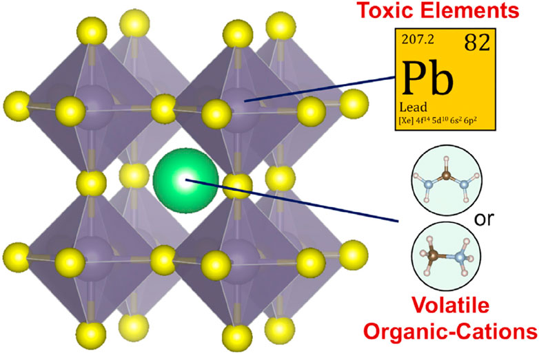
Figure 1 . The lattice assembly of common OIHPs.
Despite current research into alternate PVK materials, the B-site positive ion in modern PSCs is still lead ( Fabini, 2015 ). If all the electricity in the United States were to be generated by PSCs using the most well-deliberated OIHP, the annual consumption of lead would be 160 tons ( Fabini, 2015 ). Eliminating Pb from PSCs is the only long-term solution to the Pb-toxicity problem, even though PSCs may be managed and regulated to decrease environmental Pb discharge. The quantity of harmful components permitted in consumer or domestic niche applications, such as portable PVs, is extremely low ( Hailegnaw et al., 2015 ). The band alignment between the perovskite material and the selective materials of n-type and p-type is crucial for effective charge extraction. In particular, the electron transport layer’s conduction band edge should be lower than the perovskites, while the hole transport layer’s valence band edge should be higher. This relationship is illustrated in Figure 2 .
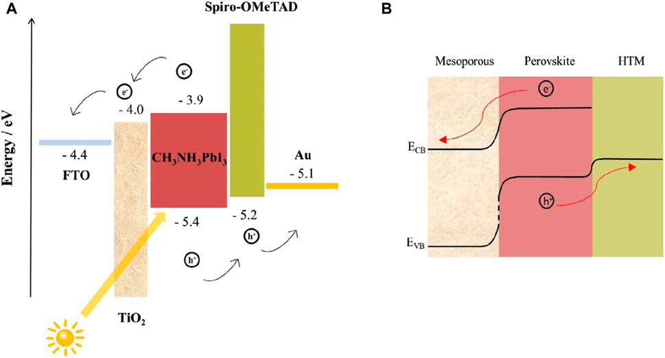
Figure 2 . (A) A typical PSC energy diagram shows the energy levels of materials in different layers and (B) the band-bending of energy levels during charge separation.
United States, Occupational Safety and Health Administration (OSHA), for instance, classifies lead and its compounds as very dangerous and has established a legally acceptable exposure limit of 0.05 mg/L for general industry ( Levin et al., 1997 ). Due to the high expense of creating Pb-based PSCs and the need to invest heavily in protecting workers’ health from the metal’s narcosis and eye/nose/throat irritation. The necessity for organic positive ions to cover the “A-site” in the PVK assembly is another major issue with existing lead-based bulk OIHPs. These organic compounds have a mild interaction with the metal-halide octahedra at PSC circumstances when the inorganic cations are present ( Brunetti et al., 2016 ).
Although PVKs include ammonia functional groups in their crystal structure, the organic species inside them are more hygroscopic, making them more susceptible to deterioration when exposed to air ( Leijtens et al., 2015 ; Rong et al., 2015 ). Lead and chemical instability of PSCs based on lead-containing bulk OIHPs are the key difficulties. To solve these issues, researchers must identify novel PVK options that are innocuous and firm while hitherto possessing sufficient PCE as an alternative to the currently employed lead-based PSCs. From this viewpoint, this paper primarily emphasizes the critical need to pinpoint the historical roots of the current PVK light-absorber materials’ inherent toxicity and instability, and then address approaches to designing new environmentally friendly, stable PVKs. Future synthesis of novel lead-free PVK compounds is discussed to round up the paper.
2 Evaluation of prospective new PVKs’ effects on the environment and their durability over time is essential
Getting the basic stuff out, synthesis/processing of cells, cell assembly, utilization, and decommissioning of cells are the usual stages in the life span of a PSC panel ( Volans, 1987 ; Babayigit et al., 2016 ), as illustrated in Figure 3A . Dangerous PVK-formed species will be released during all these phases, but it is too soon to do a full life cycle analysis of the PSC technology. Consequences like as land and water degradation are inevitable, as is the introduction of these toxins into the food chain, which ultimately reaches human people ( Florence et al., 1988 ). Possible catastrophic incidents during PSC production, transit, storage, and use, such as fire or floods, provide additional environmental concerns ( Dauvalter, 1955 ). As a result, Pb’s indirect toxicity has far-reaching effects on human health and the natural world. In addition, PVKs may be detrimental to ecosystems and people through a variety of different processes, such as acidification and nanotoxicity (see Figure 3B ).
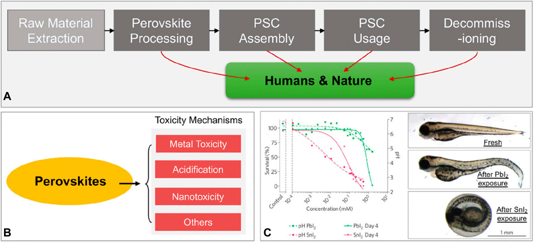
Figure 3 . Toxicity evaluation and mechanisms in PVKs (A) PVK solar panels have an average projected lifetime altered ( Babayigit et al., 2016 ). (B) Possible pathways of PVK toxicity, (C) Biological experiment outcomes utilized to evaluate Pb and Sn-based PVK toxicity ( Jellicoe et al., 2016 ).
Sn-based PSCs, for instance, have been shown to attain PCEs of 10%, making them a promising green option for PSCs. Recent research ( Babayigit et al., 2016 ), however, shows that in Sn-based PVK materials, oxidation may proceed rapidly in ambient or aqueous settings, resulting in the creation of hydroiodic acid ( Babayigit et al., 2016 ; Abate, 2017 ).
Zebrafish study shows that Strontium Iodide is more intensely lethal when compared to Lead Iodide ( Figure 2C ). The greater acidification effects of Sn2+ compared to lead ions are largely to blame for this. Nanoscale lead-based and lead-free PVKs for PSCs and optoelectronics are expected ( Im et al., 2014 ; Jellicoe et al., 2016 ). Nanoscale materials harm cells and biological systems, so, probably, nanoscale PVK materials are also dangerous.
The utilization of a mesoporous layer in the mesoporous architecture facilitates the expeditious extraction of photoinduced electrons from the perovskite material. This results in a reduction of the electron transport distance and eliminates the need for a high level of crystal quality to achieve effective light absorption ( Tétreault et al., 2010 ). Nevertheless, in comparison to other arrangements, mesoporous perovskite solar cells often exhibit a reduced V oc ( Kang et al., 2016 ) and diminished light absorption beyond 720 nm wavelengths ( Chen M. et al., 2016 ). The need for a perovskite overlayer to avoid mesoporous layer-HTL contact might cause short circuits ( Yan et al., 2016 ). Moreover, there is an ongoing dispute over the role of the mesoporous layer, especially considering the remarkable efficiencies demonstrated by two-dimensional perovskite solar cell (PSC) devices. The highest recorded efficiency, as reported in reference ( Fu et al., 2014 ), is at 20.7%. Titanium dioxide (TiO 2 ) is used as a mesoporous layer due to its broadband gap energy of 3.5 eV, chemical and thermal stability, photodegradation resistance, non-toxicity, and cost-effectiveness ( Haruyama et al., 2015 ; Sabba et al., 2015 ). Figure 4 shows that mesoporous layer thickness affects perovskite polycrystal penetration into TiO 2 pores.
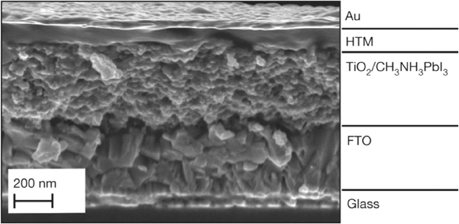
Figure 4 . Perovskite solar cell cross-sectional SEM picture ( Patrick et al., 2015 ).
In a study conducted by authors ( Wang et al., 2016 ), it was observed that a mesoporous layer of TiO 2 with a thickness ranging from 260 nm to 440 nm adequately filled the pores of mesoporous TiO 2 , as depicted in Figure 5 . These results suggest that between these bounds lies the sweet spot for maximizing light absorption while minimizing recombination due to route length. When testing the solar efficiency of each component, it was discovered that the mesoporous TiO 2 device, although thinner, performed well Figure 6 . One possible explanation is that the higher electron density in TiO 2 improves charge transfer and collecting efficiency ( Wu et al., 2017 ).
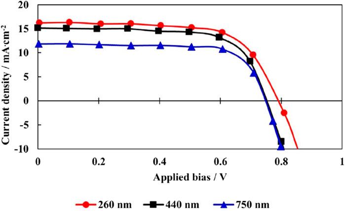
Figure 5 . Estimating the IV curves of a device, coating thickness, and pore filling in a perovskite ( Wang et al., 2016 ).
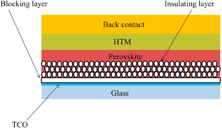
Figure 6 . Mesoporous insulating-oxide-based PSC ( Chung et al., 2012 ).
Nanoscale PVKs may be harmful due to their fibrous structure ( Xiao Z. et al., 2015 ; Ju et al., 2017a ; Ju et al., 2018 ), and radical species group ( Ming et al., 2016 ). A meaningful evaluation of the toxicity of PVK compounds, both those already in use and those that have yet to be discovered, necessitates the prompt construction and implementation of a systematic system of biological investigations. Despite the potential importance of this avenue for PSC research, nothing has been done thus far.
Figure 7 shows that water, light, heat, and oxygen are the most detrimental to the stability of a PVK material. A wide range of processes, including polymorphic transformation, hydration, ion transport ( Ke et al., 2017a ), breakdown, and oxidation, are responsible for the degradation of OIHPs by these agents. While Pb-based OIHP deterioration has been extensively investigated in recent years, our knowledge of Pb-free PVKs is still in its infancy. The stability problem of upcoming PVK materials may be much more complicated than that of Pb-based OIHPs, according to certain studies in the literature.
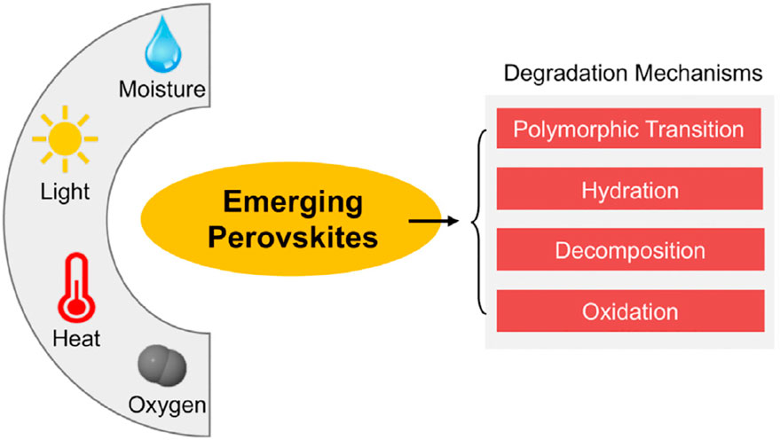
Figure 7 . Degradation of new PVK materials due to environmental variables and their processes.
Many decay pathways may be active simultaneously. CsSnI 3 , a lead-free candidate PVK material with a 1.3 eV optical bandgap and decent carrier mobility, is one such example ( Yin et al., 2017 ). CsSnI3 PVK is thermally stable because of its inorganic composition and robust covalent bonding for its lattice assembly ( Volonakis et al., 2017 ). The “black” phase of g-CsSnI 3 rapidly undergoes a “yellow” polymorph transformation when exposed to air ( Saparov et al., 2015 ; Slavney et al., 2016 ). Nevertheless, oxygen may quickly oxidize Sn (II) in CsSnI 3 to Sn (IV), turning it into Cs 2 SnI 6 ( Giustino and Snaith, 2016 ).
Moisture from the air may also penetrate CsSnI 3 PVK thin films, where it can form hydrates and break down the material into metal halides. The PCE of PSCs based on CsSnI3 may drop precipitously due to a combination of these degrading processes. Due to their extreme instability in the ambient environment, they can decay in a matter of minutes if not enclosed.
It is difficult to examine these pathways in isolation, but doing so is essential if we are to solve the PSC instability problem once and for all. In addition, there are several other Pb-free PVK possibilities whose stability and deterioration have been poorly researched ( Xiao et al., 2017a ; Xiao et al., 2017b ; Yang et al., 2017 ), including CsGeI 3 , CsSnxGe1-xI 3 , Cs2TiI 6 -xBrx, and Cs 2 AgBiBrI 6 . Research into their resistance to the major environmental variables (humidity, light, heat, oxygen) and possible breakdown mechanisms is promising. New, Pb-free, stable PVKs for PSCs can be designed using the information gleaned by studying the stability of these developing PVKs.
3 Theory and experiment required to find nontoxic, stable PVKs for PVs
Theoretical simulations screen the enormous number of PVK family compounds and derivatives to find safe and stable candidate PVKs. There are typically two phases to such materials screening processes. Finding potential elements to substitute for Pb in current Pb-based OIHPs is the first step in solving the toxicity problem. Substitutes for PVKs in PV applications must have many of the same fundamental electrical, conveyance, and ocular features. Group IV elements Sn 2+ and Ge 2+ are often cited in the literature as suitable lead ion substitutes. Lead-free Sn- and germanium-based PVKs for PV applications have emerged from this logic. Sn-based PSCs have PCE approaching 10% ( Ju et al., 2017b ), even though Sn2+ may be quickly oxidized to Sn 4+ , lowering performance. Potential Pb-free PVK candidates can also be identified using an electrical structure-based approach.
Pb-based OIHPs have high PCEs because the Pb lone-pair 6s orbital has a strong antibonding interaction with the I 5p orbital, allowing for longer carrier lifetimes and diffusion lengths ( Zhang et al., 2017 ). Several researchers have focused on PVKs made from non-traditional metals because of the presence of lone-pair ns2 ( McMeekin et al., 2016 ; Nakajima and Sawada, 2017 ; Ali et al., 2018a ). New compounds having antibonding contact between orbitals around the valence band maximum can likewise exhibit band-edge behaviour like lead-based PVKs. Skutterudite structure has been proposed, for instance ( Dai et al., 2017 ). Unlike lead-based PVKs, IrSb 3 possesses a band-edge feature indicative of p-p* antibonding interaction. The crystal structure for PVK materials provides another angle from which to hunt for promising Pb-free PVK options.
Replace Pb2+ with an aliovalent metal cation and the resulting PVK structure will have a different chemical formula from the usual AB(II/III/V) X3/X6/X9 ( Zhao et al., 2017a ; Zhao et al., 2017b ). The electrical structure of the compounds will be altered because of this structural alteration ( Sakai et al., 2017 ). Figure 8A shows how crystal structure and chemical composition can be used to create novel PVK-type compounds with favourable electrical structures for PV applications.
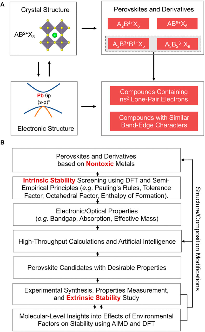
Figure 8 . Strategies for Finding Stable, Nontoxic PVKs (A) Example of the use of crystal structure and electrical structure information to potentially choose stable, non-toxic PVK candidates for PSCs. (B) A schematic showing how to find safe, stable PVK candidates to use in PSCs ( Debbichi et al., 2018 ).
To accurately anticipate the stability of PVKs, a theory-experiment integrated method is required because of the complexity of the problem ( Pang et al., 2016 ; Sun and Yin, 2017 ) To get a general idea of the stability of the PVK phase, a common empirical rule is to utilize Goldchmidt’s tolerance factor (t). Cubic assemblies are suggested by fits in the 0.9% t% one range for PVKs, whereas orthorhombic assemblies are suggested by fits in the 0.71%–0.9% t% one range. Other configurations include the hexagonal assembly, for t% 0.71 or t R 1. Figure 8B displays a flow chart for showing non-hazardous metal-based PVK intrants with PV constancy. This technique logically combines theory and experiment. Figure 8A depicts the first step of the process, which involves identifying a suitable metal-free PVK that does not include lead. Although the organic A-site positive ion is intrinsically unstable, positive ions such as caesium ions ( Castelli et al., 2012 ; Korbel et al., 2016 ) are employed as replacements ( Schmidt et al., 2017 ; Takahashi et al., 2018 ) due to their sturdier ionic interaction through unknown negative ions.
The most promising PVK candidates may be identified by combining A with B-site replacement. This method has been used to effectively anticipate and synthesise compounds like CsSnI 3 , Cs 2 AgBiBr 6 , and Cs 2 TiBr 6 , although there may be additional PVK options that are less toxic and more stable ( Lee et al., 2012 ; Burschka et al., 2013 ; Pilania et al., 2016 ; Li Z. et al., 2018 ). Several alternatives to B-site ions in PVK structures, including monovalent metals and trivalent metals ( Xiao et al., 2014 ; Chen et al., 2018a ; Ávila et al., 2017 ; Xiao J. et al., 2015 ; Elumalai et al., 2016 ), have been proposed. Substituting a tetravalence metal for the B-site ion stabilizes vacancy-order double PVKs ( Kalyanasundaram and Grätzel, 1998 ; Leijtens et al., 2013 ). The same atoms in various valence states can substitute the B-site ion in electronic double PVKs ( Xin et al., 2011 ).
Using Figure 8B’ s manufacturing cycle, first, validate new compounds’ intrinsic or thermodynamic stabilities using “density functional theory (DFT)” based design to guarantee they have the requisite PV-related electronic/optical properties. As shown in Figure 3 , when the PVKs have been synthesized, they are put through a series of experiments to determine how stable or degradable they are in the presence of various external environmental stimuli.
DFT-based mechanistic research complements experimental studies ( Bi et al., 2013 ; Leijtens et al., 2014 ). These parallel theory-experiment studies ( Son et al., 2014 ; Zuo et al., 2015 ) show how to modify PVK crystal structure/composition to find more stable PVK candidates. As can be seen in Figure 4B , the entire stability-screening procedure considers the crucial electrical assembly and the PVKs’ photovoltaic properties.
High amount simulated ingredients strategy can anticipate lead-free PVK ingredients for PV utilizing computational quantum-mechanical, thermodynamic, database development, and intelligent data-mining methods. Although useful, these calculations cannot replace a full material simulation, and they often only solve a portion of the design issue. The DFT-computed descriptors are useful for screening candidate materials and determining their important features ( Agresti et al., 2016 ).
Pauling’s principles ( Ball et al., 2013 ), are computational descriptors for intrinsic stability. Bandgap and effective mass can approximate light absorption and carrier mobility. The basic components of a material can provide an approximation of its cheap cost and nontoxicity. Descriptors screen candidates with a PVK structure and its modifications for materials with the right attributes ( Ball et al., 2013 ).
High-throughput computational design is computationally expensive due to the huge conformation space and the enormous number of acceptable descriptors for essential properties, many of which are derived by more precise functional modelling ( Wojciechowski et al., 2014 ). In divergence, a good machine learning model may be taught using existing data or data derived through computations. To find novel candidate materials with desirable qualities, this approach may be applied to the periodic table, yielding insights, and guiding experimental design ( Niu et al., 2015 ). PVK stability and bandgap have been estimated in previous works using a variety of techniques based on the identification of pertinent properties ( Christians et al., 2014 ; Qin et al., 2014 ; Jeon et al., 2015 ).
4 Advances in lead-free perovskites
4.1 tin perovskites.
Group 14 components Tin’s 5s2 electrical configuration resembles lead’s 6s2. With a similar outer electron shell structure to lead (Pb) but a smaller ionic radius, tin may be a preferable option ( Zhang Q. et al., 2018 ). Tin can replace lead in PSCs ( Wang X. et al., 2019 ). Tin is cheap, non-toxic, and electrically comparable to lead ( Shanon, 1976 ; Ke et al., 2017b ). The most researched lead-free perovskite alternative is tin-based ( Fu, 2019 ). Tin halide-based perovskites have low exciton binding energy, a tiny band gap, and excellent carrier mobility ( Liu X. et al., 2020 ). Sn-based perovskites offer several advantages for solar cells, but their unstable divalent Sn states make them extremely conductive and inefficient ( Song et al., 2017 ). Tin-based lead-free PSCs are ineffective because FASnI 3 perovskites rapidly crystallise and oxidise, resulting in rough morphology and large defect concentrations ( Liu X. et al., 2020 ). Table 1 lists current tin-based perovskites and ways to improve them.

Table 1 . Current tin-based perovskites and their fabrication methods.
4.2 Perovskites with a composition based on bismuth
The electrical configuration of group 15 element bismuth is Bi 3+ (6s2). Bismuth is less toxic than lead and has many dimensions due to its BiX 6 3 octahedron structure ( Shanon, 1976 ; Zhang et al., 2023 ). Subsalicylate and bismuth subcitrate are therapeutic ( Ganose et al., 2017 ). Chronic bismuth use can induce encephalopathy and renal failure ( Ganose et al., 2017 ). Bismuth perovskites are attractive because of their lead-like isoelectronic valence shell ( Lozhkina et al., 2018 ). Like lead (1.21 A°), Bi 3+ is stable and has an ionic radius of 1.05 A° ( Wani et al., 2015 ; Liu et al., 2022 ). Bismuth-based perovskites can replace lead-based ones due to their optoelectronic properties, environmental friendliness, and light, heat, and moisture resistance ( Dai and Tüysüz, 2019 ). Bismuth-based perovskites have the most stable optical and structural characteristics since optical parameters did not change after 3 months without surface passivation ( Kim et al., 2016 ).
Table 2 lists bismuth-based perovskites and strategies for overcoming obstacles.

Table 2 . Current bismuth-based perovskites and their fabrication methods.
4.3 Perovskites with a composition based on Sb (antimony)
Group 15 element antimony (Sb3+) has an ionic radius of 0.75 A° and an electronic configuration of 5s2 ( Wang X. et al., 2019 ). An alternative to lead, antimony is non-toxic and twice as affordable as Sn per kilogram ( Lozhkina et al., 2018 ). Irina Shtangeeva et al. found that high amounts of antimony in growth media were very hazardous to plants, resulting in a significant decrease in leaf and root biomass output ( Liu Y. et al., 2019 ). The alignment of Sb’s 5s and 5p orbitals with p-block anions makes it a lone pair effect heavy hitter, and there are several advantages to being in the 3+ oxidation state ( Lozhkina et al., 2018 ).
Trivalent Sb, which has one set of 5s2 electrons instead of lead, is an option. Therapeutics are the primary use of antimony compounds ( Lozhkina et al., 2018 ). Liu et al. (2018) performed a theoretical evaluation of Cs 3 Sb 2 X 9 ’s optoelectronic characteristics. The computed carrier mobilities of Cs 3 Sb 2 I 9 indicate an appropriate band energy gap for hydrogen production and CO2 reduction due to enhanced electronic mobilities. Cs 3 Sb 2 I 9 ’s photocatalytic activity is enhanced by the significant difference in hole and electron mobilities, which slows electron-hole recombination. The photovoltaic performance of Cs 3 Sb 2 I 9 is superior to lead-based perovskites, making it a viable replacement ( Singh et al., 2018 ). To manufacture solar cells efficiently, issues with antimony-based perovskites must be resolved. In terms of solar performance, solution-processed Sb-based perovskites are best suited for the dimer phase ( Karuppuswamy et al., 2018 ). The amorphousness and pinholes in the surface form of the zero-dimensional dimer of methylammonium antimony iodide cause its poor PCE ( Zuo and Ding, 2017 ). Table 3 lists antimony-based perovskites and strategies for overcoming obstacles.

Table 3 . Current antimony-based perovskites and their fabrication methods.
4.4 Perovskites with a composition based on germanium
The ionic radius of Ge 2+ is 0.73 Å, and its electronic configuration is 4s2 ( Wang X. et al., 2019 ; Yang et al., 2021 ). It is a group of 14 elements. Germanium is easy to find in nature, and its pure organogermanium products are safe ( Schauss, 1991 ; Ganose et al., 2017 ). Germanium has little toxicity, except for tetrahydride germane ( Gerber and Léonard, 1997 ). The covalent character and higher electronegativity of Germanium make it a possible alternative to lead PSCs ( Kopacic et al., 2018 ). Ping-Ping Sun et al. found that MAGeI 3 is theoretically very comparable to MAPbI 3 in terms of band gap, stability, outstanding optical properties, and hole and electron conductivity ( Sun et al., 2016 ). The stability and effectiveness of Ge and Sn as lead mono substitution options have been demonstrated in lead-free perovskite studies ( Ali et al., 2018b ). It is thought that perovskites based on Ge, as opposed to tin or lead, have smaller bandgaps due to the higher orbital energy of Ge (4s) as compared to Sn (5s) and Pb (6s). Perovskites based on Ge, on the other hand, exhibit larger bandgaps compared to those based on tin and lead. The fact that the [GeI 6 ] octahedral structure is structurally deformed is the primary cause of the unexpected finding ( Liu et al., 2018 ). Germanium compounds may block mutagenic activity and prevent cancer formation under certain conditions, demonstrating they are neither carcinogenic nor mutagenic ( Gerber and Léonard, 1997 ). The tumour incidence was reduced in rats that were administered 5 parts per million of sodium germanate in their drinking water during their lives ( Gerber and Léonard, 1997 ).
While germanium perovskite has several benefits, it has some problems that must be addressed for increased efficiency. Commercial usage of germanium-based perovskites in photovoltaics has been hindered by poor performance (below 0.2%) and device instability ( Kopacic et al., 2018 ). In germanium-based PSCs, Ge 2+ oxidation is the main issue. In PSCs based on germanium, this results in poor performance ( Wang X. et al., 2019 ). On the other hand, if future research can reach efficiencies beyond 10%, mixed Ge/Sn-based perovskites might be a promising material. The exorbitant price of Ge is one potential drawback of solar cells based on Ge ( Ke and Kanatzidis, 2019 ). Table 4 lists antimony-based perovskites and strategies for overcoming obstacles.

Table 4 . Current germanium-based perovskites and their fabrication methods.
4.5 Perovskites with a composition based on titanium
Titanium (IV), a non-toxic element, is abundant on Earth and has exceptional stability ( Ju et al., 2018 ). Ti 4+ has an electronic structure of 3p6 and an ionic radius of 0.53 Å ( Wang X. et al., 2019 ). Common reasons for gridlock in Pb-free perovskites include instability, undesirable defect states, and insufficient band gaps ( Bansode et al., 2015 ). Table 5 displays the titanium-based perovskites that are currently used.

Table 5 . Current titanium-based perovskites and their fabrication methods.
4.6 Perovskites with a composition based on copper
Non-toxic copper is abundant and has good charge mobility ( Sani et al., 2018 ). Cu 2+ has an ionic radius of 0.73 Å and an electronic configuration of 3d9 ( Ke et al., 2017b ; Wang X. et al., 2019 ). The transition metal copper is stable. In aerobic environments, Cu 2+ can form stable compounds with a high visible absorption coefficient ( Cortecchia et al., 2016 ). While copper’s (Cu 2+ ) stable oxidation state makes it a viable alternative to lead, the halide octahedron’s corner-sharing network is constrained by its smaller ionic radius. There are a lot of effective hole masses, a low intrinsic conductivity, and a low absorption coefficient in the perovskite layer ( Okano and Suzuki, 2017 ). Perovskites made of copper that have been used so far are listed in Table 6 .

Table 6 . Current copper-based perovskites and their fabrication methods.
4.7 Bimetallic or double perovskites
Substituting another B′ cation for half of the B site cation in the general formula of the perovskite structure ABO3 results in A 2 B 2 O 6 or A 2 BB'O 6 , two forms of double perovskites ( Saha-Dasgupta, 2020 ). Due to the nanocrystal surface energy in metastable phases, nanoscale, double perovskite materials that were limited to single monolayers exhibited quantum size effects and enhanced stability. Stable nanocrystals include Cs 2 AgBiI 6 , which cannot be mass-produced. The combinatorial compositions and quaternary nature of double perovskite materials provide them with electronic structure engineering flexibility and bandgap tunability ( Karuppuswamy et al., 2018 ; Khalfin and Bekenstein, 2019 ). Double perovskites made of lead are more environmentally friendly than other lead-free structures, and they have great chemical stability, electronic dimensions, and substitutional chemistry. LEDs, X-ray detectors, photocatalytic dye sensors, solar cells, and lead-free double perovskites are only a few examples of the many renewable energy and optoelectronic applications for these materials ( Dave et al., 2020 ; Ghrib et al., 2021 ; Grandhi et al., 2021 ). Recent lead-free perovskites Cs 2 SbAgCl 6 , Cs 2 InAgCl, Cs 2 BiAgCl 6 , and Cs 2 BiAgBr 6 exhibit outstanding optoelectronic properties because of their low carrier effective masses and detectable bandgaps ( Volonakis and Giustino, 2018 ). You may see a selection of the double perovskites that have been utilized thus far in Table 7 .

Table 7 . Current bimetallic or double perovskites and their fabrication methods.
4.8 Perovskite oxide without lead
BiMnO 3 is the sole transitional-metal perovskite oxide with unique properties including insulating and high ferromagnetism in bulk. According to a 2015 study by Di’eguez et al., solar applications might be possible using BiMnO3 films that have lower band gaps compared to ferroelectric oxides ( Diéguez and Íñiguez, 2015 ). Researchers Yuji Okamoto et al. demonstrated that dye-sensitized solar cells using perovskite oxides (SrTiO 3 , CaTiO 3 , and BaTiO 3 ) could achieve a high Voc in cells that were phase-pure ( Okamoto and Suzuki, 2014 ).
5 Possible methods for enhancing performance in lead-free perovskites
Using additives and adjustments to the solar cell fabrication process, lead-free PSCs may be made more efficient and stable. One or more of the following goals can be accomplished with the introduction of additives: control of oxidation, reduction of vacancies, alteration of the optical bandgap, increase of the fill factor (FF), or improvement of efficiency. The utilisation of appropriate techniques in fabrication can successfully address issues such as inadequate crystallisation, unfavourable morphology, and undesirable defects. This is crucial in achieving uniform and defect-free perovskite films without any pinholes ( Ke et al., 2017c ).
5.1 Tin, bismuth, Sb, and Ge-based perovskites additives overcome obstacles
Data from ( Ke et al., 2017c ; Meng et al., 2019 ) suggests that additives play a crucial role in lead-free perovskites. Few research has examined the impact of additives on optical bandgap. The power conversion efficiency (PCE) grows when the optical bandgap narrows, as demonstrated in ( Ke et al., 2017c ; Meng et al., 2019 ). Tin-based perovskites had increased efficiency with additives, whereas Bismuth and antimony-based ones had lesser efficiency. This may be because Bismuth and antimony-based perovskites have greater optical bandgaps than tin-based ones. To increase PCE in bismuth and antimony-based perovskites, chemicals that lower the optical bandgap can be utilised. When additives raise the optical bandgap of tin-based perovskites, the PCE may decrease. In experiments ( Ke et al., 2017c ) adding ethylenediammonium (ED) enhanced optical bandgaps by 1.45 eV, 1.53 eV, and 1.92 eV at 0%, 10%, and 25% concentrations. PCE decreased with loading of 0, 8, and 23%, resulting in 1.42%, 6.98%, and 2.45%, respectively. This is because 28% loading results in a greater optical bandgap ( Ke et al., 2017c ). Optimising additive amounts leads to improved efficiency by maintaining or narrowing the optical bandgaps. Results in ( Meng et al., 2019 ) found that adding poly (vinyl alcohol) PVA did not change the predicted optical bandgap of FASnI 3 at 1.39 eV. The high PCE of 8.96% might be attributed to the PVA molecule being near the grain boundary of the perovskite layer. Optimising the selection and number of additives in perovskite compound production is crucial for producing highly efficient lead-free solar cells. Table 8 shows the various effects of adding additives to the above perovskites.
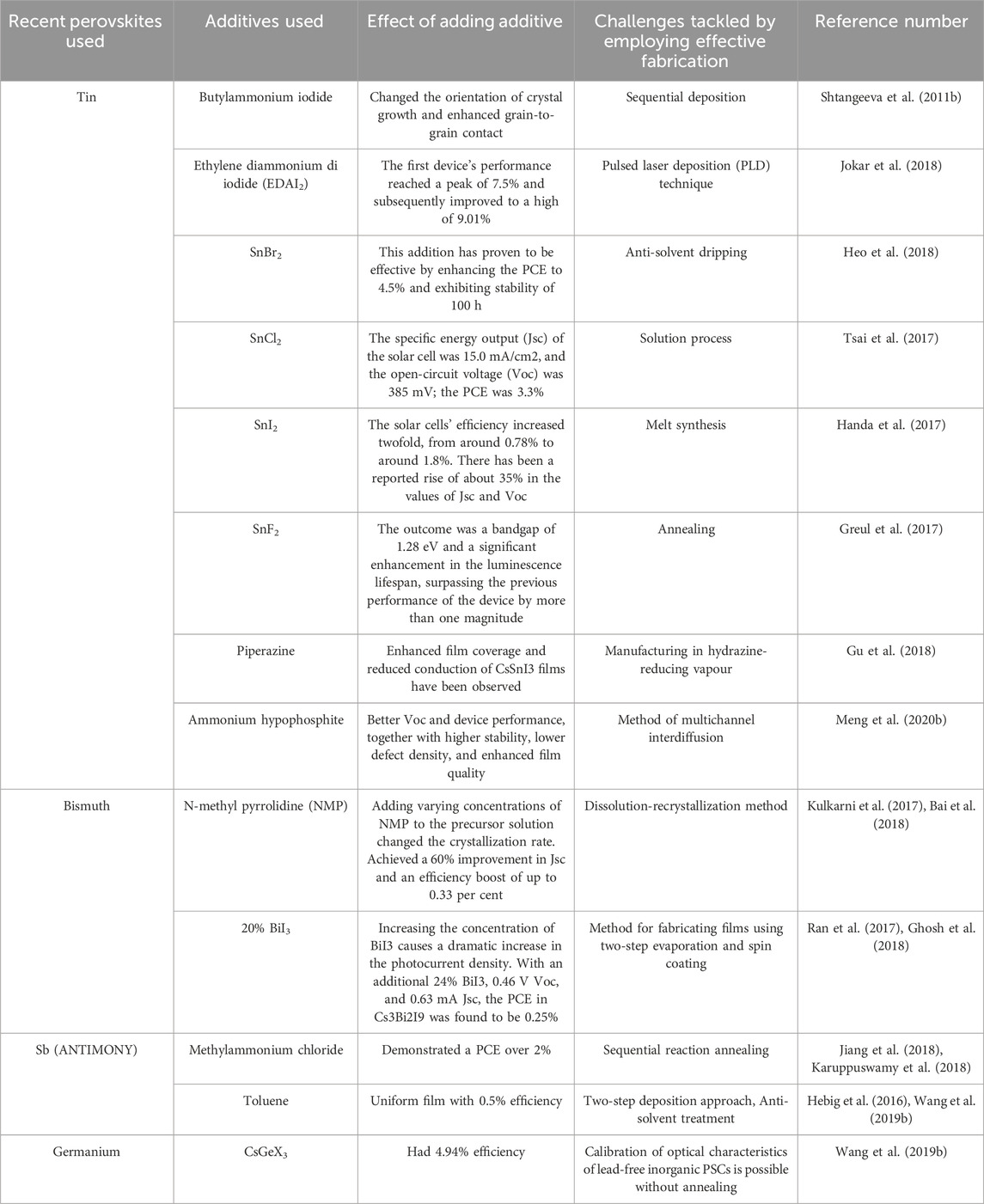
Table 8 . Challenges tackled by adding additives.
6 Recent breakthroughs/future perspectives in PSC
Recent advancements in the field of PSCs have focused on using different treatments, introducing hole transport materials, and including chiral compounds to enhance their efficiency and open-circuit voltage (Voc). These strategies will be further explored in the following discussion.
6.1 Using various hole transfer materials (HTM)
The data presented here emphasize the role that hole transport materials (HTMs) play in perovskite solar cell systems’ ability to increase efficiency and Voc. Using poly (3-hexylthiophene-2,5-diyl) (P3HT) as the hydrogen transfer material (HTM), Sagar. M. Jain et al. (2019) found that the fabrication efficiency of (CH3NH3)3Bi2I9 films was raised by 1.62%. When compared to the 1.12% efficiency attained with the conventional Spiro-OMeTAD HTL, this value is significantly greater. Min-Cherl Jung et al. (2015) used spiro-OMeTAD, C60, and P3HT, among other HTMs, to create MASnBr3 perovskite solar cell devices. In that order, the efficiencies were 0.002%, 0.221 per cent, and 0.35%. This shows that the efficiency of the solar cell devices was greatly affected by the HTM option, with P3HT showing the best efficiency out of the three HTMs that were evaluated. In addition, the lack of photocurrent and fill factor (FF) caused by spiro-OMeTAD’s high resistance is the reason for its poor efficiency. Different HTMs can cause changes in the open-circuit voltage of the solar cell devices; this is supported by the fact that devices, including C60, achieve a higher Voc than P3HT devices. Overall, these findings underscore the critical role of HTMs in achieving improved efficiency and Voc in PSC devices and highlight the potential for P3HT as an effective HTM in this context.
6.2 Antisolvent therapy
A study conducted by Jiewei Liu et al. discovered that using a hot Ph-Cl antisolvent treatment prevented the electric shunting of the Solar System and an increase in the number density of nucleation sites in the film. When the film was annealed in an atmosphere with a low concentration of dimethyl sulfoxide (DMSO) vapour, the average size of the crystal particles increased. Furthermore, according to the reference, adding DMSO vapour during annealing increased the film quality ( Song et al., 2018 ).
In 2017, Priyadharsini Karuppuswamy and colleagues produced films of (CH 3 NH 3 ) 3 Sb 2 I 9 using antimony. They improved the film’s surface morphology and device performance by employing Hydroiodic acid (HI) as an additive and treating it with Chlorobenzene (CB) Antisolvent Treatment. The alignment of the energy levels was also improved. As can be observed from the UV absorbance spectra, the increased surface coverage brought about by the combination of HI and CB treatment led to a higher absorption intensity ( Karuppuswamy et al., 2018 ).
6.3 Interfacing manufacturing
After adding a hydrophobic scaffold to (CH 3 NH 3 ) 3 Sb 2 I 9 films, several improvements were noticed. Grain size, crystallinity, crystallisation orientation, and quality all saw improvements. When compared to perovskites with a hydrophilic interlayer, those with a hydrophobic interlayer produced larger grain crystals with fewer grain boundaries, leading to better film coverage. Priyadarshini Karuppuswamy and colleagues used impedance spectroscopy to evaluate the effect of the pyrene layer on transport and recombination in PEDOT: PSS/(CH 3 NH 3 ) 3 Sb 2 I 9 and Pyrene/(CH 3 NH 3 ) 3 Sb 2 I 9 PSCs. Their discovery led them to the conclusion that pyrene prevented hysteresis in PSCs by reducing charge carrier recombination in (CH 3 NH 3 ) 3 Sb 2 I 9 . Researchers have shown that adding pyrene to solar cell materials makes them more efficient by allowing larger grains of Sb-based crystals to grow on the material. As a result, recombination near grain boundaries is less likely to occur ( Karuppuswamy et al., 2018 ).
6.4 Semiconducting molecule outline
While creating inverted tin-based FASnI 3 perovskite in 2019, Cong Liu and colleagues added a semiconducting molecule known as poly [tetraphenylethene 3,3′-(((2,2-diphenylethene-1,1-diyl) bis(4,1-phenylene)) bis(oxy)) bis (N, N-diethylpropion-1amine) tetraphenylethene] (PTN-Br) into the perovskite precursor. A medium for transporting holes was established using the semiconducting molecule PTN-Br. It achieved this by filling the gaps between the grains at the grain borders. With a maximum occupied molecular orbital energy level of −5.41 eV, this molecule was selected. Additionally, Lewis adducts were formed when the dimethylamino group of PTNBr interacted with unattached Sn atoms. By interacting with the perovskite material, the π-conjugated polymer PTNBr was able to neutralize or deactivate trap states. Consequently, an efficiency of 7.14% was achieved. Integrating PTN-Br into the device increased its stability against UV radiation, thanks to the UV barrier and PTN-Br’s passivating activity. It was able to preserve around 66% of its original efficiency even after being continuously exposed to UV light for 5 h ( Liu C. et al., 2019 ). Therefore, the incorporation of semiconducting molecules enables the production of perovskite films with exceptional electrical properties.
6.5 Passivation surface
Bin Lyu et al. (2021) capped CsSnCl 3 perovskite nanocrystals (NC) with oleic acid/oleylamine (OA/OAm) using the hot injection technique. After that, the structural stability, optical responsiveness, durability, and eco-friendliness of these nanocrystals were improved by treating them with gelatin, a natural biomass material. The nanocrystals’ performance was enhanced by the gelatin treatment, which enabled them to retain 77.4% of their photoluminescence intensity even after being distributed in water for 3 days. Polar solvents generate this effect by displacing the weaker amino-Sn coordination with the stronger carboxylate-Sn coordination. Nanocrystals bound to gelatin nevertheless exhibit mostly unaltered halogen-ammonium hydrogen bonding and carboxylate-Sn coordination. Additionally, gelatin has demonstrated anti-mildew properties that might find wider application ( Lyu et al., 2021 ).
6.6 Alteration in the physical structure of the surface
The 2017 study by Biao Shi et al. utilized textured FTO substrates to create light-absorbing perovskites. The researchers were able to increase the amount of light that could be absorbed and create larger grain-sized perovskite films with better charge transfer. Compared to smooth FTO substrates, textured FTO substrates significantly improved efficiency, reaching 22%. The short circuit density also increased significantly by 14.5 per cent. It follows that the surface form has a major impact on the enhanced efficacy of perovskites ( Shi B. et al., 2017 ).

6.7 Lacking lead, perovskite quantum dots
Research indicates that perovskite quantum dots (PVQDs) are very suitable for optoelectronic devices ( Wang et al., 2019c ). Yangyang Wang and colleagues synthesized inorganic lead-free perovskite quantum dots CsSnI 3 using a one-pot synthesis using triphenyl phosphite (TPPi). This approach resulted in a remarkable efficiency of 5.03% for the quantum dots, and the devices made from them maintained a stable power conversion efficiency (PCE) for over 25 days ( Shi B. et al., 2017 ). An improved hot-injection method was used by Hongzhe Xu et al. (2018) to create perovskite quantum dots with the composition MASnBr 3 -xIx (x = 0, 1, 2, 3). Utilized as light absorbers in mesoscopic solar cells, these quantum dots attained an efficiency of 8.79% ( Xu et al., 2018 ).
6.8 An introduction to chiral compounds
Pioneering research by Weiyin Gao et al., in 2022 showed how FASnI 3 -based PSCs might be improved in hole transportation by utilizing chiral cations α-methylbenzylamine (S-/R-/rac-MBA). Aligning energy levels and facilitating effective charge transfer at the interface were both aided by the introduction of MBAs. To facilitate the targeted transfer of accumulated holes across interfaces, the chiral R-MBA cation set off the chiral-induced spin selectivity (CISS) phenomenon in R-MBA 2 SnI 4 . As a result, as mentioned in the reference, a power conversion efficiency (PCE) of 10.73% was achieved, along with improved device stability and reduced hysteresis ( Gao et al., 2022 ).
6.9 Dion-Jacobson halide perovskites with low dimensions
Reducing Sn vacancies, improving stability with organic spacers, and perhaps increasing photo carrier transfer with divalent organic spacers were the outcomes of the synthesis of low-dimensional Dion-Jacobson Sn (II)-based halide perovskites carried out by Min Chen et al. (2018) ( Chen et al., 2018c ).
7 Conclusion
For PSCs to be extensively employed, breakthrough PVK materials that are extremely effective in light of electrical change, non-hazardous, and firm must be researched. The quest for novel PVK materials is anticipated to receive significantly expanded R&D funding during the next few years. While ongoing research towards this objective appears to develop rather randomly, we propose a reasonable roadmap that may help speed up R&D in this field. This road map is the first step toward understanding existing and future PVK noxiousness/dilapidation processes and advocates for more standardized experimental methodologies. After a comprehensive understanding of the toxicity/degradation pathways is achieved, innovative eco-friendly PVKs may be created that are particularly resistant to environmental stresses (using complementing theory-experiment techniques). Significant challenges are predicted for the future of lead-free stable PVKs thin-film manufacturing experiments in this paper. The latter is an essential step toward efficient and timely manufacturing of useful products. There are also promising new avenues for research into synthesis and processing that this opens. We anticipate that in the future, efficient and environmentally friendly PSCs will be realized because of this type of integrated scientific and technical R&D.
Author contributions
SK: Conceptualization, Investigation, Writing–original draft, Writing–review and editing. SS: Conceptualization, Methodology, Writing–original draft, Writing–review and editing. JG: Conceptualization, Data curation, Investigation, Methodology, Writing–review and editing. EM: Conceptualization, Investigation, Methodology, Resources, Validation, Writing–review and editing. TS: Conceptualization, Investigation, Methodology, Validation, Writing–review and editing. HP: Conceptualization, Data curation, Investigation, Methodology, Writing–review and editing.
The author(s) declare that no financial support was received for the research, authorship, and/or publication of this article.
Conflict of interest
The authors declare that the research was conducted in the absence of any commercial or financial relationships that could be construed as a potential conflict of interest.
Publisher’s note
All claims expressed in this article are solely those of the authors and do not necessarily represent those of their affiliated organizations, or those of the publisher, the editors and the reviewers. Any product that may be evaluated in this article, or claim that may be made by its manufacturer, is not guaranteed or endorsed by the publisher.
Abate, A. (2017). Perovskite solar cells go lead free. Joule 1, 659–664. doi:10.1016/j.joule.2017.09.007
CrossRef Full Text | Google Scholar
Agresti, A., Pescetelli, S., Taheri, B., Del Rio Castillo, A. E., Cinà, L., Bonaccorso, F., et al. (2016). Graphene–perovskite solar cells exceed 18% efficiency: a stability study. ChemSusChem 9 (18), 2609–2619. doi:10.1002/cssc.201600942
PubMed Abstract | CrossRef Full Text | Google Scholar
Ali, R., Hou, G., Zhu, Z., Yan, Q., Zheng, Q., and Su, G. (2018a). Predicted lead-free perovskites for solar cells. Chem. Mat. 30, 718–728. doi:10.1021/acs.chemmater.7b04036
Ali, R., Hou, G. J., Zhu, Z. G., Yan, Q. B., Zheng, Q. R., and Su, G. (2018b). Predicted lead-free perovskites for solar cells. J. Chem. Mater 30, 718–728. doi:10.1021/acs.chemmater.7b04036
Ávila, J., Momblona, C., Boix, P. P., Sessolo, M., and Bolink, H. J. (2017). Vapor-deposited PVKs: the route to high-performance solar cell production? Joule 1, 431–442. doi:10.1016/j.joule.2017.07.014
Babayigit, A., Ethirajan, A., Muller, M., and Conings, B. (2016). Toxicity of organometal halide perovskite solar cells. Nat. Mat. 15, 247–251. doi:10.1038/nmat4572
Bai, F., Hu, Y., Hu, Y., Qiu, T., Miao, X., and Zhang, S. (2018). Lead-free, air-stable ultrathin Cs3Bi2I9 perovskite nanosheets for solar cells. J. Sol. Energy Mater Sol. Cells 184, 15–21. doi:10.1016/j.solmat.2018.04.032
Ball, J. M., Lee, M. M., Hey, A., and Snaith, H. J. (2013). Low-temperature processed meso-superstructured to thin-film perovskite solar cells. Energy Environ. Sci. 6 (6), 1739–1743. doi:10.1039/c3ee40810h
Ban, H., Zhang, T., Gong, X., Sun, Q., Zhang, X. L., Pootrakulchote, N., et al. (2021). Fully inorganic CsSnI 3 mesoporous perovskite solar cells with high efficiency and stability via coadditive engineering. Sol. J. RRL 5, 2100069. doi:10.1002/solr.202100069
Bansode, U., Naphade, R., Game, O., Agarkar, S., and Ogale, S. (2015). Hybrid perovskite films by a new variant of pulsed excimer laser deposition: a room-temperature dry process. J. Phys. Chem. C 119, 9177–9185. doi:10.1021/acs.jpcc.5b02561
Bi, D., Boschloo, G., Schwarzmüller, S., Yang, L., Johansson, E. M. J., and Hagfeldt, A. (2013). Efficient and stable CH3NH3PbI3-sensitized ZnO nanorod array solid-state solar cells. Nanoscale 5 (23), 11686–11691. doi:10.1039/c3nr01542d
Bonomi, S., Patrini, M., Bongiovanni, G., and Malavasi, L. (2020). Versatile vapor phase deposition approach to cesium tin bromide materials CsSnBr3, CsSn2Br5 and Cs2SnBr6. J. RSC Adv. 10, 28478–28482. doi:10.1039/d0ra04680a
Brunetti, B., Cavallo, C., Ciccioli, A., Gigli, G., and Latini, A. (2016). On the thermal and thermodynamic (In)Stability of methylammonium lead halide perovskites. Sci. Rep. 6, 31896. doi:10.1038/srep31896
Burschka, J., Pellet, N., Moon, S. J., Humphry Baker, R., Gao, P., Nazeeruddin, M. K., et al. (2013). Sequential deposition as a route to high-performance perovskite-sensitized solar cells. Nature 499, 316–319. doi:10.1038/nature12340
Cao, D. H., Stoumpos, C. C., Farha, O. K., Hupp, J. T., and Kanatzidis, M. G. (2015). 2D homologous perovskites as light-absorbing materials for solar cell applications. J. Am. Chem. Soc. 137, 7843–7850. doi:10.1021/jacs.5b03796
Castelli, I. E., Olsen, T., Datta, S., Landis, D. D., Dahl, S., Thygesen, K. S., et al. (2012). Computational screening of perovskite metal oxides for optimal solar light capture. Energy Environ. Sci. 5, 5814–5819. doi:10.1039/c1ee02717d
Chen, L. J., Lee, C. R., Chuang, Y. J., Wu, Z. H., and Chen, C. (2016b). Synthesis and optical properties of lead-free cesium tin halide perovskite quantum rods with high-performance solar cell application. J. Phys. Chem. Lett. 7, 5028–5035. doi:10.1021/acs.jpclett.6b02344
Chen, M., Gu, J., Sun, C., Zhao, Y., Zhang, R., You, X., et al. (2016a). Light-driven overall water splitting enabled by a photo-Dember effect realized on 3D plasmonic structures. ACS Nano 10, 6693–6701. doi:10.1021/acsnano.6b01999
Chen, M., Ju, M., Carl, A. D., Zong, Y., Grimm, R. L., Gu, J., et al. (2018b). Cesium titanium (IV) bromide thin films based stable lead-free perovskite solar cells. J. Joule 2, 558–570. doi:10.1016/j.joule.2018.01.009
Chen, M., Ju, M. G., Carl, A. D., Zong, Y., Grimm, R. L., Gu, J., et al. (2018a). Cesium titanium(IV) bromide thin films based stable lead-free PVK solar cells. Joule 2, 1–13. doi:10.1016/j.joule.2018.01.009
Chen, M., Ju, M. G., Garces, H. F., Carl, A. D., Ono, L. K., Hawash, Z., et al. (2019). Highly stable and efficient all-inorganic lead-free perovskite solar cells with native-oxide passivation. J. Nat. Commun. 10, 16. doi:10.1038/s41467-018-07951-y
Chen, M., Ju, M. G., Hu, M., Dai, Z., Hu, Y., Rong, Y., et al. (2018c). Lead-free dion-jacobson tin halide perovskites for photovoltaics. J. ACS Energy Lett. 4, 276–277. doi:10.1021/acsenergylett.8b02051
Christians, J. A., Fung, R. C. M., and Kamat, P. V. (2014). An inorganic hole conductor for organo-lead halide perovskite solar cells. improved hole conductivity with copper iodide. J. Am. Chem. Soc. 136 (2), 758–764. doi:10.1021/ja411014k
Chung, I., Song, J. H., Im, J., Androulakis, J., Malliakas, C. D., Li, H., et al. (2012). CsSnI3: semiconductor or metal? High electrical conductivity and strong near-infrared photoluminescence from a single material. High hole mobility and phase-transitions. J. Am. Chem. Soc. 134, 8579–8587. doi:10.1021/ja301539s
Coduri, M., Strobel, T. A., Szafra´nski, M., Katrusiak, A., Mahata, A., Cova, F., et al. (2019). Band gap engineering in MASnBr3 and CsSnBr3 perovskites: mechanistic insights through the application of pressure. J. Phys. Chem. Lett. 10, 7398–7405. doi:10.1021/acs.jpclett.9b03046
Correa-Baena, J. P., Saliba, M., Buonassisi, T., Gratzel, M., Abate, A., Tress, W., et al. (2017). Promises and challenges of perovskite solar cells. Science 358, 739–744. doi:10.1126/science.aam6323
Cortecchia, D., Dewi, H. A., Yin, J., Bruno, A., Chen, S., Baikie, T., et al. (2016). Lead-free MA 2 CuCl x Br 4– x hybrid perovskites. J. Inorg. Chem. 55, 1044–1052. doi:10.1021/acs.inorgchem.5b01896
Cui, X., Jiang, K., Huang, J., Zhang, Q., Su, M., Yang, L., et al. (2015). Cupric bromide hybrid perovskite heterojunction solar cells. J. Synth. Metall. 209, 247–250. doi:10.1016/j.synthmet.2015.07.013
Dai, J., Ma, L., Ju, M., Huang, J., and Zeng, X. C. (2017). In- and Ga-based inorganic double perovskites with direct bandgaps for photovoltaic applications. Phys. Chem. Chem. Phys. 19, 21691–21695. doi:10.1039/c7cp03448b
Dai, Y., and Tüysüz, H. (2019). Lead-free Cs3Bi2Br9 perovskite as photocatalyst for ring opening reactions of epoxides. J. Chem. Eur. 12, 2587–2592. doi:10.1002/cssc.201900716
Dauvalter, V. (1955). Influence of pollution and acidification on metal concentrations in Finnish Lapland lake sediments. Water Air Soil Pollut. 85, 853–858. doi:10.1007/bf00476936
Dave, K., Fang, M. H., Bao, Z., Fu, H. T., and Liu, R. S. (2020). Recent developments in lead-free double perovskites: structure, doping, and applications. J. Chem. Asian J. 15, 242–252. doi:10.1002/asia.201901510
Debbichi, L., Lee, S., Cho, H., Rappe, A. M., Hong, K. H., Jang, M. S., et al. (2018). Mixed valence perovskite Cs 2 Au 2 I 6 : a potential material for thin-film Pb-free photovoltaic cells with ultrahigh efficiency. Adv. Mat. 30, 1707001. doi:10.1002/adma.201707001
Diéguez, O., and Íñiguez, J. (2015). Epitaxial phases of BiMnO3 from first principles. J. Phys. Rev. B 91, 184113. doi:10.1103/physrevb.91.184113
Elumalai, N., Mahmud, M., Wang, D., and Uddin, A. (2016). Perovskite solar cells: progress and advancements. Energies 9 (11), 861. doi:10.3390/en9110861
Fabini, D. (2015). Quantifying the potential for lead pollution from halide perovskite photovoltaics. J. Phys. Chem. Lett. 6, 3546–3548. doi:10.1021/acs.jpclett.5b01747
Fang, D., Tong, Y., Xu, F., Mi, B., Cao, D., and Gao, Z. (2021). Preparation of CsSnBr3 perovskite film and its all-inorganic solar cells with planar heterojunction. J. Solid State Chem. 294, 121902. doi:10.1016/j.jssc.2020.121902
Florence, T. M., Lilley, S. G., and Stauber, J. L. (1988). Skin absorption of lead. Lancet 332, 157–158. doi:10.1016/s0140-6736(88)90702-7
Fu, H. (2019). Review of lead-free halide perovskites as light-absorbers for photovoltaic applications: from materials to solar cells. J. Sol. Energy Mater Sol. Cells 193, 107–132. doi:10.1016/j.solmat.2018.12.038
Fu, P. P., Xia, Q., Hwang, H. M., Ray, P. C., and Yu, H. (2014). Mechanisms of nanotoxicity: generation of reactive oxygen species. J. Food Drug Anal. 22, 64–75. doi:10.1016/j.jfda.2014.01.005
Ganose, A. M., Savory, C. N., and Scanlon, D. O. (2017). Beyond methylammonium lead iodide: prospects for the emergent field of ns2 containing solar absorbers. J. Chemcomm. 53, 20–44. doi:10.1039/c6cc06475b
Gao, W., Dong, H., Sun, N., Chao, L., Hui, W., Wei, Q., et al. (2022). Chiral cation promoted interfacial charge extraction for efficient tin-based perovskite solar cells. J. Energy Chem. 68, 789–796. doi:10.1016/j.jechem.2021.09.019
Gerber, G. B., and Léonard, A. (1997). Mutagenicity, carcinogenicity and teratogenicity of germanium compounds. J. (MRR). 387, 141–146. doi:10.1016/s1383-5742(97)00034-3
Ghosh, B., Wu, B., Mulmudi, H. K., Guet, C., Weber, K., Sum, T. C., et al. (2018). Limitations of Cs3Bi2I9 as lead-free photovoltaic absorber materials. J. ACS Appl. Mat. Interfaces 10, 35000–35007. doi:10.1021/acsami.7b14735
Ghrib, T., Rached, A., Algrafy, E., Al-nauim, I. A., Albalawi, H., Ashiq, M. G. B., et al. (2021). A new lead free double perovskites K2Ti(Cl/Br)6; a promising materials for optoelectronic and transport properties; probed by DFT. J. Mat. Chem. Phys. 264, 124435. doi:10.1016/j.matchemphys.2021.124435
Giustino, F., and Snaith, H. J. (2016). Toward lead-free perovskite solar cells. ACS Energy Lett. 1, 1233–1240. doi:10.1021/acsenergylett.6b00499
Grandhi, G. K., Matuhina, A., Liu, M., Annurakshita, S., Ali-L¨oytty, H., Bautista, G., et al. (2021). Lead-free cesium titanium bromide double perovskite nanocrystals. J. Nanomater. 11, 1458. doi:10.3390/nano11061458
Gratzel, M. (2014). The light and shade of perovskite solar cells. Nat. Mat. 13, 838–842. doi:10.1038/nmat4065
Green, M. A., Ho-Baillie, A., and Snaith, H. J. (2014). The emergence of perovskite solar cells. Nat. Phot. 8, 506–514. doi:10.1038/nphoton.2014.134
Greul, E., Docampo, P., and Bein, T. (2017). Synthesis of hybrid tin halide perovskite solar cells with less hazardous solvents: methanol and 1,4-dioxane. J. Z Anorg. Allg. Chem. 643, 1704–1711. doi:10.1002/zaac.201700297
Gu, F., Ye, S., Zhao, Z., Rao, H., Liu, Z., Bian, Z., et al. (2018). Improving performance of lead-free formamidinium tin triiodide perovskite solar cells by tin source purification. J.Sol. RRL 2, 1800136. doi:10.1002/solr.201800136
Gupta, S., Bendikov, T., Hodes, G., and Cahen, D. (2016). CsSnBr3, A lead-free halide perovskite for long-term solar cell application: insights on SnF2 addition. J. ACS Energy Lett. 1, 1028–1033. doi:10.1021/acsenergylett.6b00402
Hailegnaw, B., Kirmayer, S., Edri, E., Hodes, G., and Cahen, D. (2015). Rain on methylammonium lead iodide based perovskites: possible environmental effects of perovskite solar cells. J. Phys. Chem. Lett. 6, 1543–1547. doi:10.1021/acs.jpclett.5b00504
Handa, T., Yamada, T., Kubota, H., Ise, S., Miyamoto, Y., and Kanemitsu, Y. (2017). Photocarrier recombination and injection dynamics in long-term stable lead-free CH3NH3SnI3 perovskite thin films and solar cells. J. Phys. Chem. C 121, 16158–16165. doi:10.1021/acs.jpcc.7b06199
Haruyama, J., Sodeyama, K., Han, L., and Tateyama, Y. (2015). First-principles study of ion diffusion in perovskite solar cell sensitizers. J. Am. Chem. Soc. 137, 10048–10051. doi:10.1021/jacs.5b03615
Hebig, J., Kühn, I., Flohre, J., and Kirchartz, T. (2016). Optoelectronic properties of (CH3NH3) 3Sb2I9 thin films for photovoltaic applications. J. ACS Energy Lett. 1, 309–314. doi:10.1021/acsenergylett.6b00170
Heo, J. H., Kim, J., Kim, H., Moon, S. H., Im, S. H., and Hong, K. H. (2018). Roles of SnX 2 (X = F, Cl, Br) additives in tin-based halide perovskites toward highly efficient and stable lead-free perovskite solar cells. J. Phys. Chem. Lett. 9, 6024–6031. doi:10.1021/acs.jpclett.8b02555
Im, J. H., Jang, I. H., Pellet, N., Gratzel, M., and Park, N. G. (2014). Growth of CH3NH3PbI3 cuboids with controlled size for high-efficiency perovskite solar cells. Nat. Nanotechnol. 9, 927–932. doi:10.1038/nnano.2014.181
Jellicoe, T. C., Richter, J. M., Glass, H. F. J., Tabachnyk, M., Brady, R., Dutton, S. E., et al. (2016). Synthesis and optical properties of lead-free cesium tin halide perovskite nanocrystals. J. Am. Chem. Soc. 138, 2941–2944. doi:10.1021/jacs.5b13470
Jeon, N. J., Noh, J. H., Kim, Y. C., Yang, W. S., Ryu, S., and Seok, S. I. (2014). Solvent engineering for high-performance inorganic–organic hybrid perovskite solar cells. Nat. Mat. 13, 897–903. doi:10.1038/nmat4014
Jeon, N. J., Noh, J. H., Yang, W. S., Kim, Y. C., Ryu, S., Seo, J., et al. (2015). Compositional engineering of perovskite materials for high-performance solar cells. Nature 517 (7535), 476–480. doi:10.1038/nature14133
Jiang, F., Yang, D., Jiang, Y., Liu, T., Zhao, X., Ming, Y., et al. (2018). Chlorine-incorporation-induced Formation of the layered phase for antimony-based lead-free perovskite solar cells. J. Am. Chem. Soc. 140, 1019–1027. doi:10.1021/jacs.7b10739
Jokar, E., Chien, C. H., Tsai, C. M., Fathi, A., and Diau, E. W. G. (2018). Robust tin-based perovskite solar cells with hybrid organic cations to attain efficiency approaching 10. J. Adv.Mater. 31, 1804835. doi:10.1002/adma.201804835
Ju, M., Chen, M., Zhou, Y., Garces, H. F., Dai, J., Ma, L., et al. (2018). Earth-abundant nontoxic titanium(IV)-based vacancy-ordered double perovskite halides with tunable 1.0 to 1.8 eV bandgaps for photovoltaic applications. J. ACS Energy Lett. 3, 297–304. doi:10.1021/acsenergylett.7b01167
Ju, M. G., Dai, J., Ma, L., and Zeng, X. C. (2017a). Lead-free mixed tin and germanium perovskites for photovoltaic application. J. Am. Chem. Soc. 139, 8038–8043. doi:10.1021/jacs.7b04219
Ju, M. G., Dai, J., Ma, L., and Zeng, X. C. (2017b). Perovskite chalcogenides with optimal bandgap and desired optical absorption for photovoltaic devices. Adv. Energy Mat. 7, 1700216. doi:10.1002/aenm.201700216
Kalyanasundaram, K., and Grätzel, M. (1998). Applications of functionalized transition metal complexes in photonic and optoelectronic devices. Coord. Chem. Rev. 177 (1), 347–414. doi:10.1016/s0010-8545(98)00189-1
Kang, D., Liu, Q., Chen, M., Gu, J., and Zhang, D. (2016). Spontaneous cross-linking for fabrication of nanohybrids embedded with size-controllable particles fabrication of nanohybrids embedded with size-controllable particles. ACS Nano 10, 889–898. doi:10.1021/acsnano.5b06022
Karuppuswamy, P., Boopathi, K. M., Mohapatra, A., Chen, H., Wong, K., Wang, P., et al. (2018). Role of a hydrophobic scaffold in controlling the crystallization of methylammonium antimony iodide for efficient lead-free perovskite solar cells. Nano Energy 45, 330–336. doi:10.1016/j.nanoen.2017.12.051
Ke, W., and Kanatzidis, M. G. (2019). Prospects for low-toxicity lead-free perovskite solar cells. J. Nat. Commun. 10, 965. doi:10.1038/s41467-019-08918-3
Ke, W., Stoumpos, C. C., Spanopoulos, I., Mao, L., Chen, M., Wasielewski, M. R., et al. (2017b). Efficient Lead-Free Solar Cells Based on Hollow {en}MASnI3 Perovskites. J. Am. Chem. Soc. 139, 14800–14806. doi:10.1021/jacs.7b09018
Ke, W., Stoumpos, C. C., Zhu, M., Mao, L., Spanopoulos, I., Liu, J., et al. (2017a). Enhanced photovoltaic performance and stability with a new type of hollow 3D perovskite {en}FASnI 3 . Sci. Adv. 3, e1701293. doi:10.1126/sciadv.1701293
Ke, W., Stoumpos, C. C., Zhu, M., Mao, L., Spanopoulos, I., Liu, J., et al. (2017c). Enhanced photovoltaic performance and stability with a new type of hollow 3D perovskite{en}FASnI 3. J. Sci.Adv. 3, 1701293. doi:10.1126/sciadv.1701293
Khalfin, S., and Bekenstein, Y. (2019). Advances in lead-free double perovskite nanocrystals, engineering band-gaps and enhancing stability through composition tunability. J. Nanoscale 11, 8665–8679. doi:10.1039/c9nr01031a
Kim, H. S., Lee, C. R., Im, J. H., Lee, K. B., Moehl, T., Marchioro, A., et al. (2012). Lead iodide perovskite sensitized all-solid-state submicron thin film mesoscopic solar cell with efficiency exceeding 9%. Sci. Rep. 2, 591. doi:10.1038/srep00591
Kim, Y., Yang, Z., Jain, A., Voznyy, O., Kim, G., Liu, M., et al. (2016). Pure cubic-phase hybrid iodobismuthates AgBi2I7 for thin-film photovoltaics. J. Angew. Chem. 128, 9738–9742. doi:10.1002/ange.201603608
Koh, T. M., Krishnamoorthy, T., Yantara, N., Shi, C., Leong, W. L., Boix, P. P., et al. (2015). Formamidinium tin-based perovskite with low Eg for photovoltaic applications. J. Mat. Chem. A 3, 14996–15000. doi:10.1039/c5ta00190k
Kojima, A., Teshima, K., Shirai, Y., and Miyasaka, T. (2009). Organometal halide perovskites as visible-light sensitizers for photovoltaic cells. J. Am. Chem. Soc. 131, 6050–6051. doi:10.1021/ja809598r
Kopacic, I., Friesenbichler, B., Hoefler, S. F., Kunert, B., Plank, H., Rath, T., et al. (2018). Enhanced performance of germanium halide perovskite solar cells through compositional engineering. ACS Appl. Energy Mat. 1, 343–347. doi:10.1021/acsaem.8b00007
Korbel, S., Marques, M. A. L., and Botti, S. (2016). Stability and electronic properties of new inorganic perovskites from high-throughput ab initio calculations. J. Mat. Chem. C 4, 3157–3167. doi:10.1039/c5tc04172d
Krishnamoorthy, T., Ding, H., Yan, C., Leong, W. L., Baikie, T., Zhang, Z., et al. (2015). Lead-free germanium iodide perovskite materials for photovoltaic applications. J. Mat. Chem. A 3, 23829–23832. doi:10.1039/c5ta05741h
Kulkarni, A., Singh, T., Ikegami, M., and Miyasaka, T. (2017). Photovoltaic enhancement of bismuth halide hybrid perovskite by N-methyl pyrrolidone-assisted morphology conversion. J. RSC Adv. 7, 9456–9460. doi:10.1039/c6ra28190g
Kumar, M. H., Dharani, S., Leong, W. L., Boix, P. P., Prabhakar, R. R., Baikie, T., et al. (2014). Lead-free halide perovskite solar cells with high photocurrents realized through vacancy modulation. J. Adv. Mat. 26, 7122–7127. doi:10.1002/adma.201401991
Lee, M. M., Teuscher, J., Miyasaka, T., Murakami, T. N., and Snaith, H. J. (2012). Efficient hybrid solar cells based on meso-superstructured organometal halide perovskites. Science 338, 643–647. doi:10.1126/science.1228604
Leijtens, T., Eperon, G. E., Noel, N. K., Habisreutinger, S. N., Petrozza, A., and Snaith, H. J. (2015). Stability of metal halide perovskite solar cells. Adv. Energy Mat. 5, 1500963. doi:10.1002/aenm.201500963
Leijtens, T., Eperon, G. E., Pathak, S., Abate, A., Lee, M. M., and Snaith, H. J. (2013). Overcoming ultraviolet light instability of sensitized TiO2 with meso-superstructured organometal tri-halide perovskite solar cells. Nat. Commun. 4, 2885. doi:10.1038/ncomms3885
Leijtens, T., Lauber, B., Eperon, G. E., Stranks, S. D., and Snaith, H. J. (2014). The importance of perovskite pore filling in organometal mixed halide sensitized TiO2-based solar cells. J. Phys. Chem. Lett. 5 (7), 1096–1102. doi:10.1021/jz500209g
Levin, S. M., Goldberg, M., and Doucette, J. T. (1997). The effect of the OSHA lead exposure in construction standard on blood lead levels among iron workers employed in bridge rehabilitation. Am. J. Ind. Med. 31, 303–309. doi:10.1002/(sici)1097-0274(199703)31:3<303::aid-ajim6>3.3.co;2-f
Li, B., Long, R., Xia, Y., and Mi, Q. (2018b). All-inorganic perovskite CsSnBr3 as a thermally stable, free-carrier semiconductor. J. Angew. Chem. Int. Ed. 57, 13154–13158. doi:10.1002/anie.201807674
Li, X., Wu, J., Wang, S., and Qi, Y. (2019). Progress of all-inorganic cesium lead-free perovskite solar cells. J. Chem.Lett. 48, 989–1005. doi:10.1246/cl.190270
Li, Z., Xu, Q., Sun, Q., Hou, Z., and Yin, W. (2018a). Stability engineering of halide PVK via machine learning. ArXiv 1803, 06042.
Google Scholar
Liao, W., Zhao, D., Yu, Y., Grice, C. R., Wang, C., Cimaroli, A. J., et al. (2016). Lead-free inverted planar formamidinium tin triiodide perovskite solar cells achieving power conversion efficiencies up to 6.22%. J. Adv. Mat. 8, 9333–9340. doi:10.1002/adma.201602992
Liu, C., Li, W., Fan, J., and Mai, Y. (2018). A brief review on the lead element substitution in perovskite solar cells. J. Energy Chem. 27, 1054–1066. doi:10.1016/j.jechem.2017.10.028
Liu, C., Tu, J., Hu, X., Huang, Z., Meng, X., Yang, J., et al. (2019b). Enhanced hole transportation for inverted tin-based perovskite solar cells with high performance and stability. J. Adv. Funct. Mat. 29, 1808059. doi:10.1002/adfm.201808059
Liu, D., Peng, H., Zeng, H., and Sa, R. (2021). A promising all-inorganic double perovskite Rb2TiBr6 for photovoltaic applications: insight from first-principles calculations. J. Solid State Chem. 303, 122473. doi:10.1016/j.jssc.2021.122473
Liu, D., Zha, W., Yuan, R., Chen, J., and Sa, R. (2020b). A first-principles study on the optoelectronic properties of mixed-halide double perovskites Cs2TiI6 xBrx. J. New J. Chem. 44, 13613–13618. doi:10.1039/d0nj02535f
Liu, M., Johnston, M. B., and Snaith, H. J. (2013). Efficient planar heterojunction perovskite solar cells by vapour deposition. Nature 501, 395–398. doi:10.1038/nature12509
Liu, X., Wu, T., Chen, J.-Y., Meng, X., He, X., Noda, T., et al. (2020a). Templated growth of FASnI 3 crystals for efficient tin perovskite solar cells. Energy Environ. Sci. 13, 2896–2902. doi:10.1039/d0ee01845g
Liu, Y., Gao, Y., Zhi, J., Huang, R., Li, W., Huang, X., et al. (2022). Allinorganic lead-free NiOx/Cs3Bi2Br9 perovskite heterojunction photodetectors for ultraviolet multispectral imaging. J. Nano Res. 15, 1094–1101. doi:10.1007/s12274-021-3608-4
Liu, Y., Yang, C., Wang, M., Ma, X., and Yi, Y. (2019a). Theoretical insight into the optoelectronic properties of lead-free perovskite derivatives of Cs3Sb2X9 (X = Cl, Br, I). J. Mat. Sci. 54, 4732–4741. doi:10.1007/s10853-018-3162-y
Lozhkina, O. A., Murashkina, A. A., Shilovskikh, V. V., Kapitonov, Y. V., Ryabchuk, V. K., Emeline, A. V., et al. (2018). Invalidity of band-gap engineering concept for Bi3+ heterovalent doping in CsPbBr3 halide perovskite. J. Phys. Chem. Lett. 9, 5408–5411. doi:10.1021/acs.jpclett.8b02178
Lyu, B., Guo, X., Gao, D., Kou, M., Yu, Y., Ma, J., et al. (2021). Highly-stable tin-based perovskite nanocrystals produced by passivation and coating of gelatin. J. Hazard Mat. 403, 123967. doi:10.1016/j.jhazmat.2020.123967
Marshall, K. P., Walker, M., Walton, R. I., and Hatton, R. A. (2016). Enhanced stability and efficiency in hole-transport-layer-free CsSnI3 perovskite photovoltaics. J. Nat.Energy 1, 16178. doi:10.1038/nenergy.2016.178
Marshall, K. P., Walton, R. I., and Hatton, R. A. (2015). Tin perovskite/fullerene planar layer photovoltaics: improving the efficiency and stability of lead-free devices. J. Mat. Chem. 3, 11631–11640. doi:10.1039/C5TA02950C
McMeekin, D. P., Sadoughi, G., Rehman, W., Eperon, G. E., Saliba, M., Ho¨ rantner, M. T., et al. (2016). A mixed-cation lead mixed-halide perovskite absorber for tandem solar cells. Science 351, 151–155. doi:10.1126/science.aad5845
Meng, X., Liu, X., He, X., Noda, T., Han, L., Yang, X., et al. (2019). Highly stable and efficient FASnI 3-based perovskite solar cells by introducing hydrogen bonding. J. Adv. Mat. 31, 1903721. doi:10.1002/adma.201903721
Meng, X., Wang, Y., Lin, J., Liu, X., He, X., Barbaud, J., et al. (2020b). Surface-Controlled oriented growth of FASnI3 crystals for efficient lead-free perovskite solar cells. Cells 4, 902–912. doi:10.1016/j.joule.2020.03.007
Meng, X., Wu, T., Liu, X., He, X., Noda, X., Wang, T., et al. (2020a). Highly reproducible and efficient FASnI3 perovskite solar cells fabricated with volatilizable reducing solvent. J. Phys. Chem. Lett. 11, 2965–2971. doi:10.1021/acs.jpclett.0c00923
Ming, W., Shi, H., and Du, M. H. (2016). Large dielectric constant, high acceptor density, and deep electron traps in perovskite solar cell material CsGeI 3 . J. Mat. Chem. A 4, 13852–13858. doi:10.1039/c6ta04685a
Moghe, D., Wang, L., Traverse, C. J., Redoute, A., Sponseller, M., Brown, P. R., et al. (2016). All vapor-deposited lead-free doped CsSnBr 3 planar solar cells. J. Nano Energy 28, 469–474. doi:10.1016/j.nanoen.2016.09.009
Nakajima, T., and Sawada, K. (2017). Discovery of Pb-free perovskite solar cells via high-throughput simulation on the K computer. J. Phys. Chem. Lett. 8, 4826–4831. doi:10.1021/acs.jpclett.7b02203
National Center for Photovoltaics (2024). Best research-cell efficiencies . U.S. Department of Energy, Office of Energy Efficiency and Renewable Energy . Available at: https://www.nrel.gov/pv/assets/images/efficiencychart.png .
Niu, G., Guo, X., and Wang, L. (2015). Review of recent progress in chemical stability of perovskite solar cells. J. Mater Chem. A 3, 8970–8980. doi:10.1039/c4ta04994b
Okamoto, Y., and Suzuki, Y. (2014). Perovskite-type SrTiO3, CaTiO3 and BaTiO3 porous film electrodes for dye-sensitized solar cells. J. Ceram. Soc. Jpn. 122, 728–731. doi:10.2109/jcersj2.122.728
Okano, T., and Suzuki, Y. (2017). Gas-assisted coating of Bi-based (CH3NH3)3Bi2I9 active layer in perovskite solar cells. J. Mat. Lett. 191, 77–79. doi:10.1016/j.matlet.2017.01.047
Pang, S., Zhou, Y., Wang, Z., Yang, M., Krause, A. R., Zhou, Z., et al. (2016). Transformative evolution of organolead triiodide perovskite thin films from strong room-temperature solid–gas interaction between HPbI 3 -CH 3 NH 2 precursor pair. J. Am. Chem. Soc. 138, 750–753. doi:10.1021/jacs.5b11824
Patrick, C. E., Jacobsen, K. W., and Thygesen, K. S. (2015). Anharmonic stabilization and band gap renormalization in the PVK CsSnI3. Phy. Rev. B 92, 201205. doi:10.1103/physrevb.92.201205
Pilania, G., Balachandran, P. V., Kim, C., and Lookman, T. (2016). Finding new perovskite halides via machine learning. Front. Mat. 3, 19. doi:10.3389/fmats.2016.00019
Qin, P., Tanaka, S., Ito, S., Tetreault, N., Manabe, K., Nishino, H., et al. (2014). Inorganic hole conductor-based lead halide perovskite solar cells with 12.4% conversion efficiency. Nat. Commun. 5, 3834. doi:10.1038/ncomms4834
Qiu, X., Cao, B., Yuan, S., Chen, X., Qiu, Z., Jiang, Y., et al. (2017). From unstable CsSnI3 to air-stable Cs2SnI6: a lead-free perovskite solar cell light absorber with bandgap of 1.48 eV and high absorption coefficient. J. Sol. Energy Mater Sol. Cells 159, 227–234. doi:10.1016/j.solmat.2016.09.022
Ran, C., Wu, Z., Xi, J., Yuan, F., Dong, H., Lei, T., et al. (2017). Construction of compact methylammonium bismuth iodide film promoting lead-free inverted planar heterojunction organohalide solar cells with open-circuit voltage over 0.8 V. J. Phys. Chem. Lett. 8, 394–400. doi:10.1021/acs.jpclett.6b02578
Rong, Y., Liu, L., Mei, A., Li, X., and Han, H. (2015). Beyond efficiency: the challenge of stability in mesoscopic perovskite solar cells. Adv. Energy Mat. 5, 1501066. doi:10.1002/aenm.201501066
Sabba, D., Mulmudi, H. K., Prabhakar, R. R., Krishnamoorthy, T., Baikie, T., Boix, P. P., et al. (2015). Impact of anionic Br – substitution on open circuit voltage in lead free perovskite (CsSnI 3-x Br x ) solar cells. J. Phys. Chem. C 119, 1763–1767. doi:10.1021/jp5126624
Saha-Dasgupta, T. (2020). Double perovskites with 3d and 4d/5d transition metals: compounds with promises. J. Mat. Res. Express 7, 014003. doi:10.1088/2053-1591/ab6293
Sakai, N., Haghighirad, A. A., Filip, M. R., Nayak, P. K., Nayak, S., Ramadan, A., et al. (2017). Solutionprocessed cesium hexabromopalladate(IV), Cs2PdBr6, for optoelectronic applications. J. Am. Chem. Soc. 139, 6030–6033. doi:10.1021/jacs.6b13258
Sanders, S., Stümmler, D., Pfeiffer, P., Ackermann, N., Schimkat, F., Simkus, G., et al. (2018). Morphology control of organic–inorganic bismuth-based perovskites for solar cell application. J. Phys. Status Solidi A 215, 1800409. doi:10.1002/pssa.201800409
Sani, F., Shafie, S., Lim, H. N., and Musa, A. O. (2018). Advancement on lead-free organic-inorganic halide perovskite solar cells: a review. J. Mat. 11, 1008. doi:10.3390/ma11061008
Saparov, B., Hong, F., Sun, J. P., Duan, H. S., Meng, W., Cameron, S., et al. (2015). Thin-film preparation and characterization of Cs 3 Sb 2 I 9 : a lead-free layered perovskite semiconductor. Chem. Mat. 27, 5622–5632. doi:10.1021/acs.chemmater.5b01989
Schauss, A. G. (1991). Nephrotoxicity and neurotoxicity in humans from organogermanium compounds and germanium dioxide. J. Biol. Trace Elem. Res. 29, 267–280. doi:10.1007/bf03032683
Schmidt, J., Shi, J., Borlido, P., Chen, L., Botti, S., and Marques, M. A. L. (2017). Predicting the thermodynamic stability of solids combining density functional theory and machine learning. Chem. Mat. 29, 5090–5103. doi:10.1021/acs.chemmater.7b00156
Shanon, R. D. (1976). Revised effective ionic radii and systematic studies of interatomic distances in halides and chalcogenides. J. Acta Cryst. 32, 751–767. doi:10.1107/s0567739476001551
Shao, S., Liu, J., Portale, G., Fang, H. H., Blake, G. R., Ten Brink, G. H., et al. (2017). Highly reproducible Sn-based hybrid perovskite solar cells with 9% efficiency. J. Adv. Energy Mat. 8, 1702019. doi:10.1002/aenm.201702019
Shi, B., Liu, B., Luo, J., Li, Y., Zheng, C., Yao, X., et al. (2017b). Enhanced light absorption of thin perovskite solar cells using textured substrates. Sol. Energy Mat. Sol. Cells 168, 214–220. doi:10.1016/j.solmat.2017.04.038
Shi, T., Zhang, H. S., Meng, W., Teng, Q., Liu, M., Yang, X., et al. (2017a). Effects of organic cations on the defect physics of tin halide perovskites. J. Mat. Chem. A 5, 15124–15129. doi:10.1039/C7TA02662E
Shtangeeva, I., Bali, R., and Harris, A. (2011a). Bioavailability and toxicity of antimony. J. Geochem. Explor. 110, 40–45. doi:10.1016/j.gexplo.2010.07.003
Shtangeeva, I., Bali, R., and Harris, A. (2011b). Bioavailability and toxicity of antimony. J. Geochem. Explor. 110, 40–45. doi:10.1016/j.gexplo.2010.07.003
Shum, K., Chen, Z., Qureshi, J., Yu, C. l., Wang, J. J., Pfenninger, W., et al. (2010). Synthesis and characterization of CsSnI3 thin films. J. Phys. Lett. 96, 221903. doi:10.1063/1.3442511
Singh, A., Boopathi, K. M., Mohapatra, A., Chen, Y. F., Li, G., and Chu, C. W. (2018). Photovoltaic performance of vapor-assisted solution-processed layer polymorph of Cs3Sb2I9. J. ACS Appl. Mat. Interfaces 10, 2566–2573. doi:10.1021/acsami.7b16349
Singh, T., Kulkarni, A., Ikegami, M., and Miyasaka, T. (2016). Effect of electron transporting layer on bismuth-based lead-free perovskite (CH3NH3)3 Bi2I9 for photovoltaic applications. J. ACS Appl. Mat. Interfaces 8, 14542–14547. doi:10.1021/acsami.6b02843
Slavney, A. H., Hu, T., Lindenberg, A. M., and Karunadasa, H. I. (2016). A bismuth-halide double perovskite with long carrier recombination lifetime for photovoltaic applications. J. Am. Chem. Soc. 138, 2138–2141. doi:10.1021/jacs.5b13294
Snaith, H. J. (2013). Perovskites: the emergence of a new era for low-cost, high-efficiency solar cells. J. Phys. Chem. Lett. 4, 3623–3630. doi:10.1021/jz4020162
Son, D. Y., Im, J. H., Kim, H. S., and Park, N. G. (2014). 11% efficient perovskite solar cell based on ZnO nanorods: an effective charge collection system. J. Phys. Chem. C 118 (30), 16567–16573. doi:10.1021/jp412407j
Song, T. B., Yokoyama, T., Aramaki, S., and Kanatzidis, M. G. (2017). Performance enhancement of lead-free tin-based perovskite solar cells with reducing atmosphere assisted dispersible additive. J. ACS Energy Lett. 2, 897–903. doi:10.1021/acsenergylett.7b00171
Song, T. B., Yokoyama, T., Logsdon, J., Wasielewski, M. R., Aramaki, S., and Kanatzidis, M. G. (2018). Piperazine suppresses self-doping in CsSnI 3 perovskite solar cells. ACS Appl. Energy Mat. 1, 4221–4226. doi:10.1021/acsaem.8b00866
Sun, P., Li, Q., Yang, L., and Li, Z. (2016). Theoretical insights into a potential lead-free hybrid perovskite: substituting Pb2+ with Ge2+. J. Nanoscale 8, 1503–1512. doi:10.1039/C5NR05337D
Sun, Q., and Yin, W. (2017). Thermodynamic stability trend of cubic perovskites. J. Am. Chem. Soc. 139, 14905–14908. doi:10.1021/jacs.7b09379
Takahashi, K., Takahashi, L., Miyazato, I., and Tanaka, Y. (2018). Searching for hidden perovskite materials for photovoltaic systems by combining data science and first principle calculations. ACS Phot. 5, 771–775. doi:10.1021/acsphotonics.7b01479
Tétreault, N., Horváth, E., Moehl, T., Brillet, J., Smajda, R., Bungener, S., et al. (2010). High efficiency solid-state dye-sensitized solar cells: fast charge extraction through self-assembled 3D fibrous network of crystalline TiO2 nanowires. ACS Nano 4, 7644–7650. doi:10.1021/nn1024434
Tsai, C. M., Mohanta, N., Wang, C. Y., Lin, Y. P., Yang, Y. W., Wang, C. L., et al. (2017). Formation of stable tin perovskites Co-crystallized with three halides for carbon-based mesoscopic lead-free perovskite solar cells. Angew. Chem. Int. Ed. 56, 13819–13823. doi:10.1002/anie.201707037
Tsai, H., Nie, W., Blancon, J. C., Stoumpos, C. C., Asadpour, R., Harutyunyan, B., et al. (2016). High-efficiency two-dimensional Ruddlesden–Popper perovskite solar cells. Nature 536, 312–316. doi:10.1038/nature18306
Umedov, S. T., Grigorieva, A. V., Lepnev, L. S., Knotko, A. V., Nakabayashi, K., hkoshi, S. I., et al. (2020). Indium doping of lead-free perovskite Cs2SnI6. J. Front. Chem. 8, 564. doi:10.3389/fchem.2020.00564
Volans, G. N. (1987). Medical toxicology, diagnosis and treatment of human poisoning. Trends Pharmacol. Sci. 9, 267. doi:10.1016/0165-6147(88)90162-9
Volonakis, G., and Giustino, F. (2018). Surface properties of lead-free halide double perovskites: possible visible-light photo-catalysts for water splitting. J. Appl. Phys. Lett. 112, 243901. doi:10.1063/1.5035274
Volonakis, G., Haghighirad, A. A., Milot, R. L., Sio, W. H., Filip, M. R., Wenger, B., et al. (2017). Cs 2 InAgCl 6 : a new lead-free halide double perovskite with direct band gap. J. Phys. Chem. Lett. 8, 772–778. doi:10.1021/acs.jpclett.6b02682
Wang, M., Zhang, T., Li, H., Ban, H., Sun, Q., and Shen, Y. (2020). Efficient CsSnI3-based inorganic perovskite solar cells based on mesoscopic metal oxide framework via incorporating donor element. J. Mat. Chem . doi:10.1039/C9TA11794F
Wang, N., Zhou, Y., Ju, M. G., Garces, H. F., Ding, T., Pang, S., et al. (2016). Heterojunction-depleted lead-free perovskite solar cells with coarse-grained B-γ-CsSnI 3 thin films. Adv. Energy Mat. 6, 1601130. doi:10.1002/aenm.201601130
Wang, X., Zhang, T., Lou, Y., and Zhao, Y. (2019a). All-inorganic lead-free perovskites for optoelectronic applications. J. Mat. Chem. Front. 3, 365–375. doi:10.1039/c8qm00611c
Wang, Y., Liu, Y., Xu, Y., Zhang, C., Bao, H., Wang, J., et al. (2019b). (CH3NH3)3Bi2I9 perovskite films fabricated via a two-stage electric-fieldassisted reactive deposition method for solar cells application. J. Electrochim. Acta 329, 135173. doi:10.1016/j.electacta.2019.135173
Wang, Y., Tu, J., Li, T., Tao, C., Deng, X., and Li, Z. (2019c). Convenient preparation of CsSnI3 quantum dots, excellent stability, and the highest performance of lead-free inorganic perovskite solar cells so far. J. Mat. Chem. A 7, 7683–7690. doi:10.1039/c8ta10901j
Wani, A. L., Ara, A., and Usmani, J. A. (2015). Lead toxicity: a review. Interdiscip. Toxicol. 8, 55–64. doi:10.1515/intox-2015-0009
Weber, S., Rath, T., Fellner, K., Fischer, R., Resel, R., Kunert, B., et al. (2019). Influence of the iodide to bromide ratio on crystallographic and optoelectronic properties of rubidium antimony halide perovskites. J. ACS Appl. Energy Mat. 2, 539–547. doi:10.1021/acsaem.8b01572
Wojciechowski, K., Saliba, M., Leijtens, T., Abate, A., and Snaith, H. J. (2014). Sub-150 °C processed meso-superstructured perovskite solar cells with enhanced efficiency. Energy Environ. Sci. 7 (3), 1142–1147. doi:10.1039/c3ee43707h
Wu, B., Zhou, Y., Xing, G., Xu, Q., Garces, H. F., Solanki, A., et al. (2017). Long minority-carrier diffusion length and low surface-recombination velocity in inorganic lead-free CsSnI 3 perovskite crystal for solar cells. Adv. Funct. Mat. 27, 1604818. doi:10.1002/adfm.201604818
Wu, C., Zhang, Q., Liu, Y., Luo, W., Guo, X., Huang, Z., et al. (2018). The dawn of lead-free perovskite solar cell: highly stable double perovskite Cs2AgBiBr6 film. J. Adv. Sci. 5, 1700759. doi:10.1002/advs.201700759
Xiao, J., Shi, J., Li, D., and Meng, Q. (2015b). Perovskite thin-film solar cell: excitation in photovoltaic science. Sci. China Chem. 58 (2), 221–238. doi:10.1007/s11426-014-5289-2
Xiao, Z., Cheng, B., Shao, Y., Dong, Q., Wang, Q., Yuan, Y., et al. (2014). Efficient, high yield perovskite photovoltaic devices grown by interdiffusion of solution-processed precursor stacking layers. Energy Environ. Sci. 7, 2619–2623. doi:10.1039/c4ee01138d
Xiao, Z., Du, K., Meng, W., Wang, J., Mitzi, D. B., and Yan, Y. (2017a). Intrinsic instability of Cs 2 In(I)M(III)X 6 (M = Bi, Sb; X = halogen) double perovskites: a combined density functional theory and experimental study. J. Am. Chem. Soc. 139, 6054–6057. doi:10.1021/jacs.7b02227
Xiao, Z., Du, K. Z., Meng, W., Mitzi, D. B., and Yan, Y. (2017b). Chemical origin of the stability difference between copper(I)- and silver(I)-Based halide double perovskites. Angew. Chem. Int. Ed. 56, 12107–12111. doi:10.1002/anie.201705113
Xiao, Z., Zhou, Y., Hosono, H., and Kamiya, T. (2015a). Intrinsic defects in a photovoltaic perovskite variant Cs 2 SnI 6 . Phys. Chem. Chem. Phys. 17, 18900–18903. doi:10.1039/c5cp03102h
Xiao, Z., Zhou, Y., Hosono, H., Kamiya, T., and Padture, N. P. (2018). Bandgap optimization of perovskite semiconductors for photovoltaic applications. Chemistry 24, 2305–2316. doi:10.1002/chem.201705031
Xin, X., Scheiner, M., Ye, M., and Lin, Z. (2011). Surface-treated TiO2 nanoparticles for dye-sensitized solar cells with remarkably enhanced performance. Langmuir 27 (23), 14594–14598. doi:10.1021/la2034627
Xu, H., Yuan, H., Duan, J., Zhao, Y., Jiao, Z., and Tang, Q. (2018). Lead-free CH3NH3SnBr3-xIx perovskite quantum dots for mesoscopic solar cell applications. Electrochim. Acta 282, 807–812. doi:10.1016/j.electacta.2018.05.143
Yan, R., Chen, M., Zhou, H., Liu, T., Tang, X., Zhang, K., et al. (2016). Bio-inspired plasmonic nanoarchitectured hybrid system towards enhanced far red-to-near infrared solar photocatalysis. Sci. Rep. 6, 20001. doi:10.1038/srep20001
Yang, B., Chen, J., Hong, F., Mao, X., Zheng, K., Yang, S., et al. (2017). Lead-free, air-stable all-inorganic cesium bismuth halide perovskite nanocrystals. Angew. Chem. Int. Ed. 56, 12471–12475. doi:10.1002/anie.201704739
Yang, D., Zhang, G., Lai, R., Cheng, Y., Lian, Y., Rao, M., et al. (2021). Germanium-lead perovskite light-emitting diodes. J. Nat. Commun. 1, 4295. doi:10.1038/s41467-021-24616-5
Yin, Y., Huang, Y., Wu, Y., Chen, G., Yin, W. J., Wei, S. H., et al. (2017). Exploring emerging photovoltaic materials beyond perovskite: the case of skutterudite. Chem. Mat. 29, 9429–9435. doi:10.1021/acs.chemmater.7b03507
Zhang, C., Gao, L., Teo, S., Guo, Z., Xu, Z., Zhao, S., et al. (2018b). Design of a novel and highly stable lead-free Cs2NaBiI6 double perovskite for photovoltaic application. J. Sustain. Energy Fuels 2, 2419–2428. doi:10.1039/c8se00154e
Zhang, M., Lyu, M., Yun, J. H., Noori, M., Zhou, X., Cooling, N. A., et al. (2016a). Low-temperature processed solar cells with formamidinium tin halide perovskite/fullerene heterojunctions. J. Nano Res. 9, 1570–1577. doi:10.1007/s12274-016-1051-8
Zhang, Q., Hao, F., Li, J., Zhou, Y., Wei, Y., and Lin, H. (2018a). Science and Technology of Advanced Materials, Perovskite solar cells: must lead be replaced – and can it be done? J. Sci. Technol. Adv. Mater 19, 425–442. doi:10.1080/14686996.2018.1460176
Zhang, R., Wang, M., Wan, Z., Wu, Z., and Xiao, X. (2023). Laser direct writing based flexible solar energy harvester. Res. Eng. 19, 101314. doi:10.1016/j.rineng.2023.101314
Zhang, T., Dar, M. I., Li, G., Xu, F., Guo, N., Gratzel, M., et al. (2017). Bication lead iodide 2D perovskite component to stabilize inorganic α-CsPbI 3 perovskite phase for high-efficiency solar cells. Sci. Adv. 3, e1700841. doi:10.1126/sciadv.1700841
Zhang, X., Wu, G., Gu, Z., Guo, B., Liu, W., Yang, S., et al. (2016b). Active-layer evolution and efficiency improvement of (CH 3 NH 3 ) 3 Bi 2 I 9 -based solar cell on TiO2-deposited ITO substrate. J. Nano Res. 9, 2921–2930. doi:10.1007/s12274-016-1177-8
Zhao, X., Yang, D., Sun, Y., Li, T., Zhang, L., Yu, L., et al. (2017b). Cu–In halide perovskite solar absorbers. J. Am. Chem. Soc. 139, 6718–6725. doi:10.1021/jacs.7b02120
Zhao, X., Yang, J., Fu, Y., Yang, D., Xu, Q., Yu, L., et al. (2017a). Design of lead-free inorganic halide perovskites for solar cells via cation-transmutation. J. Am. Chem. Soc. 139, 2630–2638. doi:10.1021/jacs.6b09645
Zhou, Y., Zhou, Z., Chen, M., Zong, Y., Huang, J., Pang, S., et al. (2016). Doping and alloying for improved perovskite solar cells. J. Mat. Chem. A 4, 17623–17635. doi:10.1039/c6ta08699c
Zuo, C., and Ding, L. (2017). Lead-free perovskite materials (NH 4 ) 3 Sb 2 I x Br 9− x . J. Angew. Chem. Int. Ed. 56, 6528–6532. doi:10.1002/anie.201702265
Zuo, L., Gu, Z., Ye, T., Fu, W., Wu, G., Li, H., et al. (2015). Enhanced photovoltaic performance of CH3NH3PbI3 perovskite solar cells through interfacial engineering using self-assembling monolayer. J. Am. Chem. Soc. 137 (7), 2674–2679. doi:10.1021/ja512518r
Keywords: perovskite, non-lead perovskite material, perovskite solar cells, chemical process, power conversion efficiency
Citation: Kumar S, Sharma SS, Giri J, Makki E, Sathish T and Panchal H (2024) Perovskite materials with improved stability and environmental friendliness for photovoltaics. Front. Mech. Eng 10:1357087. doi: 10.3389/fmech.2024.1357087
Received: 17 December 2023; Accepted: 18 March 2024; Published: 04 April 2024.
Reviewed by:
Copyright © 2024 Kumar, Sharma, Giri, Makki, Sathish and Panchal. This is an open-access article distributed under the terms of the Creative Commons Attribution License (CC BY). The use, distribution or reproduction in other forums is permitted, provided the original author(s) and the copyright owner(s) are credited and that the original publication in this journal is cited, in accordance with accepted academic practice. No use, distribution or reproduction is permitted which does not comply with these terms.
*Correspondence: Jayant Giri, [email protected]
- Open access
- Published: 15 April 2024
Machine-learning analysis reveals an important role for negative selection in shaping cancer aneuploidy landscapes
- Juman Jubran 1 na1 ,
- Rachel Slutsky 2 na1 ,
- Nir Rozenblum 2 ,
- Lior Rokach 3 ,
- Uri Ben-David 2 na2 &
- Esti Yeger-Lotem ORCID: orcid.org/0000-0002-8279-7898 1 , 4 na2
Genome Biology volume 25 , Article number: 95 ( 2024 ) Cite this article
5 Altmetric
Metrics details
Aneuploidy, an abnormal number of chromosomes within a cell, is a hallmark of cancer. Patterns of aneuploidy differ across cancers, yet are similar in cancers affecting closely related tissues. The selection pressures underlying aneuploidy patterns are not fully understood, hindering our understanding of cancer development and progression.
Here, we apply interpretable machine learning methods to study tissue-selective aneuploidy patterns. We define 20 types of features corresponding to genomic attributes of chromosome-arms, normal tissues, primary tumors, and cancer cell lines (CCLs), and use them to model gains and losses of chromosome arms in 24 cancer types. To reveal the factors that shape the tissue-specific cancer aneuploidy landscapes, we interpret the machine learning models by estimating the relative contribution of each feature to the models. While confirming known drivers of positive selection, our quantitative analysis highlights the importance of negative selection for shaping aneuploidy landscapes. This is exemplified by tumor suppressor gene density being a better predictor of gain patterns than oncogene density, and vice versa for loss patterns. We also identify the importance of tissue-selective features and demonstrate them experimentally, revealing KLF5 as an important driver for chr13q gain in colon cancer. Further supporting an important role for negative selection in shaping the aneuploidy landscapes, we find compensation by paralogs to be among the top predictors of chromosome arm loss prevalence and demonstrate this relationship for one paralog interaction. Similar factors shape aneuploidy patterns in human CCLs, demonstrating their relevance for aneuploidy research.
Conclusions
Our quantitative, interpretable machine learning models improve the understanding of the genomic properties that shape cancer aneuploidy landscapes.
Introduction
Aneuploidy, defined as an abnormal number of chromosomes or chromosome-arms within a cell, is a characteristic trait of human cancer [ 1 ]. Aneuploidy is associated with patient prognosis and with response to anticancer therapies [ 2 , 3 ], indicating that it can play a driving role in tumorigenesis. It is well established that the fitness advantage conferred by specific aneuploidies depends on the genomic, environmental, and developmental contexts [ 1 ]. One important cellular context is the cancer tissue of origin; aneuploidy patterns are cancer type-specific, and cancers that originate from related tissues tend to exhibit similar aneuploidy patterns [ 2 , 4 , 5 ]. Nonetheless, the selection pressures that shape the aneuploidy landscapes of human tumors are not fully understood, and it is not clear why some chromosome-arm gains and losses would recur in some tumor types but not in others.
Several non-mutually exclusive explanations have been previously provided in an attempt to explain the tissue selectivity of aneuploidy patterns. First, the densities of oncogenes (OGs) and tumor suppressor genes (TSGs) are enriched in chromosome-arms that tend to be gained or lost, respectively, potentially due to the cumulative effect of altering multiple such genes at the same time [ 6 ]. As cell proliferation is controlled in a tissue-dependent manner, the relative importance of OGs and TSGs varies across tissues, so that the density of tissue-specific driver genes can help predict aneuploidy patterns [ 7 ]. Second, some recurrent aneuploidies reflect the chromosome arm-wide gene expression patterns that characterize their normal tissue of origin, suggesting that chromosome-arm gains and losses may ‘hardwire’ pre-existing gene expression patterns [ 8 ]. Third, several strong cancer driver genes have been shown to underlie the recurrent aneuploidy of the chromosome-arms on which these genes reside; prominent examples are the tumor suppressors TP53 and PTEN , which have been shown to drive the recurrent loss of chromosome-arm 17p in leukemia and that of 10q in glioma, respectively [ 9 , 10 , 11 ]. Fourth, it has been recently proposed that somatic amplifications, including chromosome-arm gains, are positively selected in cancer evolution in order to buffer gene inactivation of haploinsufficient genes in mutation-prone regions [ 12 ].
Notably, each previous study focused on a separate aspect of tissue specificity; therefore, the relative contribution of each factor to shaping the overall aneuploidy landscape of human tumors is currently unknown. Furthermore, whether any additional tissue-specific traits could also play a major role in driving aneuploidy patterns remains an open question. Importantly, previous studies focused on the role of positive selection in driving the gain or the loss of specific chromosome-arms in specific tumor types. However, unlike point mutations in specific genes, aneuploidies come with a strong fitness cost [ 1 , 13 ]. Therefore, whereas positive selection greatly outweighs negative selection in shaping the landscape of point mutations in cancer, as evaluated by a refined version of the normalized ratio of non-synonymous to synonymous mutations [ 14 ], both positive selection and negative selection may be important for shaping the landscape of aneuploidy. Indeed, a recent study showed that negative selection could determine the boundaries of recurrent cancer copy number alterations [ 15 ]. It is therefore necessary to consider the balance between positive and negative selection in shaping the aneuploidy landscapes of human cancer.
Machine learning (ML) methods have been applied to study a variety of biological and medical questions where heterogeneous large-scale data are available [ 16 ]. In the context of cancer, supervised ML methods were applied to predict cancer driver genes [ 17 , 18 ], to distinguish between cancer types [ 19 , 20 ], and to predict gene dependency in tumors [ 21 ]. However, ML has not been applied to investigate the observed patterns of aneuploidy in human cancer. Whereas ML has been frequently used for prediction and often regarded as a black box, recent advancements have allowed more insight into the factors that underlie prediction. For example, Shapley Additive exPlanations algorithm (SHAP) [ 22 , 23 ] estimates the importance and relative contribution of each of the features utilized by the model to the model’s decisions.
Here, we present a novel ML approach to elucidate the factors that underlie the cancer type-specific patterns of aneuploidy. For this, we constructed separate ML models for chromosome-arm gain and loss, whereby each of 39 chromosome-arms within 24 cancer types was associated with 20 types of features corresponding to various genomic attributes of chromosome-arms, normal tissues, primary tumors, and cancer cell lines (CCLs). Our approach is focused on interpretation rather than prediction of aneuploidy recurrence patterns. Interpretation of the gain and loss models for aneuploidy in primary tumors captured known genomic features that had been previously reported to shape aneuploidy landscapes, supporting the models’ validity. Furthermore, these analyses suggested that negative selection played a greater role than positive selection in this process and revealed paralog compensation as an important contributor to cancer type-specific aneuploidy patterns, in both primary tumors and CCLs. Lastly, we experimentally validated a specific aneuploidy driver using genetically engineered isogenic human cells.
Constructing machine learning models to classify cancer aneuploidy patterns
To create a supervised classification ML model that predicts the recurrence pattern of aneuploidy across cancer types, we built a large‐scale dataset consisting of labels and features per instance of chromosome-arm and cancer type. For each instance, the label indicated whether the chromosome-arm was recurrently gained, lost, or remained neutral in that cancer. Labels were determined according to Genomic Identification of Significant Targets in Cancer (GISTIC2.0) [ 24 ]. We focused on 24 cancer types for which transcriptomic data of normal tissues of origin was available from the Genotype-Tissue Expression Consortium (GTEx) ([ 25 ] ( Methods ). In total, 199 instances of chromosome-arm and cancer type were labeled as gained, 307 were labeled as lost, and 430 were labeled as neutral (Fig. 1 A).
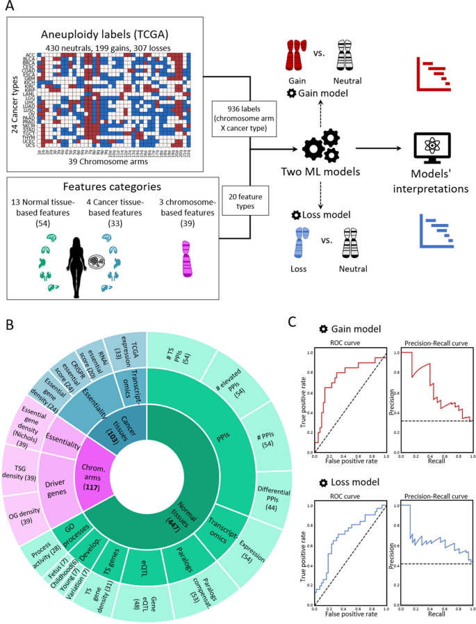
A machine learning (ML) approach for predicting aneuploidy in cancer. A Schematic view of the ML model construction. Labels represent aneuploidy status of each chromosome arm in 24 cancer types (abbreviation of cancer types detailed in Additional file 2 : Table S1), classified as gained (red, n = 199), lost (blue, n = 307), or neutral (white, n = 430). Features consist of 20 types of features pertaining to chromosome-arms, normal tissues and cancer tissues (see B ). Two separate ML models were constructed to predict gained and lost chromosome-arms (gain model and loss model). Each model was analyzed to estimate the contribution of the features to the predicted outcome. B The features analyzed by the ML model. The inner layer shows feature categories: chromosome arms (purple), cancer tissues (primary tumors and CCLs, blue), and normal tissues (green). The middle layer shows the sub-categories of the features. Chromosome-arm features include essentiality and driver genes features. Cancer-tissue features include transcriptomics and essentiality features. Normal-tissue features include protein–protein interactions (PPIs), transcriptomics, paralogs, eQTL, tissue-specific (TS) genes, development, and GO processes features. The outer layer represents all 20 feature types that were analyzed by the model. Numbers in parentheses indicate the number of tissues, organs, or cell lines from which cancer and normal tissue features were derived, or the number of chromosome-arms from which chromosome-arm features were derived. C The performance of the ML models as evaluated by the area under the receiver-operating characteristic curve (auROC, left) and the precision recall curve (auPRC, right) using tenfold cross-validation. Gain model (gradient boosting): auROC = 74% and auPRC = 63% (expected 32%). Loss model (XGBoost): auROC = 70% and auPRC = 63% (expected 42%)
Next, we defined three categories of features (Fig. 1 B; Methods ). The first category, denoted ‘chromosome-arms’, contained features of chromosome-arms that are independent of cancer type. Chromosome-arm features included the density of OGs, the density of TSGs [ 6 ], and the density of essential genes [ 26 ] per chromosome-arm. The second category, denoted ‘cancer tissues’, contained features pertaining to chromosome-arms in primary tumors and CCLs. It included features pertaining to expression of genes in primary tumors and essentiality of genes in CCLs. Expression levels of genes in each chromosome-arm per cancer type were obtained from The Cancer Genome Atlas (TCGA, https://www.cancer.gov/tcga ). Gene essentiality scores were obtained from the Cancer Dependency Map (DepMap) [ 27 ]. In total, this category included 103 omics-based readouts ( Methods ). The third category, denoted ‘normal tissues’, contained features pertaining to chromosome-arms in normal tissues from which cancer types originated (e.g., colon tissue was matched with colon adenocarcinoma, Additional file 2 : Table S1). Features of normal tissues included expression levels of genes located on each chromosome-arm in the respective normal tissue, their tissue protein–protein interactions (PPIs) [ 28 , 29 ], and their tissue-specific biological process activities [ 30 ]. It also included tissue-specific dosage relationships between paralogous genes, denoted ‘paralog compensation’ [ 31 , 32 ]. In total, this category included 447 tissue-based properties ( Methods ). To enhance our understanding of cancer and tissue selectivity, feature values of cancer and normal tissues were transformed from absolute to relative; for example, instead of indicating the absolute expression level of a gene in a given normal tissue, the expression feature was set to the expression level of the gene in the given tissue relative to its expression levels in all tissues (Additional file 1 : Fig. S1). Each chromosome-arm was then assigned with a feature value that was inferred from the values of its genes ( Methods , Additional file 1 : Fig. S2).
To fit the features dataset and the labels dataset, we further transformed the features dataset, such that each instance of chromosome-arm and cancer type was associated with features corresponding to the chromosome-arm, cancer type, and matching normal tissue ( Methods ). In total, the dataset included 20 types of features per chromosome-arm and cancer type: 3 in the chromosome-arm category, 4 in the cancer tissues category, and 13 in the normal tissues category (Fig. 1 B). We assessed the similarity between every pair of features using Spearman correlation (Additional file 1 : Fig. S3A). Most features did not correlate with each other (Additional file 1 : Fig. S3B). Among the correlated feature pairs were PPI-related features and expression in normal adult and developing tissues features (Additional file 1 : Fig. S3A). Lastly, we assessed the similarity between instances of chromosome-arm and cancer type by their feature values using principal component analysis (PCA) (Additional file 1 : Fig. S3C). Instances did not cluster by their aneuploidy pattern (gain/loss/neutral), suggesting that a more complex model is needed to classify the different patterns.
With these labels and features of each chromosome-arm and cancer type, we set out to construct two separate ML models to predict chromosome-arm gain and loss patterns across cancer types (denoted as the ‘gain model’ and the ‘loss model’, respectively; Fig. 1 A). Each model was trained and tested on data of gained (or lost) chromosome-arms versus neutral chromosome-arms. We employed five different ML methods ( Methods ) and assessed the performance of each method by using tenfold cross-validation and calculating average area under the receiver operating characteristic (auROC) and average area under the precision-recall curve (auPRC) (Additional file 1 : Fig. S4A,B). Logistic regression showed similar results to a random prediction, with auROC of 54% for each model (Additional file 1 : Fig. S4), indicating that the relationships between features and labels are non-linear. Decision tree methods that can capture such relationships [ 33 , 34 ], including gradient boosting, XGBoost, and random forest, performed better than logistic regression and similarly to each other (Additional file 1 : Fig. S4). Best performance in the gain model was achieved by gradient boosting method, with auROC of 74% and auPRC of 63% (expected: 32%) (Fig. 1 C). Best performance in the loss model was achieved by XGBoost, with auROC of 70% and auPRC of 63% (expected: 42%) (Fig. 1 C).
Revealing the top contributors to cancer aneuploidy patterns
The main purpose of our models was to identify the features that contribute the most to the recurrence patterns of aneuploidy observed in human cancer, which could illuminate the factors at play. To this aim, we used the SHAP (Shapley Additive exPlanations) algorithm [ 22 , 23 ], which estimates the importance and relative contribution of each feature to the model’s decision and ranks them accordingly. We applied SHAP separately to the gain model and to the loss model ( Methods ).
In the gain model, the topmost features were TSG density and OG density (Fig. 2 A,B). As expected, these features showed opposite directions: TSG density was low in gained chromosome-arms, whereas OG density was high, in line with previous observations [ 6 , 7 ] (Fig. 2 B). Importantly, this analysis revealed that the impact of TSGs on the gain model’s decision was twice larger than that of OGs (Fig. 2 A), highlighting the importance of negative selection for shaping cancer aneuploidy patterns. The third most important feature was TCGA expression, which quantified the expression of arm-residing genes in the given cancer type relative to their expression in other cancers. Notably, expression levels were obtained only from samples where the chromosome-arm was not gained or lost ( Methods ). This analysis revealed that, across cancer types, chromosome-arms that tend to be gained exhibit higher expression of genes even in neutral cases, consistent with a previous recent study [ 8 ]. This confirms that the genes on gained chromosome-arms are preferentially important for the specific cancer types in which these gains are recurrent. Congruently, PPIs and normal tissue expression—features of normal tissues—were also among the ten top-contributing features (Fig. 2 A). The estimated importance of all features in the gain model is shown in Additional file 1 : Fig. S5A.
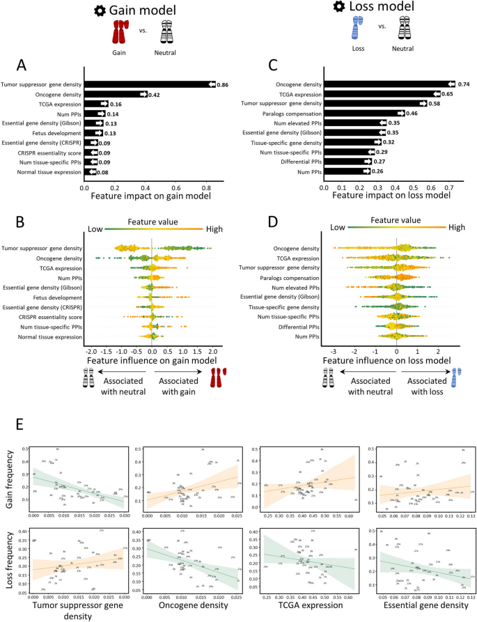
Quantitative views into the ten topmost contributing features of the gain and loss models. Features are ordered from bottom to top by their increased average absolute contribution to the model, as calculated by SHAP. A The average absolute contribution of each feature to the gain model. The directionality of the feature (i.e., whether high feature values correspond to gain or neutral) is represented by an arrow. B A detailed view of the contribution of each feature to the gain model. Per feature, each dot represents the contribution per instance of a chromosome-arm and cancer type pair. The dots are spread based on whether they were classified as neutral (left) or gain (right) by the model. Instances are colored by the feature value (green-to-orange scale denotes low-to-high value). The order (height) of each feature is the same as in A . C Same as panel A for the loss model. D Same as panel B for the loss model. E The correlations between top contributing features and the frequencies of chromosome-arm gains and losses, as measured by Spearman correlation. P -values were adjusted for multiple hypothesis testing using Benjamini–Hochberg procedure. Negative correlation between TSG density and gain frequency ( ρ = − 0.52, adjusted p = 0.006). Positive correlation between TSG density and loss frequency ( ρ = 0.3, adjusted p = 0.17). Positive correlation between OG density and gain frequency ( ρ = 0.25, adjusted p = 0.18). Negative correlation between OG density and loss frequency ( ρ = − 0.47, adjusted p = 0.01). Positive correlation between TCGA expression and gain frequency ( ρ = 0.29, adjusted p = 0.14). Negative correlation between TCGA expression and loss frequency ( ρ = − 0.33, adjusted p = 0.12). Positive correlation between essential gene density and gain frequency ( ρ = 0.16, adjusted p = 0.37). Negative correlation between essential gene density and loss frequency ( ρ = − 0.1, adjusted p = 0.5)
The loss model shared the same top three features, yet with opposite directions and different ranks (Fig. 2 C,D). OG density ranked first, was low in lost chromosome-arms, whereas TSG density ranked third, was high (Fig. 2 D), in line with previous observations [ 6 , 7 ]. In contrast to the gain model, in the loss model, the impact of OG density on the model’s decision was larger than that of TSG density, again in line with negative selection as an important force in cancer aneuploidy evolution. TCGA expression (computed from samples where the chromosome-arm was not lost or gained, see Methods ) ranked second: chromosome-arms with highly-expressed genes tended not to be recurrently lost, in line with negative selection. Another top feature that showed opposite directions between the gain and loss model was essential gene density [ 26 ]. As expected, essential gene density was low in lost chromosome-arms, in line with negative selection against losing copies of essential genes [ 26 , 27 , 35 ]. The estimated importance of all features in the loss model is shown in Additional file 1 : Fig. S5B.
To examine the direct relationships between high-ranking features and aneuploidy recurrence patterns, we assessed the correlations between these features and aneuploidy prevalence ( Methods ). In accordance with the SHAP analysis, the negative correlation between TSG density and chromosome-arm gain ( ρ = − 0.52, adjusted p = 0.0006, Spearman correlation; Fig. 2 E) was much stronger and more significant than the positive correlation between OG density and chromosome-arm gain ( ρ = 0.25, adjusted p = 0.12, Spearman correlation; Fig. 2 E). Similarly, the negative correlation between OG density and chromosome-arm loss ( ρ = − 0.47, adjusted p = 0.003, Spearman correlation; Fig. 2 E) was much stronger and more significant than the positive correlation between TSG density and chromosome-arm loss ( ρ = 0.3, adjusted p = 0.067, Spearman correlation; Fig. 2 E). TCGA expression and essential gene density were correlated with chromosome-arm gain, and anticorrelated with chromosome-arm loss, albeit to a lesser extent (Fig. 2 E, Additional file 1 : Fig. S6). Also showing positive correlations with gains and negative correlations with losses were features derived from expression levels in normal adult and developing tissues, certain PPI-related features, and additional essentiality features (Additional file 1 : Fig. S6). However, these correlations were weaker than the correlations described above. Altogether, correlation analyses supported the relationships between top features of each model and aneuploidy patterns.
The robust impact of top contributors to cancer aneuploidy patterns
Next, we asked if the above results were sensitive to our model construction schemes. We first tested the robustness of the models to internal parameters used to generate the features ( Methods ). We therefore recreated features upon modifying internal parameters and repeated model construction and interpretation ( Methods ). We found that feature importance was robust to these changes (Additional file 1 : Fig. S7, Additional file 3 : Table S2). Second, we tested the robustness of the results upon tuning the hyperparameters of each model ( Methods , Additional file 1 : Fig. S8). The top contributing features of each model were retained following hyperparameter tuning, supporting their reliability (Additional file 1 : Fig. S8C). We also checked whether the same top features would be recognized upon modeling one type of chromosome-arm event versus all other events. Applying the same approaches, we constructed two additional ML models. One model classified chromosome-arm gain versus no-gain (i.e., chromosome-arm loss or neutrality). Another model classified chromosome-arm loss versus no-loss (i.e., chromosome-arm gain or neutral). These additional models performed similarly to their respective models (Additional file 1 : Fig. S9). SHAP analysis of the two additional models revealed that feature importance was very similar between these models and the original models, which compared gained and lost chromosome-arms only to neutral chromosome-arms (Additional file 1 : Fig. S9).
We next tested whether the results were driven by a small subset of chromosome-arm and cancer type instances. For that, per model, we identified chromosome-arm and cancer type instances with the top contributions to the five topmost important features ( Methods , Additional file 4 : Table S3A,B, Additional file 5 : Table S4A,B). Most instances contributed to at least one of these features, and none of the instances contributed to all five (Additional file 5 : Table S4C). Next, we focused on chromosome-arm and cancer type instances that were top contributors to at least three of the five features (4.3% and 1.9% of the pairs in the gain and loss models, respectively). We tested their impact on the model by excluding them from the dataset and repeating the construction and interpretation of each model without them. The revised gain model retained its five topmost important features, though their ranking slightly changed (the third and fifth features switched). The revised loss model retained its four topmost important features (the fifth and seventh features switched) (Additional file 1 : Fig. S10). This suggests that the general effect of the features was not driven by a small subset of instances.
Lastly, we expanded our analyses to address whole-chromosome gains and losses. For this, we updated the features dataset to refer to whole-chromosome and cancer type instances ( Methods ). For example, the feature TSG density was updated to refer to the entire chromosome. Likewise, we updated the aneuploidy status of whole-chromosome and cancer type instances using data from GISTIC ( Methods ). This resulted in a dataset of 78 whole-chromosome gains, 151 whole-chromosome loss, and 299 neutral cases. Next, we used these data to train a whole-chromosome gain (trisomy) model and a whole-chromosome loss (monosomy) model. Model training and assessment were similar to the chromosome-arm gain and loss models. Specifically, we employed five different ML methods and assessed their performance using fivefold cross-validation. Best performance for the trisomy model was achieved by random forest, with auROC of 69% and auPRC of 47% (expected 21%; Additional file 1 : Fig. S11A). Best performance for the monosomy model was achieved by XGBoost, with auROC of 71% and auPRC of 59% (expected 34%; Additional file 1 : Fig. S11D). Performances were somewhat weaker than the chromosome-arm models, in accordance with the training data being almost twofold smaller. Lastly, we interpreted each model using SHAP. In the trisomy model, the topmost feature was TSG density and its impact was over twofold larger than the impact of other features, similarly to the chromosome-arm gain model (Additional file 1 : Fig. S11B,C). Other strong features of the chromosome-arm gain model, TCGA expression and OG density, ranked fifth and sixth, yet preserved their directionality. In the monosomy model, top features included OG density, TCGA expression, and paralogs compensation, fitting with the chromosome-arm loss model (Additional file 1 : Fig. S11E,F). The feature TSG density was ranked eight, yet preserved its directionality, similarly to the remaining features. Altogether, these results suggest that negative selection is an important factor in shaping both chromosome-arm and whole-chromosome aneuploidy patterns.
Similar features shape aneuploidy patterns in human cancer cell lines and in human tumors
Next, we aimed to test whether similar features also shape aneuploidy patterns in CCLs. We collected data of aneuploidy patterns of all chromosome-arms in CCLs [ 36 ] and analyzed 10 cancer types with matched normal tissue data from GTEx [ 25 ] ( Methods ). Similar to the analysis of cancer tissues, we labeled each instance of chromosome-arm and CCL as recurrently gained (59 instances), recurrently lost (45 instances), or neutral (286 instances) and updated the features associated with cancer types according to the CCL data ( Methods ). We then applied the gain and loss ML models, which were trained on primary tumor data, to identify determinants of aneuploidy patterns of CCLs ( Methods ). The performance of the models was at least as good as for primary tumors (gain model: auROC = 83% and auPRC = 49% (expected 15%); loss model: auROC = 76% and auPRC = 45% (expected 11%), Fig. 3 A). These results indicate that similar factors affect aneuploidy in cancers and in CCLs, consistent with the highly similar aneuploidy patterns observed in tumors and in CCLs [ 36 , 37 ].
Aneuploidy patterns in CCLs and primary tumors are shaped by similar features. A The ML scheme for analysis of aneuploidy patterns in CCLs. The gain and loss models that were trained on aneuploidy patterns in primary tumors were applied to aneuploidy patterns in CCLs. Performance was measured using tenfold cross-validation. Gain model (gradient boosting): auROC = 83%, auPRC = 49% (expected 15%). Loss model (XGBoost): auROC = 76%, auPRC = 45% (expected 11%). B The average absolute contribution of the ten topmost features to the gain model (see legend of Fig. 2 A). The order and directionality of the features generally agree with the gain model in primary tumors. C A detailed view of the contribution of the ten topmost features to the gain model (see legend of Fig. 2 B). D Same as B for the loss model. The order and directionality of the features generally agree with the loss model in primary tumors. E Same as panel C for the loss model. F The correlations between top contributing features and the frequencies of chromosome-arm gains and losses, as measured by Spearman correlation. p -values were adjusted for multiple hypothesis testing using Benjamini–Hochberg procedure. Negative correlation between TSG density and gain frequency ( ρ = − 0.37, adjusted p = 0.04). Positive correlation between TSG density and loss frequency ( ρ = 0.17, adjusted p = 0.32). Positive correlation between OG density and gain frequency ( ρ = 0.44, adjusted p = 0.012). Negative correlation between OG density and loss frequency ( ρ = − 0.28, adjusted p = 0.13). Positive correlation between CCL expression and gain frequency ( ρ = 0.53, adjusted p = 0.002). Negative correlation between CCL expression and loss frequency ( ρ = − 0.6, adjusted p = 0.0006). Positive correlation between essential gene density and gain frequency ( ρ = 0.18, adjusted p = 0.33). Negative correlation between essential gene density and loss frequency ( ρ = − 0.17, adjusted p = 0.32)
We next used SHAP to assess the contribution of each feature to each of the models. TSG density and OG density remained the top contributing features for the gain model. Consistent with our results in primary tumors, the contribution of TSG density was much stronger than that of OG density, confirming the role of negative selection (Fig. 3 B,C). In the loss model, the ranking of top features was slightly different than in primary tumors (Fig. 3 D). Expression in CCL was the top feature, such that recurrently lost chromosome-arms were associated with lower gene expression in neutral cases. OG density was one of the strongest contributing features for the loss model whereas TSG density had weaker contribution, again in line with negative selection playing an important role in shaping cancer aneuploidy landscapes (Fig. 3 D,E). Certain features of normal tissues were also highly ranked. The contribution of essential gene density was also consistent with its impact in primary tumors (Fig. 3 B,C).
As with the primary tumors, correlation analyses supported the contributions of the different features. CCL expression was highly correlated with chromosome-arm gain and anticorrelated with chromosome-arm loss ( ρ = 0.54, adjusted p = 0.02, and ρ = − 0.6, adjusted p = 0.0006, respectively; Fig. 3 F). Negative correlations were also observed between TSG density and gain frequency ( ρ = − 0.37, adjusted p = 0.04, Spearman correlation; Fig. 3 F) and between OG density and loss frequency ( ρ = − 0.28, adjusted p = 0.1, Spearman correlation; Fig. 3 F). Altogether, these results indicate that despite the continuous evolution of aneuploidy throughout CCL culture propagation [ 38 ], similar features drive aneuploidy recurrence patterns in primary tumors and in CCLs.
Chromosome 13q aneuploidy patterns are tissue-specific, and KLF5 is a driver of 13q gain in colorectal cancer
In human cancer, a chromosome-arm is either recurrently gained across cancer types or it is recurrently lost across cancer types, but rarely is a chromosome-arm both gained in some cancer types and lost in others [ 4 , 5 ]. An intriguing exception is chr13q. Of all chromosome-arms, chr13q is the chromosome-arm with the highest density of tumor suppressor genes (Fig. 2 E). It is therefore not surprising that chr13q is recurrently lost across multiple cancer types (with a median of 30% of the tumors losing one copy of 13q across cancer types) [ 4 , 5 ]. Interestingly, however, chr13q is recurrently gained in human colorectal cancer (in 58% of the samples), suggesting that it can confer a selection advantage to colorectal cells in a tissue-specific manner. Indeed, when comparing colorectal tumors and colorectal cancer cell lines against all other cancer types, chr13q was the top differentially affected chromosome-arm (Fig. 4 A,B). We therefore set out to study the basis for this unique tissue-specific aneuploidy pattern.
KLF5 is a potential driver of chromosome 13q gain in human colorectal cancer. A Comparison of the prevalence of chromosome-arm aneuploidies in colorectal tumors against all other tumors (left) and colorectal cancer cell lines against all other cancer cell lines (right). On the right side are the aneuploidies that are more common in colorectal cancer, and on the left side are the ones that are less common in colorectal cancer. Chromosome-arm 13q (in red) is the top differential aneuploidy in colorectal cancer. B Comparison of the prevalence of 13q aneuploidy between colorectal tumors and all other tumors (left) and between colorectal cancer cell lines and all other cancer cell lines (right). ****, p < 0.0001 and ****, p < 0.0001; Chi-square test. C Genome-wide comparison of differentially essential genes between colorectal cancer cell lines ( n = 85) and all other cancer cell lines ( n = 1407). On the right side are the genes that are more essential in other cancer cell lines, and on the left side are those that are more essential in colorectal cancer, based on a genome-wide CRISPR/Cas9 knockout screens [ 39 ]. The x -axis presents the effect size (i.e., the differential response between colorectal cell lines and other cell lines), and the y -axis presents the significance of the difference (-log10( p -value)). KLF5 (in red) is the second most differentially essential gene in colorectal cancer cell lines. D Comparison of the sensitivity to CRISPR knockout of KLF5 between colorectal cancer cell lines ( n = 59) and all other cancer cell lines ( n = 1041). ****, p < 0.0001; two-tailed Mann–Whitney test. E Genome-wide comparison of differentially expressed genes between colorectal tumors ( n = 434) and all other tumors (on the left, n = 11,060) and between colorectal cancer cell lines ( n = 85) and all other cancer cell lines (on the right, n = 1407). On the right side are the genes that are over-expressed in colorectal cancer and on the left side are those that are over-expressed in other cell lines. KLF5 (in red) significantly over-expressed in colorectal cancer. F Comparison of KLF5 mRNA levels between colorectal tumors ( n = 434) and all other tumors on the left ( n = 11,060) and between colorectal cancer cell lines ( n = 85) and all other cancer cell lines (on the right, n = 1407). ****, p < 0.0001; two-tailed Mann–Whitney test. G Correlation between KLF5 mRNA expression and the sensitivity to KLF5 knockdown, showing that higher KLF5 expression is associated with increased sensitivity to its RNAi-mediated knockdown. ρ = − 0.39, p = 0.01; Spearman correlation. H Comparison of KLF5 mRNA levels between DLD1-WT (without trisomy of chromosome 13) and DLD1-Ts13 (with trisomy of chromosome 13) colorectal cancer cells. **, p = 0.0025; one-sample t -test. I Representative images of DLD1-WT and DLD1-Ts13 cells treated with siRNA against KLF5 . DLD1-Ts13 cells proliferated more slowly, as previously reported, but were more sensitive to the knockdown after accounting for their basal proliferation rate. Cell masking (shown in yellow) was performed using live cell imaging (IncuCyte) following 72 h of treatment. Scale bar 400µm. J Quantification of the relative response to KLF5 knockdown between DLD1-WT and DLD1-Ts13, as evaluated by quantifying cell viability in cells treated with siRNA against KLF5 versus a control siRNA for 72 h. n = 3 independent experiments. *, p = 0.0346; one-sided paired t -test
We performed a genome-wide comparison of differentially essential genes between colorectal cell lines and all other cell lines. The two top genes, which are much more essential in colorectal cancer cells than in other cancer types, were CTNNB1 and KLF5 (Fig. 4 C). Of particular interest is KLF5 , which is located on chr13q and colorectal cancer cell lines are significantly more sensitive to its knockout (Fig. 4 D). KLF5 was reported to be tumor-suppressive in the context of several cancer types, such as breast and prostate [ 40 , 41 ]. In colon cancer, however, not only is KLF5 important for tissue identity [ 42 ], but it was also reported to be haploinsufficient [ 43 ], potentially explaining why loss of chr13q is so rare in colorectal cancer. In line with a potential driving role in the recurrence of chr13q gain in colorectal cancer, KLF5 was among the most significantly overexpressed genes in colorectal tumors and in colorectal cell lines versus all other cancer types (Fig. 4 E,F). Furthermore, KLF5 expression levels correlated with the cells’ sensitivity to its knockdown (Fig. 4 G). To confirm the association between chr13q gain and KLF5 expression and dependency, we next turned to an isogenic system of human colon cancer cells (DLD1) into which trisomy 13 had been introduced (DLD1-Ts13) [ 44 ]. Using this unique experimental system, we confirmed that trisomy 13 results in overexpression of KLF5 (Fig. 4 H) and increased sensitivity to its siRNA-mediated genetic depletion (Fig. 4 I,J and Additional file 1 : Fig. S12, Additional file 1 : Fig. S13). This differential response was specific to KLF5 , as the trisomy did not affect the sensitivity of the cells to a control siRNA (Additional file 1 : Fig. S14), to knockdown of an unrelated gene residing on chr13q ( NEK3 ; Additional file 1 : Fig. S15), or to knockdown of another transcription factor that plays a role in colon development and is located on another chromosome ( TTC7A , located on chr2p; Additional file 1 : Fig. S16). We, therefore, propose that KLF5 contributes to the uniquely variable pattern of chr13q aneuploidy across cancer types.
Paralog compensation is an important feature shaping tissue-specific aneuploidy patterns
One of the topmost contributing features to the chromosome-arm loss model in primary tumors and in CCLs, as well as to the whole-chromosome loss model, was paralog compensation. It was previously shown that while loss of genes with paralogs was less detrimental than loss of singleton genes [ 45 ], the impact of gene loss in a specific condition depends on the expression level of its paralog [ 46 ]. The paralog compensation feature was therefore designed to quantify the expression ratio between two paralogs. Specifically, higher values of this feature for a given gene correspond to a higher expression of the paralog relative to the gene ( Methods ). Previous studies of hereditary disease genes showed that lower paralog compensation in a tissue was associated with disease manifestation in that tissue [ 31 , 32 ]. Paralog compensation was also shown in cancer tissues: In CCLs, essentiality of a gene was decreased with an increased expression of its paralog [ 27 , 46 , 47 ]. In primary tumors, paralog compensation was shown to be associated with increased prevalence of non-synonymous mutations [ 48 ] and to correlate with the prevalence of homozygous gene deletion [ 49 ]. However, the contribution of paralog compensation to aneuploidy has not been studied to date.
Paralog compensation ranked fourth and sixth in the loss models of primary tumors and CCLs, respectively (Fig. 2 C, Fig. 3 D). In both, chromosome-arm loss was associated with higher paralog compensation, suggesting that loss is facilitated by higher relative expression of paralogs (Fig. 2 D, Fig. 3 E). We also analyzed the correlations between the frequency of chromosome-arm loss and paralog compensation ( Methods , Fig. 5 A). Indeed, the frequency of chromosome-arm loss was positively correlated with paralog compensation in both primary tumors and in CCLs ( ρ = 0.26 and ρ = 0.46, respectively, Spearman correlation; Fig. 5 A).
Paralog compensation is an important feature shaping tissue-specific aneuploidy patterns. A The correlation between paralog compensation values and loss frequency of chromosome arms in primary tumors (left, ρ = 0.26, adjusted p = 0.18, Spearman correlation) and in CCLs (right, ρ = 0.46, adjusted p = 0.01, Spearman correlation). B A view into the aneuploidy patterns of paralogs of recurrently lost genes. Recurrently lost genes were divided into essential, intermediate, and non-essential groups. Paralogs of essential genes were more frequently gained, whereas paralogs of non-essential genes were more frequently lost. C Genome-wide comparison of differentially essential genes in colorectal cell lines with chr13q gain ( n = 39) versus chr13q-WT colorectal cell lines ( n = 25). On the right side are the genes that are more essential in chr13q-WT cells, and on the left side those that are more essential in chr13q-gain cells, based on a genome-wide CRISPR/Cas9 knockout screens [ 39 ]. The x -axis presents the effect size (i.e., the differential response between chr1q-WT and chr13q-gain colorectal cell lines) and the y -axis presents the significance of the difference (-log10(p-value)). UCHL1 (in red) is one of the top genes identified to be more essential in chr13q-WT cells. D Comparison of the sensitivity to CRISPR knockout of UCHL1 between colorectal cell lines with ( n = 28) and without chr13q gain ( n = 16). ***, p = 0.0003; two-tailed Mann–Whitney test. E Comparison of UCHL3 mRNA expression between colorectal cell lines with ( n = 34) and without chr13q gain ( n = 23). ****, p < 0.0001; two-tailed Mann–Whitney test. F Correlation between UCHL3 mRNA expression and the sensitivity to UCHL1 knockout, showing that higher UCHL3 mRNA levels are associated with reduced sensitivity to UCHL1 knockout. ρ = 0.28, p = 0.041; Spearman correlation. G Comparison of the prevalence of chr4p loss between human primary colorectal tumors with and without chr13q gain. ****, p < 0.0001, Chi-square test. H Comparison of the prevalence of chr4p loss between human colorectal cancer cell lines with and without chr13q gain. ****, p < 0.0001, Chi-square test
Next, we tested whether paralog compensation, namely gain or overexpression of paralogs, could indeed facilitate chromosome-arm loss. We started by grouping genes in recurrently lost chromosome-arms into essential, intermediate, or non-essential, according to their essentiality in CCLs [ 27 ] ( Methods ). We then associated each gene with the aneuploidy status of the chromosome-arm of its paralog, namely whether the chromosome-arm of the paralog was gained, lost, or remained neutral in the corresponding CCL ( Methods , Additional file 1 : Fig. S17A). The fraction of genes with paralogs on neutral chromosome-arms was similar in all essentiality groups (Fig. 5 B). In contrast, the fraction of gained paralogs was highest in the group of essential genes and lowest in the group of non-essential genes. This suggests that the loss of essential genes is more likely accompanied by the gain of their paralogs. Likewise, the fraction of lost paralogs was lowest in the group of essential genes and highest in the group of non-essential genes ( p = 2.38e − 24, Chi-square test; Fig. 5 B). This suggests that the loss of essential genes is less likely to be accompanied by the loss of their paralog. The same trend was shown upon comparing the distribution of essentiality scores between genes with gained paralogs versus genes with lost paralogs ( p = 9.2e − 16, KS test; Additional file 1 : Fig. S17B). Hence, paralog compensation can facilitate chromosome-arm loss.
Next, we decided to identify a specific example. In human colon cancer, the long arm of chromosome 13 (chr13q) is commonly gained, as described above, whereas the short arm of chromosome 4 (chr4p) is commonly lost [ 5 , 37 ]. We analyzed the association between chr13q-residing genes and the essentiality of their paralogs, revealing UCHL3 (chr13q)- UCHL1 (chr 4p) as the most significant correlation (Additional file 6 : Table S5 and Fig. 5 C). Human colon cancer cell lines with chr13q gain were less sensitive to CRISPR/Cas9-mediated knockout of UCHL1 (Fig. 5 D). Consistently, chr13q-gained cell lines had significantly higher mRNA levels of UCHL3 (Fig. 5 E), and the expression of UCHL3 was significantly correlated with the essentiality of UCHL1 (Fig. 5 F). We hypothesized that the relationship between these paralogs may affect the co-occurrence patterns of the chromosome-arms on which they reside. Indeed, both in primary human colon cancer and in colon cancer cell lines, loss of chr4p was significantly more prevalent when chr13q was gained (Fig. 5 G,H). Together, these results demonstrate that paralog compensation can be affected by—and contribute to the shaping of—aneuploidy patterns.
Recurrent aneuploidy patterns are an intriguing phenomenon that is only partly understood. Several previous studies characterized the unique patterns of aneuploidy in cancer [ 4 , 5 , 50 ] or attempted to identify the driving role of a specific aberration in a specific cancer context [ 9 , 51 , 52 , 53 , 54 ]. Attempts to explain copy number patterns in cancer focused on specific pre-defined aspects, such as the specific boundaries of the alterations [ 15 ], the densities of OGs and TSGs on the aberrant chromosomes [ 6 , 7 ] or the gene expression changes that they induce [ 8 ], and these aspects were interrogated using statistical methods and correlation analyses. Here, in contrast, we studied this phenomenon using an unbiased ML-based approach. As with other ML applications, it allowed us to study multiple aspects simultaneously. Yet, unlike classical ML-based studies that mainly aim to improve prediction, for example by using deep learning to predict gene dependency in tumors [ 21 ], our focus was on interpretability. In fact, we built chromosome-arm gains and loss models only to then identify factors that shape aneuploidy patterns. Interpretable ML was recently applied to reveal genetic attributes that contribute to the manifestation of Mendelian diseases [ 55 ]. In this study, we applied interpretable ML for the first time in the context of aneuploidy and at chromosome-arm resolution.
The capability of ML to concurrently assess multiple features opened the door for assessing the relevance of features that have not been rigorously studied to date, such as paralog compensation. Yet, ML has its limitations. Mainly, the number of features that could be analyzed depends on the size of the labeled dataset [ 56 ], which, in aneuploidy, was restricted by the number of chromosome-arms and cancer types. We therefore analyzed 20 types of features and tested linear regression and tree-based ML methods, which, unlike deep learning, are suitable for this size of data. Following prediction, our main goal was to assess the relative contribution of each feature to the model’s decision and its directionality using SHAP. Nevertheless, SHAP results should be interpreted with caution. First, SHAP assumes feature independence, although features could be correlated with each other or confounded. Importantly, we found that only a small subset of features correlated with each other, and they did not include the topmost contributing features (Additional file 1 : Fig. S3A). Second, the top contributing factors could be correlated with prediction strength, rather than being causal. Lastly, due to the hierarchical nature of decision trees, features that are located low in the decision tree explain only a small fraction of the cases. To estimate feature contribution and directionality more broadly, we explicitly correlated feature values with chromosome-arm gain and loss frequency, finding support for their broad relevance (Fig. 2 E, Additional file 1 : Fig. S6). We also conducted multiple analyses that tested the robustness of the results to the models’ construction schemes (Additional file 1 : Fig. S7, S8), the modeled events (one event versus rest, Additional file 1 : Fig. S9; whole-chromosome, Additional file 1 : Fig. S11), or to a subset of the chromosome-arm and cancer type instances (Additional file 1 : Fig. S10). The different analyses repeatedly revealed the same factors at play, supporting the reliability of our results.
The features that we studied included known and previously underexplored attributes of chromosome-arms, healthy tissues and cancer cells (Fig. 1 A,B). OG and TSG densities, which have previously been observed to be enriched on gained and lost chromosome-arms, respectively [ 6 , 7 ], were top contributing features in both models, thereby supporting the validity of our approach (Fig. 2 A,C). In the gain model in particular, their contribution was over 2.6 and 5 times stronger, respectively, than any other feature (Fig. 2 A). As our TSG and OG features were cancer-independent, their importance may explain the observation that certain chromosome-arms tend to be either gained or lost across multiple cancer types [ 4 , 5 ]. Their relative contribution, however, was surprising. In both models, negative associations were much stronger than positive associations: OG density contributed to chromosome-arm loss more than TSG density, implying that it was more important to maintain OGs than to lose TSG (Fig. 2 B,D). The reciprocal relationship was true for chromosome-arm gain, as it was more important to maintain TSGs than to gain OGs (Fig. 2 A,C). These results were validated using correlation analyses (Fig. 2 E) and were recapitulated in CCLs (Fig. 3 ) and in the analysis of whole-chromosome gains and losses (Additional file 1 : Fig. S11). Together, they highlight the importance of negative selection for shaping cancer aneuploidy landscapes [ 1 , 15 ].
A known factor that contributed to both models was gene expression in primary tumors (TCGA expression, Fig. 2 ) and in CCLs (CCL expression, Fig. 3 ). This result suggests that cancers tend to gain chromosome-arms that are enriched for highly-expressed genes and tend to lose chromosome-arms that are enriched for lowly expressed genes. A Similar trend was shown recently for gene expression in normal tissues [ 8 ]. Our approach was capable of comparing the relative contributions of both features. We found that the contribution of gene expression in normal tissue was lower than that in cancer tissues, as also evident by its lower correlation with the frequencies of chromosome-arm gains and losses (Additional file 1 : Fig. S6). Nevertheless, other features that were derived from gene expression in normal tissues ranked highly, such as the number of PPIs in the gain model and paralog compensation in the loss model, and hence expression in normal tissues is also important (Fig. 2 ).
A previously under-explored feature that we considered was paralog compensation. Paralog compensation was shown to play a role in the manifestation of Mendelian and complex diseases [ 31 , 32 ] and in the dispensability of genes in tumors [ 48 , 49 ] and CCLs [ 27 , 46 , 47 ], but was not studied in the context of aneuploidy. Here, paralog compensation was among the top contributors to the loss model (Fig. 2 C, Fig. 3 D). The directionality of this feature and correlation analyses showed that, relative to genes located on neutral chromosome-arms, genes located on lost chromosome-arms tend to have higher compensation by paralogs (Fig. 5 A). This suggests that chromosome-arm loss is facilitated, or better tolerated, through paralogs’ expression. We also showed that the more essential recurrently lost genes are, the more likely they are to be associated with gains of paralog-bearing chromosome-arms (Fig. 5 B). We further demonstrated this for a specific example (the UCHL3 - UCHL1 paralog pair; Fig. 5 ). Overall, our analysis reveals that compensation between paralogs through expression or chromosome-arm gain plays an important role in shaping the landscape of chromosome-arm loss.
Combining the different results, our models reveal a previously under-appreciated role for negative selection in driving human cancer aneuploidy. This was evident by the tendency not to lose chromosome arms with high OG density, high frequency of essential genes, or low compensation by paralogs, and not to gain chromosome arms with high TSG density (Fig. 6 ). Previous studies have shown that positive selection outweighs negative selection in shaping the point mutation landscape of human tumors [ 14 ]. However, the strong fitness cost associated with aneuploidy suggests that the aneuploidy landscape of tumors might be strongly affected by negative selection as well (reviewed in [ 1 ]). Interestingly, evidence for the involvement of negative selection in shaping the copy number alteration (CNA) landscapes of tumors has been proposed in a recent study that analyzed CNA length distributions across human tumors [ 15 ]. Our study thus lends further independent support to the importance of negative selection in shaping the landscape of aneuploidy across human cancers (Fig. 6 ).
A schematic presentation of the results of the study. Cancer evolution is shaped by negative and positive selection leading to enrichment or depletion of cells with distinct aneuploidy patterns. In the gain model (left), main contributors to positive selection of gained chromosome arms are: (1) high oncogene density, (2) high expression of genes in the cancer tissue, and (3) high essential gene density. A major contributor to negative selection is high tumor suppressor gene density. Importantly, the density of TSGs is more important than the density of OGs for predicting chromosome-arm gains. In the loss model (right), a main contributor to positive selection of lost chromosome arms is high tumor suppressor gene density. Major contributors to negative selection are high oncogene density, high expression of genes in the cancer tissue, low compensation by paralogs, and high density of essential genes. In both models, the features associated with negative selection have higher overall contribution than features associated with positive selection. The thickness of the borders of the boxes reflects the relative contribution of the features to the model
Our genome-wide analysis could be expanded in future studies in several ways: (1) While we focused on the top-contributing features, other features, such as PPIs that contributed to both gain and loss models, are also relevant and remain to be studied in depth. (2) It will be interesting to consider additional types of aneuploidy, such as tetrasomies, and explore how whole-genome doubling affects the importance of the features in shaping the aneuploidy landscapes of tumors. (3) Tumors often exhibit heterogeneous (mosaic) aneuploidy patterns [ 57 , 58 , 59 , 60 ]. Our analyses were entirely based on bulk-population data, and our results therefore describe the selection pressures that shape the landscape of clonal aneuploidies. As more single-cell omics data becomes available, it will be interesting to also study the selection pressures that shape subclonal aneuploidy patterns. (4) Aneuploidies do not always arise independently, so that chromosome-arm events can co-occur or be mutually exclusive [ 37 ]. We show that only a small fraction of chromosome-arm events co-occur (Additional file 7 : Table S6), suggesting that their effect on our models would likely be small. Nonetheless, considering co-occurrence patterns could further refine the models.
Lastly, we explored one example of a unique aneuploidy pattern (chr13q) that is recurrently altered in opposite directions in different cancer types. In line with tumor suppressors and oncogenes being a major feature explaining aneuploidy patterns, we identified KLF5 as a colorectal-specific dependency gene. Using an isogenic system of colorectal cancer cells with/without gain of chr13, we experimentally demonstrated that this aneuploidy is associated with increased expression and increased essentiality of KLF5 . The finding that colorectal cells with trisomy 13 are more sensitive to KLF5 depletion suggests positive selection for its gain, on top of a potential negative selection against a deleterious loss. We therefore propose that KLF5 might explain why chr13q is commonly gained and rarely lost in colorectal cancer, unlike its recurrent loss across multiple other cancer types.
Overall, our study provides novel insights into the forces that shape the tissue-specific patterns of aneuploidy observed in human cancer and demonstrates the value of applying ML approaches to dissect this complicated question. Our results suggest that aneuploidy patterns are shaped by a combination of tissue-specific and non-tissue-specific factors. Negative selection in general and paralog compensation in particular play a major role in shaping the aneuploidy landscapes of human cancer and should therefore be computationally modeled and experimentally studied in the research of cancer aneuploidy.
Chromosome-arm aneuploidy patterns per cancer
Chromosome-arm events per cancer were defined according to GISTIC2.0 [ 24 ] for all (39) chromosome-arms in 24 cancer types for which data of the normal tissue of origin was available from GTEx [ 25 ]. GISTIC2.0 computed the probability of chromosome-arm events by comparing the observed frequency to the expected rate, while considering chromosome-arm length and other parameters [ 61 ]. A chromosome-arm was considered as gained or lost in a specific cancer if the q -value of its amplification or deletion, respectively, was lower than 0.05. Otherwise, the chromosome-arm was considered as neutral. In case the q -value of both amplification and deletion was lower than 0.05, decision was made based on the lower q -value. In case of a tie, the more frequent event was selected. GISTIC2.0 data, including q -values and frequencies, were downloaded from ref. [ 62 ]. Lastly, we analyzed co-incidence probabilities of chromosome-arm events per cancer. Co-incidence probabilities for chromosome-arms and cancers in our dataset were obtained from [ 37 ].The median fraction of chromosome-arm pairs with significant co-incidence per cancer was 2.05% (Additional file 7 : Table S6). Hence, the impact of co-incidence on the models is expected to be small.
We also carried separate analyses of gain and loss of whole-chromosomes. A whole-chromosome was considered as gained if the q -value of the amplification of its two arms was lower than 0.05. Likewise, a whole-chromosome was considered as lost if the q -value of the deletion of its two arms was lower than 0.05.
Construction of a features dataset of instances of chromosome-arm and cancer type pairs
For each chromosome-arm and cancer, we created features that were inferred from data of chromosome-arms, genes, cancer tissues and CCLs, and normal tissues (Fig. 1 B, Additional file 2 : Table S1). A schematic pipeline of the dataset construction appears in Additional file 1 : Fig. S1. The different types of features are described below.
Features of chromosome-arms
Each chromosome-arm was associated with three types of features, including oncogene density, tumor suppressor gene density, and essential gene density. Oncogene density and tumor suppressor gene density per chromosome-arm were obtained from Davoli et al. [ 6 ]. Data of essential genes was obtained from Nichols et al. [ 26 ], where a gene was considered essential if its essentiality probability was > 0.8. The density of essential genes per chromosome-arm was calculated as the fraction of essential genes out of the protein-coding genes on that chromosome-arm. Next, we associated each instance of chromosome-arm and cancer type with features of that chromosome-arm.
Features of cancer tissues
Each instance of chromosome-arm and cancer type was associated with four types of cancer-related features, including transcriptomics, essentiality by CRISPR or RNAi in CCLs, and cancer-specific density of essential genes. Transcriptomics was based on transcriptomic profiles of 33 cancer types from TCGA [ 63 ] that were obtained from GDC Xena Hub v18.0 (updated 2019–08-28). Per cancer, we associated each gene with its median expression level in samples of that cancer. To avoid expression bias due to chromosome-arm gain or loss, the median expression of each gene was computed from samples where the chromosome-arm harboring the gene was neutral according to Taylor et al. [ 5 ]. Essentiality by CRISPR was based on CRISPR screens of 24 CCLs from the DepMap portal version 21Q1. Essentiality by RNAi was based on RNAi data of 20 CCLs from DepMap [ 27 ]. In each of these datasets, the score of each gene indicated the change, relative to control, in the growth rate of the cell line upon gene inactivation via CRISPR or RNAi. Accordingly, genes with negative scores were essential for the growth of the respective cell line. We associated each gene with its median essentiality score based on either CRISPR or RNAi per cell line. To reflect gene essentiality more intuitively, we reversed the direction of the scores (multiplied them by − 1), so that more essential genes had higher scores. To avoid bias due to chromosome-arm gain or loss, the median essentially of each gene was computed from samples where the chromosome-arm harboring the gene was neutral [ 5 ]. Cancer-specific density of essential genes was calculated as the fraction of essential genes (CRISPR-based essentiality score > 0.5) in a given CCL out of the protein-coding genes residing on that chromosome-arm.
Features of normal tissues
Each instance of chromosome-arm and cancer type was associated with 13 types of features that were derived from [ 55 ]. We associated each cancer type with the normal tissue in which it originates (Additional file 2 : Table S1).
Transcriptomics
Data of normal tissues included transcriptomic profiles of 54 adult human tissues measured via RNA-sequencing from GTEx v8 [ 25 ]. Each gene was associated with its median expression in each adult human tissue. Genes with median TPM > 1 in a tissue were considered as expressed in that tissue.
Tissue-specific genes
Per gene, we measured its expression in a given tissue relative to other tissues using z -score calculation. Genes with z -score > 2 were considered tissue-specific. Lastly, we associated each chromosome-arm and tissue with the density of tissue-specific genes.
PPI features
Each gene was associated with the set of its PPI partners. We included only partners with experimentally detected interactions that were obtained from MyProteinNet web-tool [ 64 ]. Per each tissue, we associated each gene with four PPI-related features:
“Number PPIs” was set to the number of PPI partners that were expressed in that tissue.
“Number elevated PPIs” relied on preferential expression scores computed according to [ 28 ] and was set to the number of PPI partners that were preferentially expressed in that tissue (preferential expression > 2, [ 65 ].
“Number tissue-specific PPIs” was set to the number of PPI partners that were expressed in that tissue and in at most 20% of the tissues.
“Differential PPIs” relied on differential PPI scores per tissue from The DifferentialNet Database [ 28 ] and was set to gene’s median differential PPI score per tissue. If the gene was not expressed in a given tissue, its feature values in that tissue were set to 0.
Differential process activity features
Differential process activity scores per gene and tissue were obtained from [ 30 ]. The score of a gene in a given tissue was set to the median differential activity of the Gene Ontology (GO) processes involving that gene. The differential activity was relative to the activity of the same processes in other tissues.
eQTL features
eQTLs per gene and tissue were obtained from GTEx [ 25 ]. Each gene was associated with the p -value its eGene in that tissue.
Paralog compensation features
Each gene was associated with its best matching paralog according to Ensembl-BioMart. Per tissue, the gene score was set to the median expression ratio of the gene and its paralog, as described in [ 31 , 32 ]. Accordingly, high values mark genes with low paralog compensation.
Development features
Transcriptomic data of seven human organs measured at several time points during development were obtained from [ 66 ]. We united time points into time periods including fetal (4–20 weeks post-conception), childhood (newborn, infant, and toddler), and young (school, teenager and young adult). Per organ, we associated each gene with its median expression level per period. Next, we created an additional feature that reflected the expression variability of each gene across periods.
Transforming gene features into chromosome-arm features
Some of the features described above referred to genes. To create chromosome-arm-based features, we grouped together genes that were located on the same chromosome-arm [ 67 ]. Next, to highlight differences between tissues, for each feature, we associated a gene with its value in that tissue relative to other tissues. Features that were already tissue-relative, including “Differential PPIs” and “Differential process activity,” were maintained. Other features were converted into tissue-relative values via a z -score calculation (see Eq. 1 ). Lastly, per feature, we ranked genes by their tissue-relative score and associated each chromosome-arm with the median score of the genes ranking at the top 10% (Additional file 1 : Fig. S2). Transcriptomic features in the testis and whole blood were highly distinct from other tissues; we normalized all transcriptomic features per tissue. To reflect paralog compensation more intuitively, we reversed the direction of the resulting features (multiplied them by − 1), so that genes with higher compensation had higher scores.
T denotes the set of tissues, G denotes the set of genes, v denotes the value of the feature, and σ denotes the standard deviation.
Construction of the final dataset
The features described above referred to chromosome-arms in cancers, CCLs, and normal tissues. To create chromosome-arm features per cancer, we associated each cancer with the chromosome-arm features of its tissue of origin and CCL (Additional file 2 : Table S1). For features of normal tissues where multiple sub-regions were sampled (e.g., skin sun-exposed and not sun-exposed, or brain sub-regions), we set the chromosome-arm values to their median across sub-regions. The final dataset contained features for all 936 instances of 39 chromosome-arms and 24 cancers for which the cancer’s normal tissue of origin was available in GTEx [ 25 ] (Additional file 2 : Table S1). We assessed the similarity between every pair of features using Spearman correlation (Additional file 1 : Fig. S3A). We assessed whether chromosome-arm and cancer type instances had similar feature values using PCA (Additional file 1 : Fig. S3C).
ML application to model chromosome-arm and cancer aneuploidy
Below we describe the ML method used for aneuploidy classification and the SHAP (SHapley Additive exPlanations) analysis of feature importance that was used to interpret the resulting models.
Aneuploidy ML classification models
We constructed two ML models: a gain model that compared between gained and unchanged (neutral) chromosome-arms and a loss model that compared between lost and unchanged (neutral) chromosome-arm.
ML comparison and implementation
Per model, we tested several ML methods, including logistic regression, XGBoost, gradient boosting, random forest, and bagging. All ML methods were implemented using the Scikit-learn python package [ 68 ], except for XGB, which was implemented using the Scikit-learn API of the XGBoost package [ 69 ]. To assess the performance of each model, we used tenfold cross-validation. Then, we calculated the au-ROC and the au-PRC. Each point on the curve corresponded to a particular cutoff that represented a trade-off between sensitivity and specificity and between precision and recall, respectively.
SHAP analysis of feature importance
To measure the contribution and importance of the different features, we used SHAP algorithm [ 70 ]. SHAP is a game-theoretic approach to explain the output of ML models: for each feature, SHAP assigns a contribution value to each instance of chromosome-arm and cancer type. It then estimates the contribution of that feature to the model by the average absolute SHAP values of all instances. Per model, we created the SHAP plots corresponding to feature contribution and directionality. In both, features were ordered by their importance to the model (top meaning most contributing). We also visualized the directionality of each feature using arrows in the SHAP bar plot. The direction of the arrow showed whether the highest values of that feature (top 50%) corresponded to a chromosome-arm event (gain or loss, right) or to neutrality (left).
Robustness analyses
We analyzed the robustness of the models and their interpretation with respect to internal parameters used to generate the features and the hyperparameters of the ML models. For feature generation, we used top 10% of genes with highest values to calculate each gene-based chromosome-arm feature. We therefore reconstructed features by also using the top 1%, 5%, 15%, and 20% of the genes. We then assessed the performance of each method using tenfold cross-validation. In all cases, method performance was similar (Additional file 3 : Table S2). SHAP analysis of the best performing method per case showed similar results with respect to the topmost contributing features and their directionality (Additional file 1 : Fig. S7). For robustness to parameter choices, we tuned the hyperparameters per ML method separately for the gain model and for the loss model, and repeated model construction and interpretation. Tuning was optimized for precision and performed using the “RandomizedSearch” function of sklearn python package, with number of sampled parameters (iterations, n_iter) set to 200 and tenfold cross-validation. Best parameters per method and model and their performance appear in Additional file 1 : Fig. S8A,B. Performance was only slightly improved, and interpretation of the best performing models revealed similar results (Additional file 1 : Fig. S8C).
Lastly, we tested if the most important features per model were driven by a small subset of chromosome-arm and cancer type instances. For that, per model, we focused on the five most important features and identified instances with the top contributions to these features. An instance was considered a top contributor if its SHAP value for that feature that was among the 10% positive SHAP values (i.e., was a potential driver of the gain or loss) or the 10% negative SHAP values (i.e., was a potential driver of neutrality). The SHAP value for each instance and feature appears in Additional file 4 : Table S3. The list of instances and the features that they contributed to appears in Additional file 5 : Table S4. We then associated each instance with the number of features in which it was a top contributor. Next, we tested the impact of the strongest potential driver instances on the five most important features of the model. This was done by excluding from the dataset chromosome-arm and cancer type instances that were top contributors to at least three of the five features and repeating the construction and interpretation of each model using the revised dataset.
Correlation analysis
We correlated between feature values and the frequency of chromosome-arm gain or loss. The frequency of chromosome-arm gain/loss in cancers was obtained from GISTIC2.0 [ 24 ]. The frequency of chromosome-arm gain/loss in CCLs were obtained from [ 37 ]. Per chromosome-arm, its gain (loss) frequency was set to the median gain (loss) across cancers or CCLs. The feature value was set to median across cancers or CCLs. We used Spearman correlation, and p -values were adjusted using Benjamini–Hochberg procedure [ 71 ].
Paralog compensation analysis
For each cancer type and chromosome-arm, we considered all paralog pairs in which one of the genes resides on that chromosome-arm. We focused on recurrently lost genes per cancer type as defined by GISTIC2.0 [ 24 ]. We divided those genes by their minimal CRISPR essentiality score in CCLs that match the same cancer type (Additional file 2 : Table S1). Genes with a score ≤ − 0.5 were considered essential, and genes with a score ≥ − 0.3 were considered non-essential. Other genes were considered intermediate. Per gene, we checked whether its paralog was recurrently gained, lost, or neutral, in the same cancer, as detailed in Additional file 1 : Fig. S17A.
Chromosome-arm aneuploidy patterns in CCLs
Aneuploidy patterns were available for all (39) chromosome-arms in 14 CCLs from [ 37 ]. A chromosome-arm was considered as gained or lost in a CCL if the q -value of its amplification or deletion, respectively, was smaller than 0.15 (in case of ties, decision was made based on the lower q -value). In case of equal significant q -values, a chromosome-arm was considered as gained or lost based on their frequencies. Otherwise, the chromosome-arm was considered as neutral.
Construction of a feature dataset of instances of chromosome-arm and CCL pairs
The features dataset was similar to the dataset created for cancers, with the following exceptions. In features of cancer tissues, we replaced the transcriptomic features of cancers with transcriptomic features of CCLs. We obtained transcriptomic data of 25 CCLs from DepMap [ 27 ] and constructed the feature values per chromosome-arm and CCL as described above per chromosome-arm and cancer. Development features were removed since only a small number of CCLs had a matching organ. The final dataset contained features for all instances of 39 chromosome-arms and 10 CCLs for which the cancer’s normal tissue of origin was available in GTEx.
Cell culture
DLD1-WT cells and DLD1-Ts13 cells were cultured in RPMI-1640 (Life Technologies) with 10% fetal bovine serum (Sigma-Aldrich) and 1% penicillin–streptomycin-glutamine (Life Technologies). Cells were incubated at 37 °C with 5% CO2 and passaged twice a week using Trypsin–EDTA (0.25%) (Life Technologies). Cells were tested for mycoplasma contamination using the MycoAlert Mycoplasma Detection Kit (Lonza), according to the manufacturer’s instructions.
Cells were harvested using Bio-TRI® (Bio-Lab) and RNA was extracted following manufacturer’s protocol. cDNA was amplified using GoScript™ Reverse Transcription System (Promega) following manufacturer’s protocol. qRT-PCR was performed using Sybr® green, and quantification was performed using the ΔCT method. The following primer sequences were used: human KLF5 , forward, 5' ACACCAGACCGCAGCTCCA 3' and reverse 5' TCCATTGCTGCTGTCTGATTTGTAG 3', human NEK3 , forward, 5’ TACCCAAATGTGCCTTGGAG 3’, reverse 5’ ATCGGATTGGAGAGAAGACG 3’, human TTC7A , forward 5’ CTCGTGACCTGCAGACAAG 3’, reverse 5’ GGCTCCTAAAGTCTCCCAGC 3’.
siRNA transfection
For siRNA experiments, cells were plated in 96-well plates at 6000 cells per well and treated with compounds 24 h later. The cells were transfected with 15 nM siRNA against KLF5 (ONTARGETplus SMART-POOL®, Dharmacon) or with a control siRNA at the respective concentration (ONTARGETplus SMART-POOL®, Dharmacon) using Lipofectamine® RNAiMAX (Invitrogen) following the manufacturer’s protocol. Alternatively, for siRNA experiments against NEK3 and TTC7A , and for additional KLF5 experiments, cells were plated in 6-well plates at 400,000 cells per well and treated with compounds 24 h later. The cells were transfected with 30 nM against NEK3 and TTC7A or with 5 nM and 10 nM against KLF5 ; 48 h post seeding, the cells were split and plated in 96-wells at 10,000 cells per well. The effect of the knockdown against KLF5 , NEK3 , or TTC7A on cell viability/proliferation was measured by live cell imaging using Incucyte® (Satorius) or by the MTT assay (Sigma M2128) at 72 h (or at the indicated time point) post-transfection; 500 µg/mL MTT salt was diluted in complete medium and incubated at 37°C for 2 h. Formazan crystals were extracted using 10% Triton X-100 and 0.1 N HCl in isopropanol, and color absorption was quantified at 570 nm and 630 nm (Alliance Q9, Uvitec).
Cancer cell line and tumor data analysis
mRNA gene expression values, arm-level CNAs, CRISPR, and RNAi dependency scores (Chronos and DEMETER2 scores, respectively) were obtained from DepMap 22Q4 release ( www.depmap.org ). Effect size, p -values, and q -values (Fig. 4 A,C,E, Fig. 5 C) were taken directly from DepMap and were calculated as described in Tsherniak et al. TCGA mRNA gene expression values were obtained using the Xena browser [ 63 ]. Tumor arm-level alterations were retrieved from Taylor et al. 2018, Cancer Cell. Effect size, Spearman’s R and p -values in Fig. 4 G and Fig. 5 F were calculated using R functions. All colorectal cancer cell lines ( n = 85) and colorectal tumors ( n = 434) were included in the analyses.
The analyses that led to our choice of the paralog pair UCHL3 - UCHL1 are summarized in Additional file 6 : Table S5. In the left column are the paralogs that reside on chr-13q, which is frequently gained; in the adjacent column are the respective paralogs that reside on commonly lost chromosomes. The following columns describe the Spearman correlation between each paralog pair and the respective p -value. The right-hand columns describe the effect size of chr-13q paralogs’ gene expression between CRC cell lines with and without chr13q gain. Our criteria for finding appropriate paralog pairs for further analysis were as follows: firstly, to have a high expression of the chr-13q paralogs in CRC cell lines. Secondly, we aimed to reach a significant correlation between chr13q-residing genes and the essentiality of their paralogs.
Statistical analyses
Statistical analysis was performed using GraphPad PRISM® 9.1 software. Details of the statistical tests were reported in figure legends. Error bars represent SD. All experiments were performed in at least three biological replicates.
Availability of data and materials
The code for all the analyses is available on GitHub [ 72 ]. The datasets that were processed to build the dataset for the ML methods are available on Zenodo [ 73 ]. This includes features of normal tissues that were extracted from TRACE [ 74 ], TCGA expression data of the different cancer types that were obtained from Xena [ 75 ], and CRISPR and RNAi datasets that were obtained from DepMap [ 76 ].
Ben-David U, Amon A. Context is everything: aneuploidy in cancer. Nat Rev Genet. 2020;21(1):44–62.
Article CAS PubMed Google Scholar
Shukla A, Nguyen THM, Moka SB, Ellis JJ, Grady JP, Oey H, et al. Chromosome arm aneuploidies shape tumour evolution and drug response. Nat Commun. 2020;11(1):449.
Article CAS PubMed PubMed Central Google Scholar
Vasudevan A, Baruah PS, Smith JC, Wang Z, Sayles NM, Andrews P, et al. Single-Chromosomal gains can function as metastasis suppressors and promoters in colon cancer. Dev Cell. 2020;52(4):413–28 e6.
Ben-David U, Ha G, Tseng YY, Greenwald NF, Oh C, Shih J, et al. Patient-derived xenografts undergo mouse-specific tumor evolution. Nat Genet. 2017;49(11):1567–75.
Taylor AM, Shih J, Ha G, Gao GF, Zhang X, Berger AC, et al. Genomic and functional approaches to understanding cancer aneuploidy. Cancer Cell. 2018;33(4):676–89 e3.
Davoli T, Xu AW, Mengwasser KE, Sack LM, Yoon JC, Park PJ, et al. Cumulative haploinsufficiency and triplosensitivity drive aneuploidy patterns and shape the cancer genome. Cell. 2013;155(4):948–62.
Sack LM, Davoli T, Li MZ, Li Y, Xu Q, Naxerova K, et al. Profound tissue specificity in proliferation control underlies cancer drivers and aneuploidy patterns. Cell. 2018;173(2):499–514 e23.
Patkar S, Heselmeyer-Haddad K, Auslander N, Hirsch D, Camps J, Bronder D, et al. Hard wiring of normal tissue-specific chromosome-wide gene expression levels is an additional factor driving cancer type-specific aneuploidies. Genome Med. 2021;13(1):93.
Liu Y, Chen C, Xu Z, Scuoppo C, Rillahan CD, Gao J, et al. Deletions linked to TP53 loss drive cancer through p53-independent mechanisms. Nature. 2016;531(7595):471–5.
Zhou XP, Li YJ, Hoang-Xuan K, Laurent-Puig P, Mokhtari K, Longy M, et al. Mutational analysis of the PTEN gene in gliomas: molecular and pathological correlations. Int J Cancer. 1999;84(2):150–4.
Verhaak RG, Hoadley KA, Purdom E, Wang V, Qi Y, Wilkerson MD, et al. Integrated genomic analysis identifies clinically relevant subtypes of glioblastoma characterized by abnormalities in PDGFRA, IDH1, EGFR, and NF1. Cancer Cell. 2010;17(1):98–110.
Alfieri F, Caravagna G, Schaefer MH. Cancer genomes tolerate deleterious coding mutations through somatic copy number amplifications of wild-type regions. Nat Commun. 2023;14(1):3594.
Sheltzer JM, Amon A. The aneuploidy paradox: costs and benefits of an incorrect karyotype. Trends Genet. 2011;27(11):446–53.
Martincorena I, Raine KM, Gerstung M, Dawson KJ, Haase K, Van Loo P, et al. Universal patterns of selection in cancer and somatic tissues. Cell. 2017;171(5):1029–41 e21.
Shih J, Sarmashghi S, Zhakula-Kostadinova N, Zhang S, Georgis Y, Hoyt SH, et al. Cancer aneuploidies are shaped primarily by effects on tumour fitness. Nature. 2023;619(7971):793–800.
Zitnik M, Nguyen F, Wang B, Leskovec J, Goldenberg A, Hoffman MM. Machine learning for integrating data in biology and medicine: principles, practice, and opportunities. Inf Fusion. 2019;50:71–91.
Article PubMed Google Scholar
Han Y, Yang J, Qian X, Cheng WC, Liu SH, Hua X, et al. DriverML: a machine learning algorithm for identifying driver genes in cancer sequencing studies. Nucleic Acids Res. 2019;47(8): e45.
Luo P, Ding Y, Lei X, Wu FX. deepDriver: predicting cancer driver genes based on somatic mutations using deep convolutional neural networks. Front Genet. 2019;10:13.
Mostavi M, Chiu YC, Chen Y, Huang Y. CancerSiamese: one-shot learning for predicting primary and metastatic tumor types unseen during model training. BMC Bioinformatics. 2021;22(1):244.
Article PubMed PubMed Central Google Scholar
Ramirez R, Chiu YC, Hererra A, Mostavi M, Ramirez J, Chen Y, et al. Classification of cancer types using graph convolutional neural networks. Front Phys. 2020;8:203.
Chiu Y-C, Zheng S, Wang L-J, Iskra BS, Rao MK, Houghton PJ, et al. Predicting and characterizing a cancer dependency map of tumors with deep learning. Science Advances. 2021;7(34):eabh1275.
Lundberg SM, Lee S-I. A unified approach to interpreting model predictions. Adv Neural Inf Process Syst. 2017;30(9):4768–77.
Google Scholar
Rodriguez-Perez R, Bajorath J. Interpretation of compound activity predictions from complex machine learning models using local approximations and Shapley values. J Med Chem. 2020;63(16):8761–77.
Mermel CH, Schumacher SE, Hill B, Meyerson ML, Beroukhim R, Getz G. GISTIC2.0 facilitates sensitive and confident localization of the targets of focal somatic copy-number alteration in human cancers. Genome Biol. 2011;12(4):R41.
GTEx Consortium. The GTEx Consortium atlas of genetic regulatory effects across human tissues. Science. 2020;369(6509):1318–30.
Article Google Scholar
Nichols CA, Gibson WJ, Brown MS, Kosmicki JA, Busanovich JP, Wei H, et al. Loss of heterozygosity of essential genes represents a widespread class of potential cancer vulnerabilities. Nat Commun. 2020;11(1):2517.
Tsherniak A, Vazquez F, Montgomery PG, Weir BA, Kryukov G, Cowley GS, et al. Defining a cancer dependency map. Cell. 2017;170(3):564–76 e16.
Basha O, Argov CM, Artzy R, Zoabi Y, Hekselman I, Alfandari L, et al. Differential network analysis of multiple human tissue interactomes highlights tissue-selective processes and genetic disorder genes. Bioinformatics. 2020;36(9):2821–8.
Greene CS, Krishnan A, Wong AK, Ricciotti E, Zelaya RA, Himmelstein DS, et al. Understanding multicellular function and disease with human tissue-specific networks. Nat Genet. 2015;47(6):569–76.
Sharon M, Vinogradov E, Argov CM, Lazarescu O, Zoabi Y, Hekselman I, et al. The differential activity of biological processes in tissues and cell subsets can illuminate disease-related processes and cell-type identities. Bioinformatics. 2022;38(6):1584–92.
Barshir R, Hekselman I, Shemesh N, Sharon M, Novack L, Yeger-Lotem E. Role of duplicate genes in determining the tissue-selectivity of hereditary diseases. PLoS Genet. 2018;14(5): e1007327.
Jubran J, Hekselman I, Novack L, Yeger-Lotem E. Dosage-sensitive molecular mechanisms are associated with the tissue-specificity of traits and diseases. Comput Struct Biotechnol J. 2020;18:4024–32.
Kingsford C, Salzberg SL. What are decision trees? Nat Biotechnol. 2008;26(9):1011–3.
Kotsiantis SB. Decision trees: a recent overview. Artif Intell Rev. 2013;39:261–83.
McFarland JM, Ho ZV, Kugener G, Dempster JM, Montgomery PG, Bryan JG, et al. Improved estimation of cancer dependencies from large-scale RNAi screens using model-based normalization and data integration. Nat Commun. 2018;9(1):4610.
Cohen-Sharir Y, McFarland JM, Abdusamad M, Marquis C, Bernhard SV, Kazachkova M, et al. Aneuploidy renders cancer cells vulnerable to mitotic checkpoint inhibition. Nature. 2021;590(7846):486–91.
Prasad K, Bloomfield M, Levi H, Keuper K, Bernhard SV, Baudoin NC, et al. Whole-genome duplication shapes the aneuploidy landscape of human cancers. Cancer Res. 2022;82(9):1736–52.
Ben-David U, Siranosian B, Ha G, Tang H, Oren Y, Hinohara K, et al. Genetic and transcriptional evolution alters cancer cell line drug response. Nature. 2018;560(7718):325–30.
Dempster JM, Boyle I, Vazquez F, Root DE, Boehm JS, Hahn WC, et al. Chronos: a cell population dynamics model of CRISPR experiments that improves inference of gene fitness effects. Genome Biol. 2021;22(1):343.
Chen C, Bhalala HV, Qiao H, Dong JT. A possible tumor suppressor role of the KLF5 transcription factor in human breast cancer. Oncogene. 2002;21(43):6567–72.
Ma J-B, Bai J-Y, Zhang H-B, Jia J, Shi Q, Yang C, et al. KLF5 inhibits STAT3 activity and tumor metastasis in prostate cancer by suppressing IGF1 transcription cooperatively with HDAC1. Cell Death Dis. 2020;11(6):466.
Luo Y, Chen C. The roles and regulation of the KLF5 transcription factor in cancers. Cancer Sci. 2021;112(6):2097–117.
McConnell BB, Bialkowska AB, Nandan MO, Ghaleb AM, Gordon FJ, Yang VW. Haploinsufficiency of Kruppel-like factor 5 rescues the tumor-initiating effect of the Apc(Min) mutation in the intestine. Cancer Res. 2009;69(10):4125–33.
Rutledge SD, Douglas TA, Nicholson JM, Vila-Casadesus M, Kantzler CL, Wangsa D, et al. Selective advantage of trisomic human cells cultured in non-standard conditions. Sci Rep. 2016;6:22828.
Chen WH, Zhao XM, van Noort V, Bork P. Human monogenic disease genes have frequently functionally redundant paralogs. PLoS Comput Biol. 2013;9(5): e1003073.
Wang T, Birsoy K, Hughes NW, Krupczak KM, Post Y, Wei JJ, et al. Identification and characterization of essential genes in the human genome. Science. 2015;350(6264):1096–101.
Ito T, Young MJ, Li R, Jain S, Wernitznig A, Krill-Burger JM, et al. Paralog knockout profiling identifies DUSP4 and DUSP6 as a digenic dependence in MAPK pathway-driven cancers. Nat Genet. 2021;53(12):1664–72.
Zapata L, Pich O, Serrano L, Kondrashov FA, Ossowski S, Schaefer MH. Negative selection in tumor genome evolution acts on essential cellular functions and the immunopeptidome. Genome Biol. 2018;19(1):1–17.
de Kegel B, Ryan CJ. Paralog dispensability shapes homozygous deletion patterns in tumor genomes. Mol Syst Biol. 2023;19(12):e11987. https://doi.org/10.15252/msb.202311987 .
Zack TI, Schumacher SE, Carter SL, Cherniack AD, Saksena G, Tabak B, et al. Pan-cancer patterns of somatic copy number alteration. Nat Genet. 2013;45(10):1134–40.
Cai Y, Crowther J, Pastor T, Abbasi Asbagh L, Baietti MF, De Troyer M, et al. Loss of chromosome 8p governs tumor progression and drug response by altering lipid metabolism. Cancer Cell. 2016;29(5):751–66.
Girish V, Lakhani AA, Thompson SL, Scaduto CM, Brown LM, Hagenson RA, et al. Oncogene-like addiction to aneuploidy in human cancers. Science. 2023;381(6660):eadg4521.
Zhao X, Cohen EEW, William WN Jr, Bianchi JJ, Abraham JP, Magee D, et al. Somatic 9p24.1 alterations in HPV(-) head and neck squamous cancer dictate immune microenvironment and anti-PD-1 checkpoint inhibitor activity. Proc Natl Acad Sci U S A. 2022;119(47):e2213835119.
Ben-David U, Ha G, Khadka P, Jin X, Wong B, Franke L, et al. The landscape of chromosomal aberrations in breast cancer mouse models reveals driver-specific routes to tumorigenesis. Nat Commun. 2016;7:12160.
Simonovsky E, Sharon M, Ziv M, Mauer O, Hekselman I, Jubran J, et al. Predicting molecular mechanisms of hereditary diseases by using their tissue-selective manifestation. Mol Syst Biol. 2023;19(8):e11407. https://doi.org/10.15252/msb.202211407 .
Hua J, Xiong Z, Lowey J, Suh E, Dougherty ER. Optimal number of features as a function of sample size for various classification rules. Bioinformatics. 2005;21(8):1509–15.
Bakker B, Taudt A, Belderbos ME, Porubsky D, Spierings DC, de Jong TV, et al. Single-cell sequencing reveals karyotype heterogeneity in murine and human malignancies. Genome Biol. 2016;17(1):115.
Gao R, Bai S, Henderson YC, Lin Y, Schalck A, Yan Y, et al. Delineating copy number and clonal substructure in human tumors from single-cell transcriptomes. Nat Biotechnol. 2021;39(5):599–608.
Gao R, Davis A, McDonald TO, Sei E, Shi X, Wang Y, et al. Punctuated copy number evolution and clonal stasis in triple-negative breast cancer. Nat Genet. 2016;48(10):1119–30.
Gavish A, Tyler M, Greenwald AC, Hoefflin R, Simkin D, Tschernichovsky R, et al. Hallmarks of transcriptional intratumour heterogeneity across a thousand tumours. Nature. 2023;618(7965):598–606.
Beroukhim R, Getz G, Nghiemphu L, Barretina J, Hsueh T, Linhart D, et al. Assessing the significance of chromosomal aberrations in cancer: methodology and application to glioma. Proc Natl Acad Sci U S A. 2007;104(50):20007–12.
Center BITGDA. SNP6 copy number analysis (GISTIC2). Broad Institute of MIT and Harvard. 2016. https://gdac.broadinstitute.org/runs/analyses__latest/reports/cancer/STAD-TP/CopyNumber_Gistic2/nozzle.html .
Goldman MJ, Craft B, Hastie M, Repecka K, McDade F, Kamath A, et al. Visualizing and interpreting cancer genomics data via the Xena platform. Nat Biotechnol. 2020;38(6):675–8.
Basha O, Flom D, Barshir R, Smoly I, Tirman S, Yeger-Lotem E. MyProteinNet: build up-to-date protein interaction networks for organisms, tissues and user-defined contexts. Nucleic Acids Res. 2015;43(W1):W258–63.
Sonawane AR, Platig J, Fagny M, Chen C-Y, Paulson JN, Lopes-Ramos CM, et al. Understanding tissue-specific gene regulation. Cell Rep. 2017;21(4):1077–88.
Cardoso-Moreira M, Halbert J, Valloton D, Velten B, Chen C, Shao Y, et al. Gene expression across mammalian organ development. Nature. 2019;571(7766):505–9.
Cunningham F, Allen JE, Allen J, Alvarez-Jarreta J, Amode MR, Armean IM, et al. Ensembl 2022. Nucleic Acids Res. 2022;50(D1):D988–95.
Pedregosa F, Varoquaux G, Gramfort A, Michel V, Thirion B, Grisel O, et al. Scikit-learn: machine learning in Python. The Journal of machine Learning research. 2011;12:2825–30.
Chen T, Guestrin C, editors. Xgboost: a scalable tree boosting system. Proceedings of the 22nd acm sigkdd international conference on knowledge discovery and data mining; 2016. p. 785–94. https://doi.org/10.1145/2939672.2939785 .
Lundberg SM, Erion G, Chen H, DeGrave A, Prutkin JM, Nair B, et al. From local explanations to global understanding with explainable AI for trees. Nat Mach Intell. 2020;2(1):56–67.
Benjamini Y, Hochberg Y. Controlling the false discovery rate: a practical and powerful approach to multiple testing. J Roy Stat Soc: Ser B (Methodol). 1995;57(1):289–300.
Jubran J, Yeger-Lotem E. Machine-learning analysis of factors that shape cancer aneuploidy landscapes reveals an important role for negative selection. GitHub https://github.com/JumanJubran/AneuploidyML .
Jubran J, Yeger-Lotem E. Machine-learning analysis of factors that shape cancer aneuploidy landscapes reveals an important role for negative selection. Zenodo. https://zenodo.org/records/8199048 .
Simonovsky E, Yeger-Lotem E. Predicting molecular mechanisms of hereditary diseases by using their tissue-selective manifestation. Datasets. Zenodo. https://zenodo.org/records/10115922 .
Goldman MJ, Craft B, Hastie M, Repecka K, McDade F, Kamath A, et al. Visualizing and interpreting cancer genomics data via the Xena platform. Datasets. Xena. https://xenabrowser.net/datapages/?hub=https://gdc.xenahubs.net:443 .
Tsherniak A, Vazquez F, Montgomery P, Weir B, Kryukov G, Cowley G. Defining a cancer dependency map. Datasets. DepMap. https://depmap.org/portal/download/all/ .
Download references
Acknowledgements
The authors would like to thank Jason Sheltzer for providing DLD1-WT and DLD1 Ts13 cell lines.
J.J. wishes to thank the Baroness Ariane de Rothschild Women Doctoral Program.
Peer review information
Andrew Cosgrove was the primary editor of this article and managed its editorial process and peer review in collaboration with the rest of the editorial team.
Review history
The review history is available as Additional File 8 .
This study was funded by the Israel Science Foundation [401/22 to E.Y.-L.] and by a Ben-Gurion University grant [to E.Y.-L.]. Work in the Ben-David lab is supported by the European Research Council Starting Grant (grant #945674 to U.B.-D.), the Israel Science Foundation (grant #1805/21 to U.B.-D.), the Israel Cancer Research Fund (Project Grant to U.B.-D.), and the BSF Project Grant (grant #2019228 to U.B.-D.), and by the EMBO Young Investigator Program (to U.B.-D.).
Author information
Juman Jubran and Rachel Slutsky are equally contributing first authors.
Uri Ben-David and Esti Yeger-Lotem are equally contributing last authors.
Authors and Affiliations
Department of Clinical Biochemistry and Pharmacology, Ben-Gurion University of the Negev, 84105, Beer Sheva, Israel
Juman Jubran & Esti Yeger-Lotem
Department of Human Molecular Genetics and Biochemistry, Faculty of Medicine, Tel Aviv University, Tel Aviv, Israel
Rachel Slutsky, Nir Rozenblum & Uri Ben-David
Department of Software & Information Systems Engineering, Ben-Gurion University of the Negev, 84105, Beer Sheva, Israel
Lior Rokach
The National Institute for Biotechnology in the Negev, Ben-Gurion University of the Negev, 84105, Beer Sheva, Israel
Esti Yeger-Lotem
You can also search for this author in PubMed Google Scholar
Contributions
U.B.-D. and E.Y.-L. conceived and oversaw the study. J.J. designed and performed the computational analyses and developed and interpreted the ML models. R.S. designed and performed the UCHL1 and KLF5 DepMap data analyses and the in vitro experiments. N.R. assisted with the in vitro experiments. L.R. advised on the ML analyses. J.J., R.S., U.B.-D., and E.Y.-L. analyzed and interpreted the data and wrote the manuscript. All authors reviewed and approved the manuscript.
Authors’ Twitter handles
Twitter handles: @yegerlotemlab (Esti Yeger-Lotem), @BenDavidLab (Uri Ben-David).
Corresponding authors
Correspondence to Uri Ben-David or Esti Yeger-Lotem .
Ethics declarations
Ethics approval and consent to participate.
Ethics approval is not applicable.
Competing interests
The authors declare that they have no competing interests.
Additional information
Publisher’s note.
Springer Nature remains neutral with regard to jurisdictional claims in published maps and institutional affiliations.
Supplementary Information
Additional file 1: supplementary figures..
This file contains Supplementary Figures S1-S17.
Additional file 2: Table S1.
Association of TCGA cancer types with normal tissues-of-origin and matching cell lines.
Additional file 3: Table S2.
The auROC and auPRC performance of ML models whose features were calculated using distinct percentages of genes.
Additional file 4: Table S3.
SHAP value per feature of each instance of chromosome-arm and tumor type in the gain and loss models.
Additional file 5: Table S4.
Potential driver instances of each feature in the gain and loss models, and their frequencies.
Additional file 6: Table S5.
Correlations between chr-13q residing genes and the essentiality of their paralogs.
Additional file 7: Table S6.
Co-incidence of arm-level events in the different cancer types, and their frequencies.
Additional file 8.
Rights and permissions.
Open Access This article is licensed under a Creative Commons Attribution 4.0 International License, which permits use, sharing, adaptation, distribution and reproduction in any medium or format, as long as you give appropriate credit to the original author(s) and the source, provide a link to the Creative Commons licence, and indicate if changes were made. The images or other third party material in this article are included in the article's Creative Commons licence, unless indicated otherwise in a credit line to the material. If material is not included in the article's Creative Commons licence and your intended use is not permitted by statutory regulation or exceeds the permitted use, you will need to obtain permission directly from the copyright holder. To view a copy of this licence, visit http://creativecommons.org/licenses/by/4.0/ . The Creative Commons Public Domain Dedication waiver ( http://creativecommons.org/publicdomain/zero/1.0/ ) applies to the data made available in this article, unless otherwise stated in a credit line to the data.
Reprints and permissions
About this article
Cite this article.
Jubran, J., Slutsky, R., Rozenblum, N. et al. Machine-learning analysis reveals an important role for negative selection in shaping cancer aneuploidy landscapes. Genome Biol 25 , 95 (2024). https://doi.org/10.1186/s13059-024-03225-7
Download citation
Received : 05 July 2023
Accepted : 26 March 2024
Published : 15 April 2024
DOI : https://doi.org/10.1186/s13059-024-03225-7
Share this article
Anyone you share the following link with will be able to read this content:
Sorry, a shareable link is not currently available for this article.
Provided by the Springer Nature SharedIt content-sharing initiative
Genome Biology
ISSN: 1474-760X
- Submission enquiries: [email protected]
- General enquiries: [email protected]
Advertisement
Desiccation and crack behavior of modified waste materials–clay mixture as landfill liner: a systematic review
- Published: 07 January 2024
- Volume 21 , pages 5231–5246, ( 2024 )
Cite this article
- A. S. Puspita 1 , 2 ,
- M. A. Budihardjo ORCID: orcid.org/0000-0002-1256-3076 3 &
- B. P. Samadikun 3
164 Accesses
Explore all metrics
Cracking behavior can reduce soil hydraulic and mechanical properties and is a preferential pathway for water flow and pollutant transportation, resulting in polluted environment, such as application to landfill liners and capping. Recently, researchers have advocated the use of waste materials for clay mixtures using various measurement and analysis methods. Therefore, this study aims to conduct a bibliometric analysis of the scientific literature published between 2002 and 2021 obtained from Scopus to quantitatively identify research trends, key research areas, and future research paths in this field on desiccation and crack behavior using waste materials as landfill liners. The VOS viewer software was used to analyze 41 articles in which the paper selection process was filtered. The results showed that the fly ash mixture's application as a landfill liner could reduce cracking significantly. Furthermore, fractal analysis and X-ray computed tomography measurements have proven to be good candidates for measuring cracks because they are the most accurate for calculating the crack value. Waste materials such as fly ash can be applied as landfill liners with other materials, such as bentonite and coconut coir fibers. This study is beneficial for improving the design and selecting the appropriate materials for landfill liners.
Geotechnical properties of materials used in landfill clay liner: A critical review
Rajiv Kumar & Sunita Kumari
A feasibility study of fly ash and bentonite composite mix for assessing its suitability as landfill liner material
Cracking Behavior and Hydraulic Conductivity of Amended Soils Used in Landfill Cover Under Wetting–Drying Cycles
Sanoop Giresh, Sobha Cyrus & Benny Mathews Abraham
Avoid common mistakes on your manuscript.
Introduction
The world's main challenges in meeting sustainable energy needs are energy accessibility, energy security, and environmental sustainability, commonly known as the world's energy trilemma (Khan et al. 2022 ). The world's energy trilemma policies can impact the choice of materials used as landfill liners. Transformative energy developments can also be essential in creating new technologies and solutions to produce more environmentally friendly and sustainable materials (Ahmad et al. 2023 ; Azam et al. 2023 ). Waste management and environmental protection are important issues for global energy policy and sustainable economic growth (Awan et al. 2022 ).The problem of cracks due to soil shrinkage has become a concern in recent years. Cracking behavior can reduce soil hydraulic and mechanical properties and is a preferential pathway for water flow and pollutant transportation, resulting in polluted environment, such as application to landfill liners and capping (Greve et al. 2010 ; Puspita et al. 2023 ). Cracks in the soil used as landfill liners can affect the stability and shear strength of the soil (Mukhlisin and Khiyon 2018 ; Xu et al. 2020 ). Soil with significant cracks indicates that the soil has low permeability and stability and vice versa (Syafrudin et al. 2022 ). This can lead to erosion and landslides. Cracks in landfill liners deepen when the water content decreases during the dry season (Pei et al. 2020 ). Thus, leachate can seep, be toxic to the groundwater, and cause biomagnification. This event is predicted to worsen in the future when extreme climate change occurs, such as a high summer temperature, increasing the frequency of cracks (Mehmanparast and Vidament 2021 ).
The impact of this problem on cracking and desiccation behavior in landfill liners has prompted researchers to experiment with various methods adapted to field conditions. Experimental research has been conducted on compacted soil on plates of different thicknesses (Basson and Ayothiraman 2020 ), material variations (Chaduvula et al. 2017 ; Narani et al. 2020 ), shapes (Priyankara et al. 2016 ; Saleh-Mbemba et al. 2016 ), and temperatures (Kumar et al. 2020 ). The purpose of the experiment was to observe the cycle of crack and desiccation behaviors under control conditions and determine the effect of different treatments (soil type, thickness, and temperature) on the morphology of the cracked tissue occurring in the landfill liner. Field studies have been conducted to determine the depth and spacing of cracks that develop according to environmental conditions (Dyer et al. 2009 ; Li and Zhang 2010 ). Furthermore, laboratory-scale tests have been conducted to simulate changing environmental conditions using various measurement and analysis methods that indirectly study dry crack morphology (Gui et al. 2016 ; Jones et al. 2012 ; Lu et al. 2016 ; Sánchez et al. 2014 ; Tang et al. 2019 ) to obtain the best solution.
Over the last 20 years (2002–2021), 28 research articles, 12 conference proceedings and book chapters, and 1 review article relating to the utilization of waste material as landfill liner in various methods have been published on accredited journal sites. However, there are limited article about the use of waste material for comprehensive waste management. Priyankara et al. ( 2016 ); Budihardjo et al. ( 2021a ); Ehrlich et al. ( 2019 ) have advocated the use of waste materials for clay mixtures, such as fly ash, construction waste, bentonite, and other residues, as landfill liners. Thus, wide-scale adoption needs to be conducted with proper waste material treatment before it is applied as a landfill liner. The waste material for clay mixtures as landfill liners is determined by the increase in predicted soil requirements that need to be implemented as landfill liners. This scenario predicts that soil will become scarce in the future. Therefore, the focus of waste materials has shifted from residue to the utilization of modified clay as landfill liner mixtures. Currently, recycling waste materials such as fly ash and construction waste from different sources has been developed in various countries (Marieta et al. 2021 ). Researchers are developing an environmentally friendly approach to achieve maximum results (Chaduvula et al. 2017 ; Rubinos and Spagnoli 2018 ; Ali et al. 2023 ). Bosmans et al. ( 2013 ) showed that using waste material as mixed materials in landfill liners is a hot topic in waste management.
Various metrics, such as informetrics, scientometrics, and bibliometrics, have been developed to examine changes in different research fields related to science growth (Wong et al. 2020 ). The three matrices have different subject backgrounds, but have similarities in method, theory, application, and technology. However, Siluo and Qingli ( 2017 ) stated that scientific publications using the literature studies are better than the other two types of matrices. Numerous researchers argue that bibliometric analysis cannot determine the impact and productivity of publishers, researchers, or countries related to this research. However, bibliometric analysis remains the most practical, accessible, and recognized method in academia (Aleixandre-Tudo et al. 2019 ).
Thus, recent developments in the field have been reviewed by several scientists, as have been conducted by Rubinos and Spagnoli ( 2018 ); Wu et al. ( 2017 ); and Garg et al. ( 2020 ). Rubinos and Sapgnoli ( 2018 ) found that using waste materials as landfill liners and cover materials can be effectively implemented and has several advantages, such as reducing landfill construction costs, increasing stability, and reducing the need for conventional materials. However, the effectiveness of waste materials depends on their physical and chemical properties, site-specific conditions, and regulatory requirements. Overall, Rubinos and Spagnoli suggest that using waste materials such as fly ash, bottom ash, and recycled plastics as alternative landfill linings and covering materials has the potential to be a cost-effective and sustainable option. However, more research is needed to assess this material's long-term performance (Liu et al. 2022 ). Wu et al. ( 2017 ) investigated the possibility of using coal gangue, a byproduct of coal mining, as a landfill liner material. Through laboratory tests, the authors determined that coal gangue is a suitable liner material, including low hydraulic conductivity, high compressibility, and sufficient shear strength. The study concluded that coal gangue could prevent leachate contamination and provide cost savings compared to traditional clay liners. Furthermore, using coal gangue as a landfill liner can reduce the environmental impact of coal mining and promote waste recycling. However, the study recommends further research to evaluate its long-term performance and safety and explore its potential use with other waste materials in landfill construction. Garg et al. ( 2020 ) modeled the migration of contaminants in a landfill liner made of fly ash and bentonite. The authors studied the transport mechanisms of various types of ions in the liner and assessed their potential to cause environmental contamination. The study used laboratory experiments and computer simulations to analyze the transport of ions in the composite liner. The findings indicated that the characteristics of the ions, such as their size and charge, and the properties of the liner, such as hydraulic conductivity and diffusion coefficient, influenced the transport of ions. The study revealed that divalent cations had a higher chance of migrating through the liner than monovalent cations due to greater mobility. The study emphasized the importance of considering the diffusion process in contaminant transport modeling. The study's results offer insights into the behavior of ions in fly ash-bentonite composite liners and could aid in designing effective landfill liners to prevent environmental contamination.
Nevertheless, it is necessary to examine the latest research and publication trends regarding the cracking behavior and desiccation of modified waste materials–clay mixtures as landfill liners, which will be valuable for researchers in this field as it can provide important information on how the mixture can be used effectively as a durable cover for landfills and can prevent environmental pollution. By understanding the behavior and characteristics of waste-clay mixtures, researchers can design and develop better technologies to manage waste and prevent adverse environmental impacts. Therefore, recent research and trending publications on this topic are of great value to researchers in this field. In addition, although multiple researchers have examined the development of landfill liners, the phenomenon of desiccation cracking is not widely known or discussed. Therefore, this paper reviews recent developments in desiccation and crack behavior using a modified waste material–clay mixture as a landfill liner through a bibliometric analysis of scientific publications from 2002 to 2021. The documents processed in this study are analyzed in terms of publishers, document citations, keywords, crack measurement methods, waste materials used, and the most productive authors in researching the main topic in this review paper. To the best of our knowledge, this is the first review paper to combine bibliometric studies of desiccation and crack behavior by modifying mixed waste materials as landfill liners to provide readers with an overview of current research trends and publications related to the topic.
This paper is organized as follows. The first step is the paper selection process, the main document. The second step is explaining the data processing; after that, the third step shows the data that have been analyzed in terms of publishers, citations, keywords, and authors using VOSviewer software. The fourth step introduces several methods used to process crack data and shows crack reduction from a mixture of different waste materials. The last step is close to this study, with the conclusion of this work.
Review methodology
Systematic review.
Systematic reviews consist of domain-based, theory-based, and review-based methods (Budihardjo et al. 2021c ). There are four types of domain-based reviews: structured reviews, bibliometric frameworks, hybrid-structured reviews, bibliometric studies, and studies on theory development. A literature review was combined with the preferred reporting items for systematic reviews and meta-analyses (PRISMA) process as a systematic qualitative review and scientific mapping as a quantitative approach to determine the desiccation and crack behavior of a mixture of waste material and clay (Barrot 2021 ). Science mapping was used to determine trends in desiccation and crack behavior by utilizing waste materials and clay. In addition, map science identifies keywords that are frequently used, writers and publishers who are productive in researching and publishing the topic, papers cited the most, and various crack measurements that can be performed. According to the guidelines Page et al. ( 2021a ) merging with the PRISMA process consists of four steps to complete this systematic review: identification, screening, eligibility, and inclusion. Using the PRISMA method in systematic reviews has several advantages, such as ensuring the accuracy of the information, minimizing bias, providing a comprehensive overview, and increasing credibility (Page et al. 2021b ). This method involves using reliable and valid academic sources, ensuring the reliability of the information presented. PRISMA method helps in providing a comprehensive and objective overview of the topic (O'Dea et al. 2021 ; Budihardjo et al., 2023). Authors can easily analyze and synthesize data from the multiple literature sources, providing a comprehensive and in-depth overview. The PRISMA method ensures that a paper is conducted with good methodology and follows existing academic standards (Lockwood et al. 2019 ). This will help readers evaluate the review paper's quality more objectively and provide confidence in the findings and conclusions produced.The Scopus database accessed on December 28, 2021, collected, searched, and filtered metadata and required information (Gui et al. 2021 ). A similar search on different dates and with different results may be possible because Scopus is one of the most trusted and widely used databases for the scientific literature (Tennant 2020 ). The three keywords were entered into the search box. Some of the study results were obtained, filtered, and excluded from the study dataset because they had ambiguous and low-quality methods. The article screening stage is limited to research articles, review articles, conference proceedings, and book chapters published in English between 2002 and 2021. The selection of papers according to these criteria was conducted to ensure that the information obtained from these articles is relevant to the current context and represents the results of the latest research and thinking in the appropriate fields. This criterion also makes it possible to ensure that the resources used for screening are focused on the most actual and relevant articles and the quality and reliability of information sources. Screening of articles published in English also makes it possible to gain access to publications that are globally accessible and understood by the majority of researchers and practitioners in the relevant fields. At the eligibility stage, documents that were appropriate for the selected criteria, such as the implementation of research results as landfill liner, were obtained, and the study emphasizes the measurement of desiccation and crack behavior and explores a mixture of waste material with clay to produce the best variety for the landfill liner. Finally, data were obtained at the inclusion stage and were ready for further analysis.
Qualitative content analysis
The next stage involved qualitative data analysis using bibliometric analysis to obtain appropriate keywords from the research area (Corallo et al. 2019 ). The data exported from Scopus include citation information, bibliographical information, abstracts and keywords, and references saved in Excel CSV form. Data cleaning is then conducted to avoid double words in the VOSviewer process, which can interfere with the data analysis process. The data are then processed using VOSviewer, which creates a correlated relationship in a single image, called a scientific map. This study was selected because it effectively conducts the literature surveys, such as data mining, scientific measurements, information analysis, and graph plots. VOSviewer provides a scientific map offering interconnected labels. This scientific map displays visualizations at different densities, colors, and depths, indicating the meaning of the relationship and the number of related discussions. Multiple scientific maps have been created in VOSviewer for analysis, either from co-authorship, keyword co-occurrence, citation, bibliographic coupling, or co-citation maps based on bibliographic data. The 41 selected documents were summarized and discussed in this paper based on predetermined criteria (see Supplementary Materials). The steps in determining and identifying the desiccation and cracking behavior of modified waste materials with clay mixtures as landfill liners are presented in Fig. 1 .
Flow diagram of paper selection process
Desiccation and crack data collection
The selection of desiccation and cracking as the main topic is based on data availability, the current problem of landfill liners, and many researchers uncovering the use of waste materials as landfill liners. Various crack-behavior measurements have been widely discussed and applied in several countries (Zhang et al. 2014 ; Bittner and Oettel 2022 ; Aliha et al. 2020 ). From 2002 to 2021, several researchers discussing this problem have contributed to solving the landfill liner problem related to crack and desiccation behavior in which leachate did not seep into the soil, affecting soil pollution. Data were generated from relevant articles obtained by accredited journal sites, with publications in the past 20 years. Therefore, it was possible to compare and summarize the entire study.
Results and discussion
Research trends in the use of waste materials as landfill liners from 2002 to 2021, data processing.
The first search, performed on Scopus, used filters from keywords, titles, and abstracts with the “TITLE-ABS-KEY” function.
TITLE-ABS-KEY (Crack AND Using AND Waste AND Material).
There, 2025 papers were found. The data were then screened according to the criteria, resulting in 1671 after-screened documents. The eligibility stage was entered from the data screening stage, where the title and abstract were read to obtain documents that were appropriate to the topic. Thus, 1557 papers were reduced from the documents produced during the screening stage. Subsequently, the documents from the eligibility process were filtered again by reading the full text, which generated 41 papers. The selection process for this study is illustrated in Fig. 1 . As shown in Fig. 2 , 28 articles, 12 conference proceedings, book chapters, and 1 review identified this trend. This classification shows an interest in discussing this topic in articles, proceedings, reviews, and books. The data were then analyzed and processed using VOSviewer software.
Trend of total documents, reviews, or conference proceedings and book chapters published from 2002 to 2021
Popular journal publications and citations from 2002 to 2021
The different colors in Fig. 3 a indicate that the Canadian Geotechnical Journal and lecturer notes in civil engineering have the most publications on landfill liners from 2002 to 2021 (4). The second highest position is for Water, Air, and Soil Pollution and the Journal of Cleaner Production with the number of publications (2). The relationship between published documents is also shown in Fig. 3 a, where the connecting lines have different thicknesses, indicating a strong relationship.
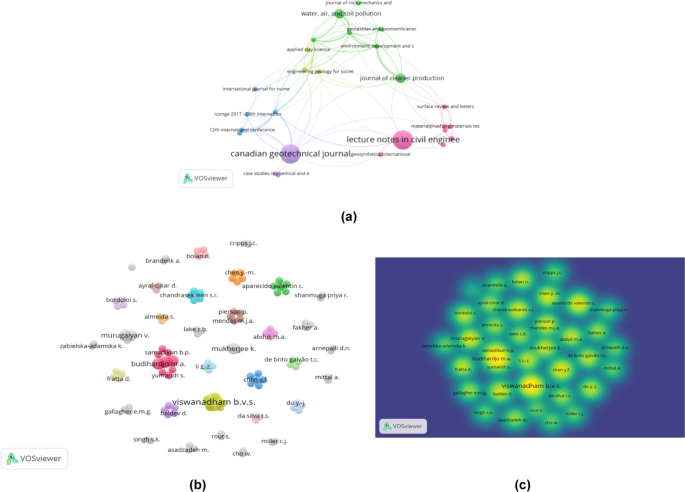
a Bibliometric map of journal publications discussed about using waste material as landfill liners. b VOSviewer bibliographic map of the authors reporting on using waste material as landfill liners and from 2002 to 2021. c The density of authors reporting on using waste material as landfill liners
The data processed in VOSviewer can also be seen in the citations in Table 1 . The table shows the highest citation ranking of articles published by a publisher. Publisher Canadian Geotechnical Journal obtained the highest citations from published journals on landfill liners (195); “Desiccation-Induced Crack and Its Effect On The Hydraulic Conductivity of Clayey Soil” written by Rayhani et al. ( 2007 ) had 93 citations; “Evaluation of An HDPE Geomembrane After 14 Years As A Leachate Lagoon Liner” written by Rowe et al. ( 2003 ) had 54 citations; “Modeling Deformation Behavior of Clay Liner in A Small Centrifuge” written by Viswanadham and Mahesh ( 2002 ) had 35 citations; and “Centrifuge Modeling of Municipal Solid Waste Landfill Failures Induced by Rising Water Levels” written Chen et al. ( 2017 ) had 13 citations.
The data obtained from Scopus show that the article entitled “Fiber Reinforcement for Waste Containment Soil Liner” written Miller and Rifai ( 2004 ) in the Journal of Environmental Engineering received the highest number of citations (144). This article discusses the impact of a mixture of clay and fiber materials on the fiber strength and the development of drying cracks to determine the soil's workability, hydraulic conductivity, and compaction characteristics.
Keywords analysis
There are prominent nodes in bold and large letters, as shown in Fig. 4 . This indicates that landfill liners and cracks are often mentioned in the literature. These two keywords will be discussed and become the main topic of this study. The interconnected nodes of the main keywords include shrinkage, wetting, mixtures, and other words that frequently appear and are discussed in the literature. These words support the discussion of the main topics raised in this paper.
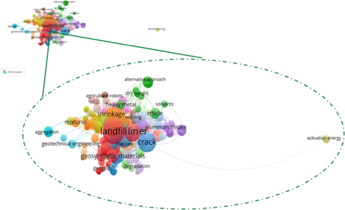
VOSviewer bibliographic map of the most used keywords regarding using waste material as landfill liners and other keywords from 2011 to 2021
As shown in Fig. 4 , the keywords were divided into several categories based on colors, such as red, green, orange, and blue, and strings related to each other. In blue, the words crack, flow rate, aggregation, and geotechnical engineering appear in node map science. This indicates that cracks are related to the flow rate of water seepage into the soil. The crack size and soil strength depend on the aggregation between particles, all of which are discussed in geotechnical engineering. Geotechnical engineering applies engineering technology to several aspects of soil and rock. Geotechnical engineering discusses issues regarding seepage, effective stress, soil compaction, slope stability, and various other problems (Fang and Daniels 2017 ). In addition, red indicates that the landfill liner contains materials that bind to each other and form networks, such as geosynthetic materials and geogrids. According to ASTM D-4439, geosynthetics are products made from polymer materials in sheets, using soil, rock, and geotechnical materials. A geogrid is a polymer material arranged in a net that crosses filaments at the connection (Shukla 2017 ). The geogrid is made of polypropylene as a base material, which is a type of geosynthetic in the form of a net with openings that allow interlocking between the gravel embankment materials to provide a layer of reinforcement for embankment construction with heavy loads (Bieliatynskyi et al. 2021 ). Orange indicates that the soil mixture used as a landfill liner can shrink in the dry state during the dry season. This is because of the reduction in soil moisture (Budihardjo et al. 2021a ).
Discussions on fly ash, landfill liners, clay, and compressive strength became a trending topic from 2018 to 2020. Multiple studies have discussed the characteristics of the best waste materials used as landfill liners that year. This is reinforced from 2021 to 2022, which shows the trend of research topics regarding alternative materials used as landfill liners and leachate migration. Researchers can use this study’s development as a research topic. The data processed in VOSviewer show that the keyword landfill liner was mentioned 733 times in 41 papers that had been filtered. Another keyword is crack, which was mentioned 559 times. These numbers make these two keywords the main topics of this review. It can be concluded that keywords are related because the strength of the soil used as landfill liner can be seen from the size of the crack that appears, which is associated with other keywords, such as those found on the map.
Analysis of journal article authors
The 41 papers published in Scopus over the last 20 years were analyzed by the most productive authors discussing landfill liners using VOSviewer software. The bibliographic map illustrated in Fig. 3 b shows that Budihardjo M.A has the most relationships, such as Yumaroh and Samadikun et al. Furthermore, Viswanadham B.V.S is the author who published the most papers (4), which were cited in 102 documents. The thickness of the corresponding line at each node indicates a strong relationship with each author.
The bibliometric map (Fig. 3 c) shows that the depth of color indicates that Viswanadham B.V.S frequently discusses landfill liners. The results of the data processed in VOSviewer from 41 selected papers showed that 123 authors discussed this theme. The most cited paper, in 144 documents, is “Fiber Reinforcement for Waste Containment Soil Liner” written by Miller and Rifai (Miller and Rifai 2004 ).
Measurement of cracking behavior
In several studies, various types of cracking behavior measurements have been conducted. In this paper, we summarize the most effective measurement techniques and additional information in Table 2 .
Sánchez et al. ( 2014 ) said that the mesh fragmentation technique was used to calculate the cracks. Owing to many numerical models, the selection of the mesh fragmentation technique in this study made it difficult to correctly simulate the 3D crack pattern in dry soil (Paraskevoulakos et al. 2022 ). The element ratio was inserted from high to standard aspects that were analyzed in several case studies on soil drying cracks developed on a laboratory scale and under field conditions. Sánchez et al. ( 2014 ) revealed that this technique could satisfactorily simulate the main patterns typically observed in fractured soils. In addition, this technique can also model the discontinuities associated with the pooling degradation between dissimilar materials. However, a limitation of the fragmentation technique is that it is unable to detect and stimulate crack thickness, and it is difficult to correctly simulate a 3D crack pattern on dry soil.
There are some differences in previous studies that used the crack intensity factor (CIF) method to calculate the crack value (Li et al. 2016 ; Safari et al. 2014 ; Tang et al. 2019 ). CIF is the most frequently used measurement because it is easy to use by comparing the crack and total soil surface area (Li et al. 2016 ). The output of this calculation using the CIF is the size of cracks in the soil. Thus, whether the soil type has low or high permeability will be determined. However, the limitation of this method is that it cannot be used to measure vertical cracks and cannot show 3D simulations.
Moreover, it was reported by Gui et al. ( 2016 ), who performed crack measurements using the Universal Distinct Element Code (UDEC). In this study, the soil was modeled using a cohesive fault model by combining the compressive, tensile, and shear behaviors of the material. The approach was conducted through numerical simulation of a laboratory-scale linear boundary drying test. The advantage of using UDEC is that modeling can be performed on thick soils and simulated under actual conditions in the field (Wang et al. 2022 ). However, it is necessary to recheck the field conditions to produce accurate results. The output of the UDEC obtains the values of cohesion, tensile strength, and shear angle of the crack pattern, which are essential factors in determining desiccation and crack behavior in soil. This was related to research conducted by Amarasiri et al. ( 2011 ) and Wei et al. ( 2020 ), who stated that the UDEC could realistically demonstrate and treat the phenomenon of soil drying cracking.
Studies conducted by Horgan and Young ( 2000 ); Lu et al. ( 2016 ); Shokri et al. ( 2015 ); and Wang et al. ( 2017 ) used fractal analysis to determine desiccation patterns. This study demonstrated crack testing using the wetting drying cycle method, which was then processed using fractal analysis. The concept of fractal dimensions can provide new findings on the effect of freeze–thaw cycles on cracking behavior in clays. Another fact-based research is that fractal analysis can be valuable for describing drying kinetics and characterizing images of dehydrated samples (García-García et al. 2019 ). In addition, the advantage of fractal analysis is that it can evaluate the spatial distribution of cracks, crack density of crack, and tendency of the crack trace to fill the area where the crack is embedded (Lu et al. 2016 ).
Another finding using X-ray computed tomography (CT) has also been used to investigate the evolution of draining crack networks in soils. Tang et al. ( 2019 ) applied an approach that integrated X-ray CT with digital image processing techniques to measure the morphological evolution of the drying crack pattern. The results representing the resulting crack ratio, soil area, average crack width, crack length, shrinkage strain, and crack segment number will be generated. Zaidi et al. ( 2021 ) revealed that this approach is essential for characterizing 3D soil drying crack patterns and provides a new perspective for studying the hydromechanical behavior of clays. However, a limitation of this technique is that it is necessary to perform manual calculations to determine the value of the cracks.
Chaduvula et al. ( 2022 ) used centrifuge modeling to observe the cracking behavior and permeability of clay layers on a large scale. The results of the centrifuge modeling test showed the potency of the intense clay layer to maintain integrity without significant cracking and crack propagation (Chaduvula et al. 2022 ). Centrifuge modeling can make a small model into centripetal acceleration, which is preponderant to Earth's gravity (Meng et al. 2021 ). In addition, the resulting crack value is made more accurate by adding the unit weight of the soil under test so that the stresses at the appropriate points in the model and prototype become identical. However, this technique requires manual calculations to determine the value of the crack and requires more time for analysis (Chaduvula et al. 2022 ).
Based on the various measurement techniques above, fractal analysis and X-ray CT measurements are the most effective and accurate to calculate the crack value. Although fractal analysis requires more time, this technique is the only one that can investigate crack evolution and water loss. Fractal analysis is effective and efficient for calculating and analyzing cracks when combined with the X-ray CT method, which can calculate cracks horizontally and vertically in 3D to produce an accurate crack value.
Crack reduction of different waste mixtures
Every type of soil material has a different main element content and resistance (Yu et al. 2016 ). It is necessary to mix soil with other waste materials to complement each other to obtain the best results for landfill liners. Multiple researchers have conducted experiments with various types of waste materials. This is similar to the research performed by Budihardjo et al. ( 2021b ), who conducted an investigation using a composite of fly ash, bentonite, and lime with variations in bentonite. The results showed that the higher the bentonite content, the smaller the permeability value. This is proven by a mixture of fly ash and 25% bentonite, which produces a permeability value of 1.584 × 10 –7 cm/s, with the most significant being 1.674, which saturates the standard safety factor. Bentonite can be used as a fly ash mixture to minimize the occurrence of collapse. Budihardjo et al. ( 2021a ) stated that fly ash could be applied as a landfill liner with other materials, such as bentonite, thereby reducing the permeability value in Table 3 .
Priyankara et al. ( 2016 ) reported the shrinkage behavior of liner materials using a mixture of soil, bentonite, oleic acid, and coconut fiber as the test materials. The results show that the sample with a mixture of bentonite relies on the fact that the thickness of the sample affects the appearance of cracks, and the higher the bentonite content, the higher the CIF value. After mixing bentonite, an experiment was conducted by adding oleic acid to reduce shrinkage. However, the results showed that a higher oleic acid content resulted in a significant increase in shrinkage. In addition, coconut fiber was used as a mixture to control the shrinkage behavior in this study. The results showed that the development of shrinkage behavior decreased significantly with the addition of coconut fiber. This is because the coir fibers function as reinforcement and resist tensile stresses that develop during the soil–water drying process, as a result of which the position of cracks can be reduced (Islam 2016 ).
Akbarimehr et al. ( 2020 ) used a mixture of granular rubber and clay as the fill material because of its low density compared to clay. The results showed that clay containing crumb rubber experienced a 10–25% stronger increase than the mixture containing rubber powder in a limited pressure range. Furthermore, it was shown that rubber fibers have a higher strength than other rubber forms. Akbarimehr et al. ( 2020 ) stated that the addition of rubber to clay could fill the cavities contained in clay. This is the perfect approach for reducing waste.
Several studies have shown that the principles for selecting clay soils for landfill liners are based on their stability and hydraulic conductivity. According to the study conducted by Akinwumi et al. ( 2016 ) in stabilizing clay to be used as a landfill finer, sawdust provided excellent results that met the standard hydraulic conductivity requirement. Their study utilized different percentages of sawdust: 0, 5, 10, 15, 20, 25, and 30% of the soil's dry weight. They concluded that rates of less than 10% sawdust of the dry unit weight are recommended as an amendment for clay soils to be used as landfill liners. However, the nature of soils from different areas has different features. Sawdust from the other three species also shows different properties. For instance, the study conducted by Niyomukiza et al. ( 2020b ) using Keruing sawdust to stabilize the expansive soils in Grobogan, Indonesia, found out that the composition having 3% sawdust + 97% soil provided high unconfined compressive results and lower free swell index. Sawdust beyond 3% of the dry weight of soil showed a reduction in unconfined compressive strength and increased free swell index.
Conversely, the higher percentages of sawdust (7% sawdust + 93% soil) reduced the liquid limit and increased the plasticity index, thus overall reduced plasticity index. This phenomenon, in turn, increased the workability of the expansive soil. Therefore, there is no clearly defined percentage of sawdust that applies to the soils. As they differ in different areas, research from various scholars showed that small portions of sawdust provide better performance results (El Halim and El Baroudy 2014 ; Jasim 2016 ; Niyomukiza et al. 2020a ). Sawdust has the capability of removing toxic substances, for instance, lead (Pb) and cadmium (Cd), and absorb water in the clays (Naiya et al. 2009 ; Yasemin and Zeki 2007 ). High percentages of sawdust could weaken the bond formed between the clay soil, silica, and alumina from sawdust, thus using small portions of sawdust.
Based on the above research summary, the waste material tends to have a hollow shape (Bahrami et al. 2016 ). Therefore, it is necessary to add clay to fill the cavities in waste materials. However, the addition of a large amount of clay results in additional cracks (Olgun 2013 ). Therefore, other compositions and materials are required to produce complementary structures. The formation of tight mixtures can reduce cracking (Liu et al. 2016 ). As shown in Fig. 5 regarding the schematic mechanism, the waste material can reduce cracks.
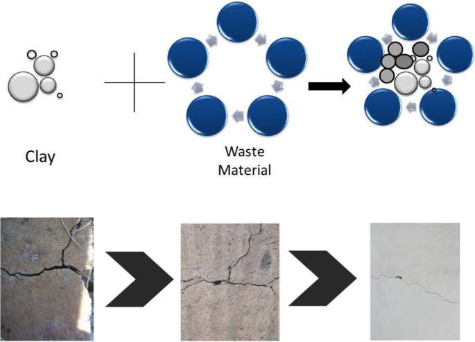
Schematic mechanism of crack reduction of different waste mixtures as landfill liners
Future perspectives
As this review highlights, the gaps in current knowledge are in our understanding of soil pollution in landfills, especially cracking and desiccation behavior in landfill liners and their impact. In the world of research, the knowledge of the material as landfill liner is rapidly growing. However, in many cases, this knowledge has not been linked to the effects of hazardous waste because waste materials such as fly ash are one of the categories of hazardous waste. The types of coal waste such as fly ash and bottom ash removed from the hazardous waste category are fly ash and bottom ash (Febriyani and Hartiwiningsih 2022 ; Jayaranjan et al. 2014 ; Park et al. 2020 ). Therefore, only fly ash and bottom ash from the power plant can be used as a landfill liner mixture. The lack of quantitative data is an obstacle to assessing the hazardous nature of the hazardous waste used as a landfill liner mixture and how the concentration of the presence of hazardous waste used as mixed material. There is uncertainty regarding the volume, composition, and type of waste material, and both are included in the hazardous and non-hazardous waste categories (Hassan et al. 2023 ). In addition, information on the composition of the waste material used as a landfill liner mixture is available, but the level of leachate seepage into the soil is not calculated. Seepage from this accidental discharge is one of the most significant uncertainties for emission predictions (Xiang et al. 2019 ; Mallants et al. 2020 ). It also shows that this information can predict what material waste and its composition is used as a landfill liner and provides insight into cracking and desiccation behavior (Wang et al. 2018 ). Furthermore, further research can still be conducted using another bibliometric method with different software or even meta-analysis using forest plots. It will be more useful to identify the current research gaps.
The number of articles discussing desiccation and crack behavior using waste materials as landfill liners has increased significantly from 2002 to 2021. The most productive publisher on this topic is the Canadian Geotechnical Journal, which has published four documents with 195 documents. According to the analysis, the keywords "landfill liner" and “crack” were the most frequently used keywords during the 20-year research period. This indicates that keywords are related because the strength of soil used as landfill liners can be seen from the size of the crack that appears, which is associated with other keywords, such as those found on the map. Various measurement techniques have been explored to determine desiccation and crack behavior. Fractal analysis and X-ray CT measurements proved to be good candidates for measuring cracks because they were the most accurate for calculating the crack value. Fractal analysis will be effective in analyzing cracks when combined with the X-ray CT method, which can calculate cracks horizontally and vertically simulated in 3D to produce an accurate crack value. Based on these findings, it is recommended that it be further developed using variations in waste material with different compositions calculated from the liner area using fractal analysis and X-ray CT measurements. This research has a significant impact in selecting materials for landfill liners and cracks behavior due to desiccation. It provides important information about trends, key areas, and future research directions. The study also includes information about effective measurement techniques to analyze crack behavior using waste materials such as fly ash, which can be applied as landfill liners with other materials such as bentonite and coconut coir fibers. Therefore, this research can be used as a reference for designing and selecting appropriate landfill liner materials. The limitations of this study include the bibliometric analysis being restricted to papers published until 2021 and the use of Scopus as the data source for research. It means that the potential of other topics can still be explored.
Ahmad M, Khan I, Khan MQS et al (2023) Households’ perception-based factors influencing biogas adoption: innovation diffusion framework. Energy 263:126155. https://doi.org/10.1016/j.energy.2022.126155
Article Google Scholar
Akbarimehr D, Eslami A, Aflaki E (2020) Geotechnical behaviour of clay soil mixed with rubber waste. J Clean Prod 271:122632. https://doi.org/10.1016/j.jclepro.2020.122632
Akinwumi II, Ojuri OO, Edem D, Adebanji SO (2016) Sawdust stabilization of lateritic clay as a landfill liner to retain heavy metals. Geo-Chicago 2016 GSP 271 81, 2005, 978–988. https://doi.org/10.1300/J202vO7n02_05
Aleixandre-Tudo JL, Castelló-Cogollos L, Aleixandre JL, Aleixandre-Benavent R (2019) Unravelling the scientific research on grape and wine phenolic compounds: a bibliometric study. Scientometrics 119(1):119–147. https://doi.org/10.1007/s11192-019-03029-8
Ali S, Yan Q, Razzaq A et al (2023) Modeling factors of biogas technology adoption: a roadmap towards environmental sustainability and green revolution. Environ Sci Pollut Res 30:11838–11860. https://doi.org/10.1007/s11356-022-22894-0
Article CAS Google Scholar
Aliha M, Ziari H, Mojaradi B et al (2020) Modes I and II stress intensity factors of semi-circular bend specimen computed for two-phase aggregate/mastic asphalt mixtures. Theoret Appl Fract Mech 106:102437. https://doi.org/10.1016/j.tafmec.2019.102437
Amarasiri AL, Kodikara JK, Costa S (2011) Numerical modelling of desiccation cracking. Int J Numer Anal Methods Geomech 35(1):82–96. https://doi.org/10.1002/nag.894
Awan A, Sadiq M, Hassan ST et al (2022) Combined nonlinear effects of urbanization and economic growth on CO 2 emissions in Malaysia. An application of QARDL and KRLS. Urban Climate 46:101342. https://doi.org/10.1016/j.uclim.2022.101342
Azam W, Khan I, Ali SA (2023) Alternative energy and natural resources in determining environmental sustainability: a look at the role of government final consumption expenditures in France. Environ Sci Pollut Res 30:1949–1965. https://doi.org/10.1007/s11356-022-22334-z
Bahrami A, Soltani N, Pech-Canul MI, Gutiérrez CA (2016) Development of metal-matrix composites from industrial/agricultural waste materials and their derivatives. Crit Rev Environ Sci Technol 46(2):143–208. https://doi.org/10.1080/10643389.2015.1077067
Barrot JS (2021) Social media as a language learning environment: a systematic review of the literature (2008–2019). Comput Assist Lang Learn. https://doi.org/10.1080/09588221.2021.1883673
Basson MS, Ayothiraman R (2020) Effect of human hair fiber reinforcement on shrinkage cracking potential of expansive clay. Bull Eng Geol Environ 79(4):2159–2168. https://doi.org/10.1007/s10064-019-01685-x
Bieliatynskyi A, Krayushkina K, Breskich V (2021) Basalt fiber geomats–modern material for reinforcing the motor road embankment slopes. Transp Res Rec 54:744–757. https://doi.org/10.1016/j.trpro.2021.02.128
Bittner CM, Oettel V (2022) Fiber reinforced concrete with natural plant fibers—investigations on the application of bamboo fibers in ultra-high performance concrete. Sustainability 14:12011
Bosmans A, Vanderreydt I, Geysen D, Helsen L (2013) The crucial role of waste-to-energy technologies in enhanced landfill mining: a technology review. J Clean Prod 55:10–23. https://doi.org/10.1016/j.jclepro.2012.05.032
Budihardjo M, Hadiwidodo M, Wardhana IW, Tuasykal MRNA, Samadikun BP, Ramadan BS (2021a) A study on desiccation cracking behavior of landfill liner developed from construction waste. IOP Conf Ser Earth Environ Sci 802:012004. https://doi.org/10.1088/1755-1315/802/1/012004
Budihardjo MA, Syafrudin S, Priyambada IB, Ramadan BS (2021) Hydraulic stability of fly ash-bentonite mixtures in landfill containment system. J Ecol Eng 22(7):132–141. https://doi.org/10.12911/22998993/139064
Budihardjo MA, Ramadan BS, Putri SA, Wahyuningrum IFS, Muhammad FI (2021c) Towards sustainability in higher-education institutions: analysis of contributing factors and appropriate strategies. Sustain 13(12):6562. https://doi.org/10.3390/su13126562
Chaduvula U, Viswanadham BVS, Kodikara J (2017) A study on desiccation cracking behavior of polyester fiber-reinforced expansive clay. Appl Clay Sci 142:163–172. https://doi.org/10.1016/j.clay.2017.02.008
Chaduvula U, Viswanadham B, Kodikara J (2022) Centrifuge model studies on desiccation cracking behaviour of fiber-reinforced expansive clay. Geotext Geomembr 50:480–497
Chen YM, Li JC, Yang CB, Zhu B, Zhan LT (2017) Centrifuge modeling of municipal solid waste landfill failures induced by rising water levels. Can Geotech J 54(12):1739–1751. https://doi.org/10.1139/cgj-2017-0046
Corallo A, Latino ME, Menegoli M, De DB, Viscecchia R (2019) Human factor in food label design to support consumer healthcare and safety: a systematic literature review. Sustain 11(15):4019. https://doi.org/10.3390/su11154019
Dyer M, Utili S, Zielinski M (2009) Field survey of desiccation fissuring of flood embankments. P I Civil Eng-Wat M 162(3):221–232. https://doi.org/10.1680/wama.2009.162.3.221
Ehrlich M, Almeida MSS, Curcio D (2019) Hydro-mechanical behavior of a lateritic fiber-soil composite as a waste containment liner. Geotext Geomembr 47(1):42–47. https://doi.org/10.1016/j.geotexmem.2018.09.005
El Halim AA, El Baroudy A (2014) Influence addition of Fine Sawdust on the physical properties of expansive soil in the middle Nile delta. Egypt J Soil Sci Plant Nutr 14(2):483–490. https://doi.org/10.4067/S0718-95162014005000038
Fang HY, Daniels JL (2017) Introductory geotechnical engineering: an environmental perspective. CRC Press, Boca Raton
Book Google Scholar
Febriyani NH, Hartiwiningsih H (2022) Corporate criminal liability post elimination of coal faba waste status from b3 waste category in indonesia. J Huk 38(1):12–31. https://doi.org/10.26532/jh.v38i1.20971
García-García AB, Fernández-Valle ME, Castejón D, Escudero R, Cambero MI (2019) Use of MRI as a predictive tool for physicochemical and rheologycal features during cured ham manufacturing. Meat Sci 148:171–180. https://doi.org/10.1016/j.meatsci.2018.10.015
Article CAS PubMed Google Scholar
Garg A, Reddy NG, Huang H (2020) Modelling contaminant transport in fly ash–bentonite composite landfill liner: mechanism of different types of ions. Sci Rep 10(1):1–8. https://doi.org/10.1038/s41598-020-68198-6
Greve A, Andersen MS, Acworth RI (2010) Investigations of soil cracking and preferential flow in a weighing lysimeter filled with cracking clay soil. J Hydrol 393(1–2):105–113. https://doi.org/10.1016/j.jhydrol.2010.03.007
Gui Y, Zhao ZY, Kodikara J (2016) Numerical modelling of laboratory soil desiccation cracking using UDEC with a mix-mode cohesive fracture model. Eng Geol 202:14–23. https://doi.org/10.1016/j.enggeo.2015.12.028
Gui Y, Wong WY, Gallage C (2021) Reinforced clayey soil: effectiveness and sensitivity of fibre inclusion on desiccation cracking behaviour of clayey soil. Int J Geomech. https://doi.org/10.1061/(ASCE)GM.1943-5622.0002278
Hassan ST, Wang P, Khan I et al (2023) The impact of economic complexity, technology advancements, and nuclear energy consumption on the ecological footprint of the USA: towards circular economy initiatives. Gondwana Res 113:237–246
Article ADS Google Scholar
Horgan G, Young I (2000) An empirical stochastic model for the geometry of two-dimensional crack growth in soil (with discussion). Geoderma 96(4):263–276. https://doi.org/10.1016/S0016-7061(00)00015-X
Islam NN (2016) Performance of fiber reinforced soft clay as a subgrade material of highway. Dissertation, Dhaka University
Jasim OH (2016) Effect of Sawdust Usage on the Shear Strength Behavior of Clayey Silt Soil. Sigma J Eng Nat Sci Sigma Mühendis Fen Bilimleri Dergisi Res 34(1):31–34
Google Scholar
Jayaranjan MLD, Van Hullebusch ED, Annachhatre AP (2014) Reuse options for coal fired power plant bottom ash and fly ash. Rev Environ Sci Biotechnol 13(4):467–486. https://doi.org/10.1007/s11157-014-9336-4
Jones G, Zielinski M, Sentenac P (2012) Mapping desiccation fissures using 3-D electrical resistivity tomography. J Appl Geophy 84:39–51. https://doi.org/10.1016/j.jappgeo.2012.06.002
Khan I, Zakari A, Dagar V et al (2022) World energy trilemma and transformative energy developments as determinants of economic growth amid environmental sustainability. Energy Econ 108:105884
Kumar H, Cai W, Lai J, Chen P, Ganesan SP, Bordoloi S, Liu X, Wen YP, Mei G (2020) Influence of in-house produced biochars on cracks and retained water during drying-wetting cycles: comparison between conventional plant, animal, and nano-biochars. J Soils Sedim 20(4):1983–1996. https://doi.org/10.1007/s11368-020-02573-8
Li JH, Zhang L (2010) Geometric parameters and REV of a crack network in soil. Comput Geotech 37(4):466–475. https://doi.org/10.1016/j.compgeo.2010.01.006
Li JH, Li L, Chen R, Li DQ (2016) Cracking and vertical preferential flow through landfill clay liners. Eng Geol 206:33–41. https://doi.org/10.1016/j.enggeo.2016.03.006
Liu H, Zhang Q, Gu C, Su H, Li VC (2016) Influence of micro-cracking on the permeability of engineered cementitious composites. Cem Concr 72:104–113. https://doi.org/10.1016/j.cemconcomp.2016.05.016
Liu H, Alharthi M, Atil A et al (2022) A non-linear analysis of the impacts of natural resources and education on environmental quality: green energy and its role in the future. Resour Policy 79:102940
Lockwood C, Dos Santos KB, Pap R (2019) Practical guidance for knowledge synthesis: scoping review methods. Asian Nurs Res 13:287–294
Lu Y, Liu S, Weng L, Wang L, Li Z, Xu L (2016) Fractal analysis of cracking in a clayey soil under freeze–thaw cycles. Eng Geol 208:93–99. https://doi.org/10.1016/j.enggeo.2016.04.023
Mallants D, Bekele E, Schmid W, Miotlinski K, Taylor A, Gerke K, Gray B (2020) A generic method for predicting environmental concentrations of hydraulic fracturing chemicals in soil and shallow groundwater. Water 12(4):941. https://doi.org/10.3390/w12040941
Marieta C, Guerrero A, Leon I (2021) Municipal solid waste incineration fly ash to produce eco-friendly binders for sustainable building construction. Waste Manage 120:114–124. https://doi.org/10.1016/j.wasman.2020.11.034
Mehmanparast A, Vidament A (2021) An accelerated corrosion-fatigue testing methodology for offshore wind applications. Eng Struct 240:112414. https://doi.org/10.1016/j.engstruct.2021.112414
Meng F, Chen R, Xu Y et al (2021) Centrifuge modeling of effectiveness of protective measures on existing tunnel subjected to nearby excavation. Tunn Undergr Space Technol 112:103880
Miller CJ, Rifai S (2004) Fiber reinforcement for waste containment soil liners. J Environt Eng 130(8):891–895. https://doi.org/10.1061/(ASCE)0733-9372(2004)130:8(891)
Mukhlisin M, Khiyon KN (2018) The effects of cracking on slope stability. J Geol Soc India 91(6):704–710. https://doi.org/10.1007/s12594-018-0927-5
Naiya TK, Chowdhury P, Bhattacharya AK, Das SK (2009) Sawdust and neem bark as low-cost natural biosorbent for adsorptive removal of Zn(II) and Cd(II) ions from aqueous solutions. Chem Eng J 148(1):68–79. https://doi.org/10.1016/j.cej.2008.08.002
Narani SS, Abbaspour M, Hosseini SMM, Aflaki E, Nejad FM (2020) Sustainable reuse of waste tire textile fibers (WTTFS) as reinforcement materials for expansive soils: with a special focus on landfill liners/covers. J Clean Prod 247:119151. https://doi.org/10.1016/j.jclepro.2019.119151
Niyomukiza, J. B., Wardani, S. P. R., & Setiadji, B. H. (2020a). The Effect of Curing Time on the Engineering Properties of Sawdust and Lime Stabilized Expansive Soils. In: 2nd international symposium on transportation studies in developing countries (ISTSDC 2019), 193(Istsdc 2019), pp 157–161. https://doi.org/10.2991/aer.k.200220.033
Niyomukiza JB, Wardani SPR, Setiadji BH (2020b) The influence of Keruing Sawdust on the geotechnical properties of expansive Soil. In: The 1st international conference on environment, sustainability issues and community development, pp 0–10. https://doi.org/10.1088/1755-1315/448/1/012040
O’Dea RE, Lagisz M, Jennions MD et al (2021) Preferred reporting items for systematic reviews and meta-analyses in ecology and evolutionary biology: a PRISMA extension. Biol Rev 96:1695–1722
Article PubMed Google Scholar
Olgun M (2013) Effects of polypropylene fiber inclusion on the strength and volume change characteristics of cement-fly ash stabilized clay soil. Geosynth Int 20(4):263–275. https://doi.org/10.1680/gein.13.00016
Page MJ, McKenzie JE, Bossuyt PM et al (2021) The PRISMA 2020 statement: an updated guideline for reporting systematic reviews. Bmj 372. https://doi.org/10.1016/j.ijsu.2021.105906
Page MJ, Moher D, Bossuyt PM et al (2021) PRISMA 2020 explanation and elaboration: updated guidance and exemplars for reporting systematic reviews. Bmj 372. https://doi.org/10.1136/bmj.n160
Paraskevoulakos C, Forna-Kreutzer JP, Hallam KR et al (2022) Investigating the mechanical behaviour of Fukushima MCCI using synchrotron Xray tomography and digital volume correlation. npj Mater Degrad 6:55. https://doi.org/10.1038/s41529-022-00264-y
Park JH, Eom JH, Lee SL, Hwang SW, Kim SH, Kang SW, Yun JJ, Lee YH, Seo DC (2020) Exploration of the potential capacity of fly ash and bottom ash derived from wood pellet-based thermal power plant for heavy metal removal. Sci Total Environ 740:140205. https://doi.org/10.1016/j.scitotenv.2020.140205
Article CAS PubMed ADS Google Scholar
Pei P, Zhao Y, Ni P, Mei G (2020) A protective measure for expansive soil slopes based on moisture content control. Eng Geol 269:105527. https://doi.org/10.1016/j.enggeo.2020.105527
Priyankara NH, Taud T, Odl K, Kawamoto K, Amn A (2016) Shrinkage behaviour of landfill clay liner materials in dry zone. Jpn Geotech Soc Spec Publ 2(61):2090–2095. https://doi.org/10.3208/jgssp.LKA-02
Puspita AS, Budihardjo MA, Samadikun BP (2023) Evaluating coconut fiber and fly ash composites for use in landfill retention layers. Global Nest J. https://doi.org/10.30955/gnj.004554
Rayhani MH, Yanful EK, Fakher A (2007) Desiccation-induced cracking and its effect on the hydraulic conductivity of clayey soils from Iran. Can Geotech J 44(3):276–283. https://doi.org/10.1139/t06-125
Rowe RK, Sangam HP, Lake CB (2003) Evaluation of an HDPE geomembrane after 14 years as a leachate lagoon liner. Can Geotech J 40(3):536–550. https://doi.org/10.1139/t03-019
Rubinos DA, Spagnoli G (2018) Utilization of waste products as alternative landfill liner and cover materials–A critical review. Crit Rev Environ Sci Technol 48(4):376–438. https://doi.org/10.1080/10643389.2018.1461495
Safari E, Ghazizade MJ, Abduli MA, Gatmiri B (2014) Variation of crack intensity factor in three compacted clay liners exposed to annual cycle of atmospheric conditions with and without geotextile cover. Waste Manage 34(8):1408–1415. https://doi.org/10.1016/j.wasman.2014.03.029
Saleh-Mbemba F, Aubertin M, Li L (2016) Experimental characterization of the shrinkage and water retention behaviour of tailings from hard rock mines. Geotech Geol Eng 34(1):251–266. https://doi.org/10.1007/s10706-015-9942-0
Sánchez M, Manzoli OL, Guimãraes LJ (2014) Modeling 3-D desiccation soil crack networks using a mesh fragmentation technique. Comput Geotech 62:27–39. https://doi.org/10.1016/j.compgeo.2014.06.009
Shokri N, Zhou P, Keshimiri A (2015) Patterns of desiccation cracks in saline bentonite layers. Transp Porous Media 110(2):333–344. https://doi.org/10.1007/s11242-015-0521-x
Shukla SK (2017) An introduction to geosynthetic engineering. CRC Press, Boca Raton
Siluo Y, Qingli Y (2017) Are scientometrics, informetrics, and bibliometrics different. In: 2017 the 16th international conference on scientometrics & informetrics. https://doi.org/10.4236/dsi.2020.11003
Syafrudin HN, Samadikun BP, Budihardjo M et al (2022) Geostability of dewatered sludge as landfill cover material. Global NEST Journal. https://doi.org/10.30955/gnj.004498
Tang CS, Cui YJ, Shi B, Tang AM, Liu C (2011) Desiccation and cracking behaviour of clay layer from slurry state under wetting–drying cycles. Geoderma 166(1):111–118. https://doi.org/10.1016/j.geoderma.2011.07.018
Tang CS, Zhu C, Leng T, Shi B, Cheng Q, Zeng H (2019) Three-dimensional characterization of desiccation cracking behavior of compacted clayey soil using X-ray computed tomography. Eng Geol 255:1–10. https://doi.org/10.1016/j.enggeo.2019.04.014
Tennant J (2020) Web of science and scopus are not global databases of knowledge. Eur Sci Editing. https://doi.org/10.31235/osf.io/qhvgr
Viswanadham BV, Mahesh K (2002) Modeling deformation behaviour of clay liners in a small centrifuge. Can Geotech J 39(6):1406–1418. https://doi.org/10.1139/t02-075
Wang C, Zhang ZY, Liu Y, Fan SM (2017) Geometric and fractal analysis of dynamic cracking patterns subjected to wetting-drying cycles. Soil Tillage Res 170:1–13. https://doi.org/10.1016/j.still.2017.02.005
Wang LL, Tang CS, Shi B, Cui YJ, Zhang GQ, Hilary I (2018) Nucleation and propagation mechanisms of soil desiccation cracks. Eng Geol 238:27–35. https://doi.org/10.1016/j.enggeo.2018.03.004
Wang J, Apel DB, Dyczko A et al (2022) Analysis of the damage mechanism of strainbursts by a global-local modeling approach. J Rock Mech Geotechn Eng 14:1671–1696. https://doi.org/10.1016/j.jrmge.2022.01.009
Wei X, Gao C, Liu K (2020) A review of cracking behavior and mechanism in clayey soils related to desiccation. Adv Civ Eng. https://doi.org/10.1155/2020/8880873
Wong S, Mah AXY, Nordin AH, Nyakuma BB, Ngadi N, Mat R, Amin NAS, Ho WS, Lee TH (2020) Emerging trends in municipal solid waste incineration ashes research: a bibliometric analysis from 1994 to 2018. Environ Sci Pollut Res 27(8):7757–7784. https://doi.org/10.1007/s11356-020-07933-y
Wu H, Wen Q, Hu L, Gong M, Tang Z (2017) Feasibility study on the application of coal gangue as landfill liner material. Waste Manage 63:161–171. https://doi.org/10.1016/j.wasman.2017.01.016
Xiang R, Xu Y, Liu YQ, Lei GY, Liu JC, Huang QF (2019) Isolation distance between municipal solid waste landfills and drinking water wells for bacteria attenuation and safe drinking. Sci Rep 9(1):1–11. https://doi.org/10.1038/s41598-019-54506-2
Xu J, Li Y, Wang S, Wang Q, Ding J (2020) Shear strength and mesoscopic character of undisturbed loess with sodium sulfate after dry-wet cycling. Bull Eng Geol Environ 79(3):1523–1541. https://doi.org/10.1007/s10064-019-01646-4
Yasemin B, Zeki T (2007) Removal of heavy metals from aqueous solution by sawdust adsorption. J Environ Sci 19(2):160–166
Yu KQ, Zhao YR, Liu F, He Y (2016) Laser-induced breakdown spectroscopy coupled with multivariate chemometrics for variety discrimination of soil. Sci Rep 6(1):1–10. https://doi.org/10.1038/srep27574
Zaidi M, Ahfir ND, Alem A, Taibi S, Mansouri BE, Zhang Y, Wang H (2021) Use of X-ray computed tomography for studying the desiccation cracking and self-healing of fine soil during drying–wetting paths. Eng Geol 292:106255. https://doi.org/10.1016/j.enggeo.2021.106255
Zhang B, Ye C, Liang B et al (2014) Ductile failure analysis and crack behavior of X65 buried pipes using extended finite element method. Eng Fail Anal 45:26–40. https://doi.org/10.1016/j.engfailanal.2014.06.009
Download references
Acknowledgements
The authors acknowledge John Bosco Niyomukiza and Nurani Ikhlas for their review of this work.
Author information
Authors and affiliations.
Master of Environmental Engineering, Diponegoro University, Semarang, 50275, Indonesia
A. S. Puspita
Environmental Sustainability Research Group, Universitas Diponegoro, Semarang, 50275, Indonesia
Department of Environmental Engineering, Diponegoro University, Semarang, 50275, Indonesia
M. A. Budihardjo & B. P. Samadikun
You can also search for this author in PubMed Google Scholar
Corresponding author
Correspondence to M. A. Budihardjo .
Ethics declarations
Conflict of interest.
The authors declare that there is no conflict of interest exists.
Human and animal rights
This article does not contain any studies with human participants or animal performed by any of the authors.
Additional information
Editorial responsibility: Senthil Kumar Ponnusamy.
Supplementary Information
Below is the link to the electronic supplementary material.
Supplementary file1 (DOCX 18 kb)
Rights and permissions.
Springer Nature or its licensor (e.g. a society or other partner) holds exclusive rights to this article under a publishing agreement with the author(s) or other rightsholder(s); author self-archiving of the accepted manuscript version of this article is solely governed by the terms of such publishing agreement and applicable law.
Reprints and permissions
About this article
Puspita, A.S., Budihardjo, M.A. & Samadikun, B.P. Desiccation and crack behavior of modified waste materials–clay mixture as landfill liner: a systematic review. Int. J. Environ. Sci. Technol. 21 , 5231–5246 (2024). https://doi.org/10.1007/s13762-023-05425-3
Download citation
Received : 06 August 2022
Revised : 04 September 2023
Accepted : 12 December 2023
Published : 07 January 2024
Issue Date : March 2024
DOI : https://doi.org/10.1007/s13762-023-05425-3
Share this article
Anyone you share the following link with will be able to read this content:
Sorry, a shareable link is not currently available for this article.
Provided by the Springer Nature SharedIt content-sharing initiative
- Bibliometric analysis
- Fractal analysis
- X-ray computed tomography (CT)
- Waste material
- Find a journal
- Publish with us
- Track your research

IMAGES
VIDEO
COMMENTS
The figures should be indicated within parentheses in their first mention in the "Materials and Methods" section. Headings and as a prevalent convention legends of the figures should be indicated at the end of the manuscript. If a different method is used in the study, this should be explained in detail.
A reader would need to know which search engine and what key words you used. Open this section by describing the overall approach you took or the materials used. Then describe to the readers step-by-step the methods you used including any data analysis performed. See Fig. 2.5 below for an example of materials and methods section. Writing tips: Do:
The methods section of a research paper typically constitutes materials and methods; while writing this section, authors usually arrange the information under each category. The materials category describes the samples, materials, treatments, and instruments, while experimental design, sample preparation, data collection, and data analysis are ...
To structure your methods section, you can use the subheadings of "Participants," "Materials," and "Procedures.". These headings are not mandatory—aim to organize your methods section using subheadings that make sense for your specific study. Note that not all of these topics will necessarily be relevant for your study.
2. Functions of the Materials and Methods Section. The M&M section of a paper has two main functions (): To allow readers to repeat the work and to convince them that the work has been done in an appropriate way.For hypothesis-testing papers, the most important function of the M&M section is to provide information on "what procedures were used to answer the main question(s) stated in the ...
The Materials and Methods section is a vital component of any formal lab report. This section of the report gives a detailed account of the procedure that was followed in completing the experiment (s) discussed in the report. Such an account is very important, not only so that the reader has a clear understanding of the experiment, but a well ...
The methods section should describe what was done to answer the research question, describe how it was done, justify the experimental design, and explain how the results were analyzed. Scientific writing is direct and orderly. Therefore, the methods section structure should: describe the materials used in the study, explain how the materials ...
Your Methods Section contextualizes the results of your study, giving editors, reviewers and readers alike the information they need to understand and interpret your work. Your methods are key to establishing the credibility of your study, along with your data and the results themselves. A complete methods section should provide enough detail for a skilled researcher to replicate your process ...
The Materials and Methods section of a hypothesis-testing paper is essentially a cookbook. Thus, the main content of the Materials and Methods section is a detailed description of the materials and methods you used. In addition, in hypothesis-testing papers in which all the experiments are designed in advance, the Materials and ...
Materials and methods. The study's methods are one of the most important parts used to judge the overall quality of the paper. In addition the Methods section should give readers enough information so that they can repeat the experiments. Reviewers should look for potential sources of bias in the way the study was designed and carried out ...
The Materials and methods section of a research paper is oftentimes the first and easiest part to write. It details the steps taken to answer a research hypothesis, the success of which determines whether or not the study can be replicated. Arranging the section in chronological order, writing succinctly, and consistently using the third-person ...
When writing scientific papers to share their research findings with their peers, it is not enough for researchers to just communicate the results of their study; it is equally important to explain the process by which they arrived at their results, so that the study can be replicated to validate the observations. The materials and methods ...
To test whether striations are weapon type specific, this study aimed to examine whether different sharpening materials and methods produced different striations on the kerf walls of cut marks on bone produced by machetes. Or, if they produce the previously identified 'rolling hill' striation pattern.
If the authors provide sufficient detail, other scientists can repeat their experiments to verify their findings. It is generally recommended that the materials and methods should be written in the past tense, either in active or passive voice. In this section, ethical approval, study dates, number of subjects, groups, evaluation criteria ...
The Materials and methods section of a research paper is oftentimes the first and easiest part to write. It details the steps taken to answer a research hypothesis, the success of which determines whether or not the study can be replicated. Arranging the section in chronological order, writing succinctly, and consistently using the third-person ...
Materials and Methods. The Methods and Materials section of a paper often seems the least interesting to read, or to write, but it serves several essential purposes. First, it demonstrates to readers that the research was designed appropriately and conducted competently. Scientists are skeptical readers.
Right. Just as a recap, these are the things that you should be alert of when you're writing the materials and methods section in scientific writing: Provide the details of your materials and chemicals. Organize your methodology that bests tell your discussion. Get your experimental design right.
7 Tips for Writing an Effective Materials and Methods Section in Your Research Manuscript: 1. Begin writing the Materials and Methods while you are performing your experiments. 2. Start with general information that applies to the entire manuscript and then move on to specific experimental details. 3.
In any research article, the detailed description and process of an experiment is provided in the section termed as "Materials and Method.". The Materials and Method section is also called Method section in few journals. This section describes how the experiment was conducted to arrive at the results. The aim of this section in any research ...
Methods / Materials Overview. These sections of the research paper should be concise. The audience reading the paper will always want to know what materials or methods that were used. The methods and materials may be under subheadings in the section or incorporated together. The main objective for these sections is to provide specialized ...
This free white paper tackles the best ways to write the Materials and Methods section of a scientific manuscript. The Materials and Methods (or "Methods section") is the section of a research paper that provides the reader. with all the information needed to understand your work and how the reported results were produced.
4. Use subheadings: Dividing the Methods section in terms of the experiments helps the reader to follow the section better. You may write the specific objective of each experiment as a subheading. Alternatively, if applicable, the name of each experiment can also be used as subheading. 5.
Abstract. We investigated whether deep reinforcement learning (deep RL) is able to synthesize sophisticated and safe movement skills for a low-cost, miniature humanoid robot that can be composed into complex behavioral strategies. We used deep RL to train a humanoid robot to play a simplified one-versus-one soccer game.
To write your methods section in APA format, describe your participants, materials, study design, and procedures. Keep this section succinct, and always write in the past tense. The main heading of this section should be labeled "Method" and it should be centered, bolded, and capitalized. Each subheading within this section should be bolded ...
We will look at some examples of materials and methods structure in different disciplines. 2.1. Materials & methods example #1 (Engineering paper) If you are writing an engineering sciences research paper in which you are introducing a new method, your materials and methods section would typically include the following information.
Despite current research into alternate PVK materials, the B-site positive ion in modern PSCs is still lead (Fabini, 2015).If all the electricity in the United States were to be generated by PSCs using the most well-deliberated OIHP, the annual consumption of lead would be 160 tons (Fabini, 2015).Eliminating Pb from PSCs is the only long-term solution to the Pb-toxicity problem, even though ...
A machine learning (ML) approach for predicting aneuploidy in cancer. A Schematic view of the ML model construction. Labels represent aneuploidy status of each chromosome arm in 24 cancer types (abbreviation of cancer types detailed in Additional file 2: Table S1), classified as gained (red, n = 199), lost (blue, n = 307), or neutral (white, n = 430).
The PRISMA method ensures that a paper is conducted with good methodology and follows existing academic standards ... Based on the above research summary, the waste material tends to have a hollow shape (Bahrami et al. 2016). Therefore, it is necessary to add clay to fill the cavities in waste materials.
Introduction. This article has resulted from the initial consultations and continued collaboration between some of the partners in the earlier networking and research project entitled "Indigenous and non-indigenous residents of the Nordic-Russian region: Best practices for equity in healthy ageing" (NORRUS-AGE) in 2020-2022 [Citation 1].In this review, the Nordic Arctic has been defined ...