- Biology Article

Human Respiratory System
Respiratory system of humans.
Breathing involves gaseous exchange through inhalation and exhalation. The human respiratory system has the following main structures – Nose, mouth, pharynx, larynx, trachea, bronchi, and lungs. Explore in detail.
Table of Contents
- What Is Respiratory System
Respiratory Tract
Respiratory system definition.
“Human Respiratory System is a network of organs and tissues that helps us breathe. The primary function of this system is to introduce oxygen into the body and expel carbon dioxide from the body.”
What is the Respiratory System?
As defined above, the human respiratory system consists of a group of organs and tissues that help us to breathe. Aside from the lungs, there are also muscles and a vast network of blood vessels that facilitate the process of respiration.
Also Read: Mechanism of Breathing
Human Respiratory System Diagram
To gain a clearer understanding, we have illustrated the human respiratory system and its different parts involved in the process.
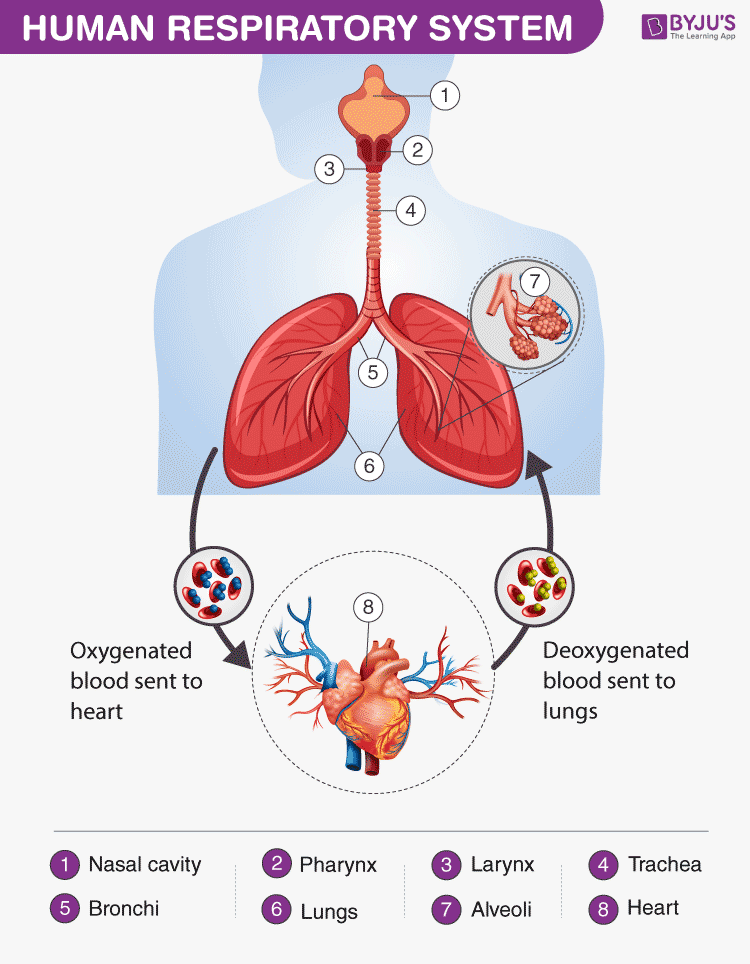
Human Respiratory System Diagram showing different parts of the Respiratory Tract
Features of the Human Respiratory System
The respiratory system in humans has the following important features:
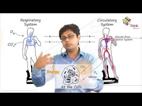
- The energy is generated by the breakdown of glucose molecules in all living cells of the human body.
- Oxygen is inhaled and is transported to various parts and are used in the process of burning food particles (breaking down glucose molecules) at the cellular level in a series of chemical reactions.
- The obtained glucose molecules are used for discharging energy in the form of ATP- (adenosine triphosphate)
Also Read: Difference between trachea and oesophagus
Respiratory System Parts and Functions
Let us have a detailed look at the different parts of the respiratory system and their functions.
Humans have exterior nostrils, which are divided by a framework of cartilaginous structure called the septum. This is the structure that separates the right nostril from the left nostril. Tiny hair follicles that cover the interior lining of nostrils act as the body’s first line of defence against foreign pathogens . Furthermore, they provide additional humidity for inhaled air.
Two cartilaginous chords lay the framework for the larynx. It is found in front of the neck and is responsible for vocals as well as aiding respiration. Hence, it is also informally called the voice box. When food is swallowed, a flap called the epiglottis folds over the top of the windpipe and prevents food from entering into the larynx.
Also check: What is the role of epiglottis and diaphragm in respiration?
The nasal chambers open up into a wide hollow space called the pharynx. It is a common passage for air as well as food. It functions by preventing the entry of food particles into the windpipe. The epiglottis is an elastic cartilage, which serves as a switch between the larynx and the oesophagus by allowing the passage of air into the lungs, and food in the gastrointestinal tract .
Have you ever wondered why we cough when we eat or swallow?
Talking while we eat or swallow may sometimes result in incessant coughing. The reason behind this reaction is the epiglottis. It is forced to open for the air to exit outwards and the food to enter into the windpipe, triggering a cough.
The trachea or the windpipe rises below the larynx and moves down to the neck. The walls of the trachea comprise C-shaped cartilaginous rings which give hardness to the trachea and maintain it by completely expanding. The trachea extends further down into the breastbone and splits into two bronchi, one for each lung.
The trachea splits into two tubes called the bronchi, which enter each lung individually. The bronchi divide into secondary and tertiary bronchioles, and it further branches out into small air-sacs called the alveoli. The alveoli are single-celled sacs of air with thin walls. It facilitates the exchange of oxygen and carbon dioxide molecules into or away from the bloodstream.
Lungs are the primary organs of respiration in humans and other vertebrates. They are located on either side of the heart, in the thoracic cavity of the chest. Anatomically, the lungs are spongy organs with an estimates total surface area between 50 to 75 sq meters. The primary function of the lungs is to facilitate the exchange of gases between the blood and the air. Interestingly, the right lung is quite bigger and heavier than the left lung.
Also Read: Respiration
The respiratory tract in humans is made up of the following parts:
- External nostrils – For the intake of air.
- Nasal chamber – which is lined with hair and mucus to filter the air from dust and dirt.
- Pharynx – It is a passage behind the nasal chamber and serves as the common passageway for both air and food.
- Larynx – Known as the soundbox as it houses the vocal chords, which are paramount in the generation of sound.
- Epiglottis – It is a flap-like structure that covers the glottis and prevents the entry of food into the windpipe.
- Trachea – It is a long tube passing through the mid-thoracic cavity.
- Bronchi – The trachea divides into left and right bronchi.
- Bronchioles – Each bronchus is further divided into finer channels known as bronchioles.
- Alveoli – The bronchioles terminate in balloon-like structures known as the alveoli.
- Lungs – Humans have a pair of lungs, which are sac-like structures and covered by a double-layered membrane known as pleura.
Air is inhaled with the help of nostrils, and in the nasal cavity, the air is cleansed by the fine hair follicles present within them. The cavity also has a group of blood vessels that warm the air. This air then passes to the pharynx, then to the larynx and into the trachea.
The trachea and the bronchi are coated with ciliated epithelial cells and goblet cells (secretory cells) which discharge mucus to moisten the air as it passes through the respiratory tract. It also traps the fine bits of dust or pathogen that escaped the hair in the nasal openings. The motile cilia beat in an ascending motion, such that the mucus and other foreign particles are carried back to the buccal cavity where it may either be coughed out (or swallowed.)
Once the air reaches the bronchus, it moves into the bronchioles, and then into the alveoli.
Respiratory System Functions
The functions of the human respiratory system are as follows:
Inhalation and Exhalation
The respiratory system helps in breathing (also known as pulmonary ventilation.) The air inhaled through the nose moves through the pharynx, larynx, trachea and into the lungs. The air is exhaled back through the same pathway. Changes in the volume and pressure in the lungs aid in pulmonary ventilation.
Exchange of Gases between Lungs and Bloodstream
Inside the lungs, the oxygen and carbon dioxide enter and exit respectively through millions of microscopic sacs called alveoli. The inhaled oxygen diffuses into the pulmonary capillaries, binds to haemoglobin and is pumped through the bloodstream. The carbon dioxide from the blood diffuses into the alveoli and is expelled through exhalation.
Also read: Exchange Of Gases in Plants
Exchange of Gases between Bloodstream and Body Tissues
The blood carries the oxygen from the lungs around the body and releases the oxygen when it reaches the capillaries. The oxygen is diffused through the capillary walls into the body tissues. The carbon dioxide also diffuses into the blood and is carried back to the lungs for release.
The Vibration of the Vocal Cords
While speaking, the muscles in the larynx move the arytenoid cartilage. These cartilages push the vocal cords together. During exhalation, when the air passes through the vocal cords, it makes them vibrate and creates sound.
Olfaction or Smelling
During inhalation, when the air enters the nasal cavities, some chemicals present in the air bind to it and activate the receptors of the nervous system on the cilia. The signals are sent to the olfactory bulbs via the brain.
Also Read: Respiratory System Disorders
Respiration is one of the metabolic processes which plays an essential role in all living organisms. However, lower organisms like the unicellular do not “breathe” like humans – intead, they utilise the process of diffusion. Annelids like earthworms have a moist cuticle which helps them in gaseous exchange. Respiration in fish occurs through special organs called gills. Most of the higher organisms possess a pair of lungs for breathing.
Also Read: Amphibolic Pathway
To learn more about respiration, check out the video below:

Frequently Asked Questions
What is the human respiratory system.
The human respiratory system is a system of organs responsible for inhaling oxygen and exhaling carbon dioxide in humans. The important respiratory organs in living beings include- lungs, gills, trachea, and skin.
What are the important respiratory system parts in humans?
The important human respiratory system parts include- Nose, larynx, pharynx, trachea, bronchi and lungs.
What is the respiratory tract made up of?
The respiratory tract is made up of nostrils, nasal chamber, larynx, pharynx, epiglottis, trachea, bronchioles, bronchi, alveoli, and lungs.
What are the main functions of the respiratory system?
The important functions of the respiratory system include- inhalation and exhalation of gases, exchange of gases between bloodstream and lungs, the gaseous exchange between bloodstream and body tissues, olfaction and vibration of vocal cords.
What are the different types of respiration in humans?
The different types of respiration in humans include- internal respiration, external respiration and cellular respiration. Internal respiration includes the exchange of gases between blood and cells, external respiration is the breathing process, whereas cellular respiration is the metabolic reactions taking place in the cells to produce energy.
What are the different stages of aerobic respiration?
Aerobic respiration is the process of breaking down glucose to produce energy. It occurs in the following different stages- glycolysis, pyruvate oxidation, citric acid cycle or Krebs cycle, and electron transport system.
Why do the cells need oxygen?
Our body cells require oxygen to release energy. The oxygen inhaled during respiration is used to break down the food to release energy.
What is the main difference between breathing and respiration in humans?
Breathing is the physical process of inhaling oxygen and exhaling carbon dioxide in and out of our lungs. On the contrary, respiration is the chemical process where oxygen is utilized to break down glucose to generate energy to carry out different cellular processes.
Explore more details about the human respiratory system or other related topics by registering at BYJU’S Biology

Put your understanding of this concept to test by answering a few MCQs. Click ‘Start Quiz’ to begin!
Select the correct answer and click on the “Finish” button Check your score and answers at the end of the quiz
Visit BYJU’S for all Biology related queries and study materials
Your result is as below
Request OTP on Voice Call
Leave a Comment Cancel reply
Your Mobile number and Email id will not be published. Required fields are marked *
Post My Comment
Wow!!!!! Great notes . Thankyou byjus
Wow! this is really helpful. Thank you
Greet notes
Very helpful
Very nice information
good answers!
Thank you so much for the notes
Great notes helpful for students
The notes are really amazing. It helped me a lot. Thank you BYJU’S
The notes are amazing. It helped me a lot. Thank you Byju’s.
Thank you for the notes helpful in the revision
Very good notes. I love Byjus learning application
thank you byjus
These notes are very good for study, thank u so much Byjus.
Helpful for studying. Informative about the topic. Great notes!
Great job you are great Byjus and your notes is superb ❤❤❤❤❣❣❣❣❣🙏🙏🙏🙏
Register with BYJU'S & Download Free PDFs
Register with byju's & watch live videos.
Respiratory System Anatomy and Physiology
Breathe life into your understanding with our guide on the respiratory system anatomy and physiology. Nursing students, immerse yourself in the intricate dance of inhalation and exhalation that fuels every living moment.
Table of Contents
Functions of the respiratory system, main bronchi, the respiratory membrane, respiration, mechanics of breathing, respiratory volumes and capacities, respiratory sounds, external respiration, gas transport, and internal respiration, control of respiration, age-related physiological changes in the respiratory system.
The functions of the respiratory system are:
- Oxygen supplier. The job of the respiratory system is to keep the body constantly supplied with oxygen.
- Elimination. Elimination of carbon dioxide.
- Gas exchange. The respiratory system organs oversee the gas exchanges that occur between the blood and the external environment.
- Passageway. Passageways that allow air to reach the lungs.
- Humidifier. Purify, humidify, and warm incoming air.
Anatomy of the Respiratory System
The organs of the respiratory system include the nose, pharynx , larynx, trachea, bronchi, and their smaller branches, and the lungs, which contain the alveoli.
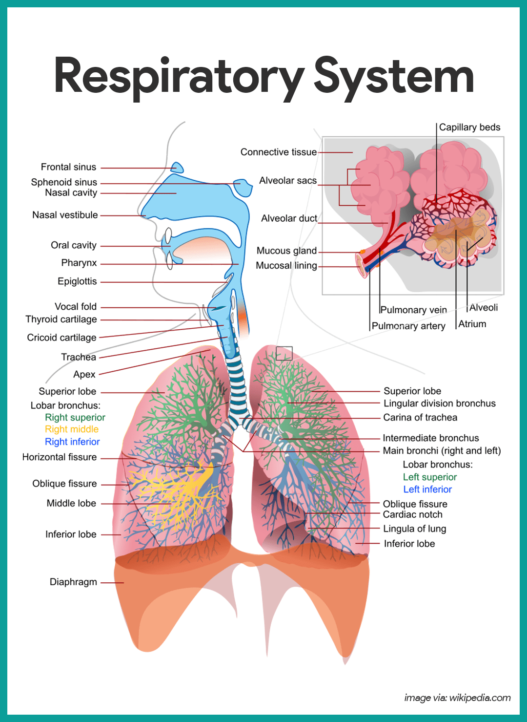
The nose is the only externally visible part of the respiratory system.
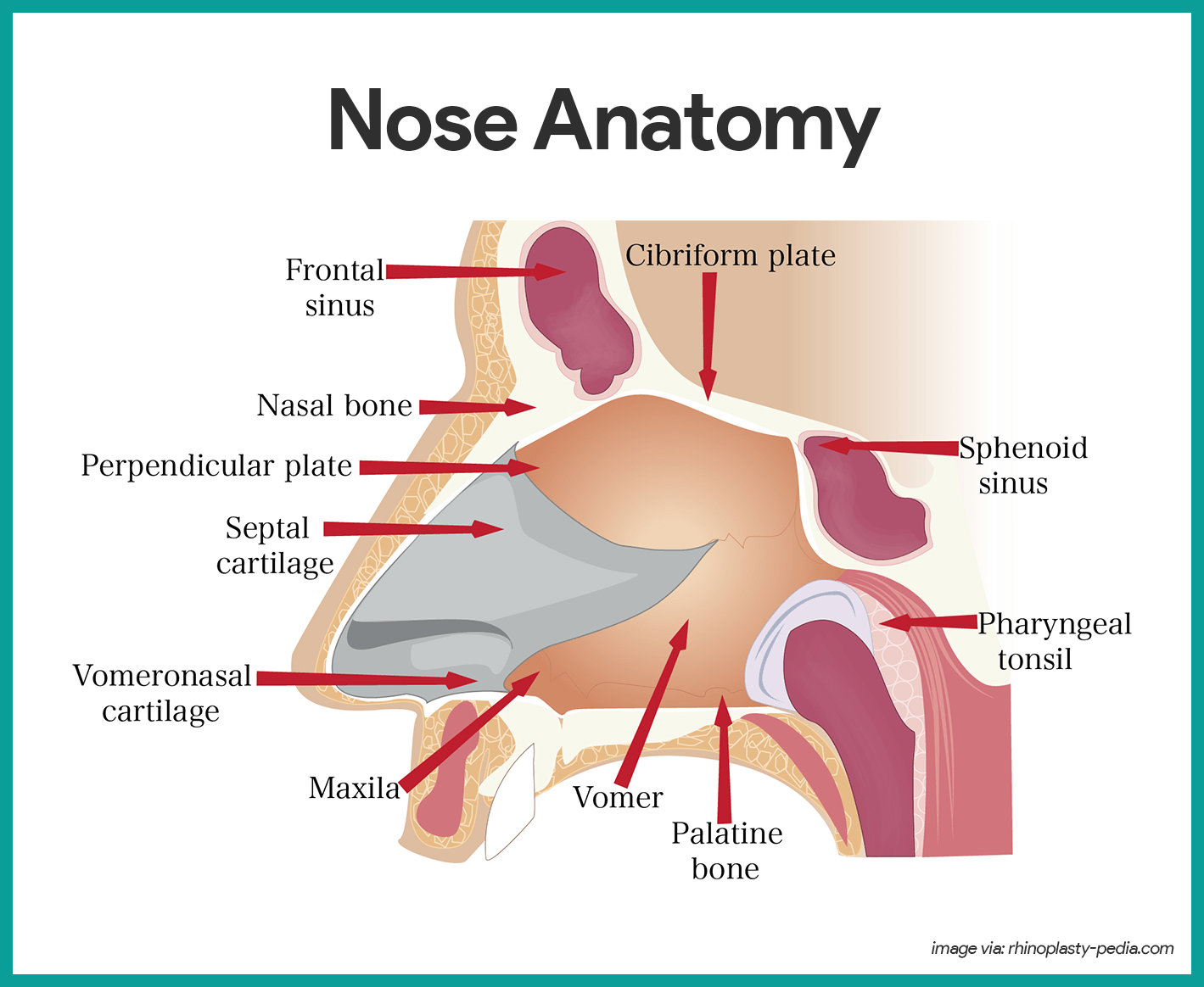
- Nostrils. During breathing, air enters the nose by passing through the nostrils, or nares.
- Nasal cavity. The interior of the nose consists of the nasal cavity, divided by a midline nasal septum .
- Olfactory receptors. The olfactory receptors for the sense of smell are located in the mucosa in the slitlike superior part of the nasal cavity, just beneath the ethmoid bone .
- Respiratory mucosa. The rest of the mucosal lining, the nasal cavity called the respiratory mucosa, rests on a rich network of thin-walled veins that warms the air as it flows past.
- Mucus. In addition, the sticky mucus produced by the mucosa’s glands moistens the air and traps incoming bacteria and other foreign debris, and lysozyme enzymes in the mucus destroy bacteria chemically.
- Ciliated cells. The ciliated cells of the nasal mucosa create a gentle current that moves the sheet of contaminated mucus posteriorly toward the throat, where it is swallowed and digested by stomach juices.
- Conchae. The lateral walls of the nasal cavity are uneven owing to three mucosa-covered projections, or lobes called conchae, which greatly increase the surface area of the mucosa exposed to the air, and also increase the air turbulence in the nasal cavity.
- Palate. The nasal cavity is separated from the oral cavity below by a partition, the palate; anteriorly, where the palate is supported by bone, is the hard palate; the unsupported posterior part is the soft palate .
- Paranasal sinuses. The nasal cavity is surrounded by a ring of paranasal sinuses located in the frontal, sphenoid, ethmoid, and maxillary bones ; theses sinuses lighten the skull , and they act as a resonance chamber for speech.
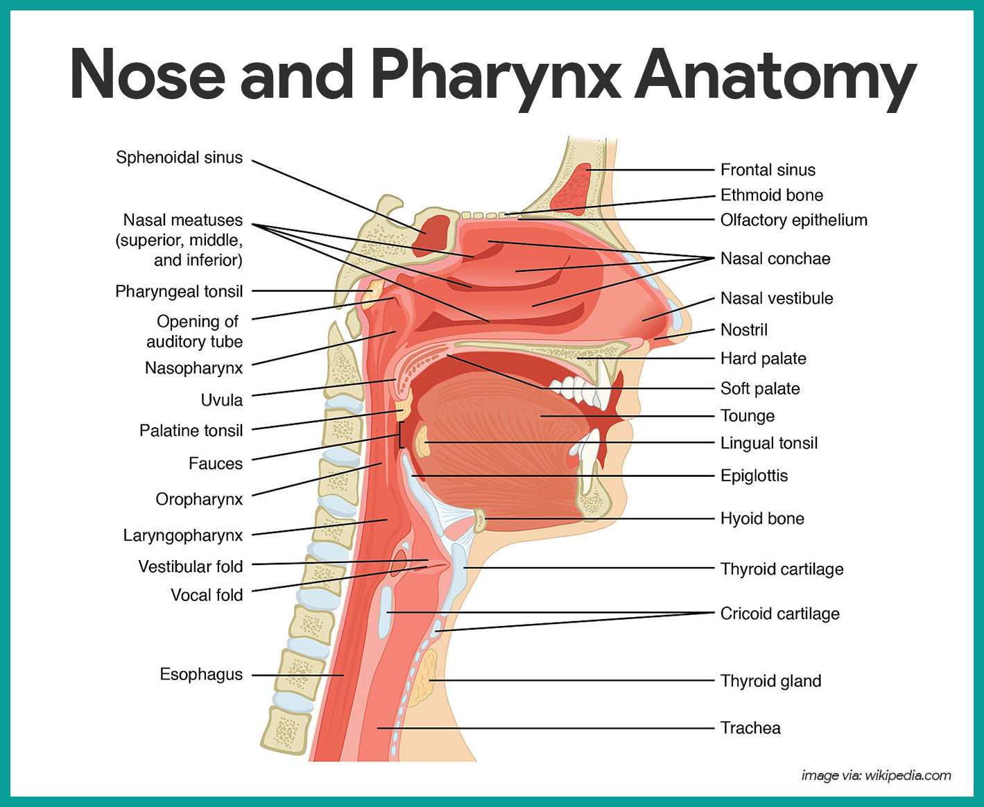
- Size. The pharynx is a muscular passageway about 13 cm (5 inches) long that vaguely resembles a short length of red garden hose.
- Function. Commonly called the throat , the pharynx serves as a common passageway for food and air.
- Portions of the pharynx. Air enters the superior portion, the nasopharynx , from the nasal cavity and then descends through the oropharynx and laryngopharynx to enter the larynx below.
- Pharyngotympanic tube. The pharyngotympanic tubes, which drain the middle ear open into the nasopharynx.
- Pharyngeal tonsil. The pharyngeal tonsil, often called adenoid is located high in the nasopharynx.
- Palatine tonsils . The palatine tonsils are in the oropharynx at the end of the soft palate.
- Lingual tonsils . The lingual tonsils lie at the base of the tongue.
The larynx or voice box routes air and food into the proper channels and plays a role in speech.
- Structure. Located inferior to the pharynx, it is formed by eight rigid hyaline cartilages and a spoon-shaped flap of elastic cartilage, the epiglottis .
- Thyroid cartilage. The largest of the hyaline cartilages is the shield-shaped thyroid cartilage, which protrudes anteriorly and is commonly called Adam’s apple .
- Epiglottis. Sometimes referred to as the “guardian of the airways” , the epiglottis protects the superior opening of the larynx.
- Vocal folds. Part of the mucous membrane of the larynx forms a pair of folds, called the vocal folds, or true vocal cords , which vibrate with expelled air and allows us to speak.
- Glottis. The slitlike passageway between the vocal folds is the glottis.
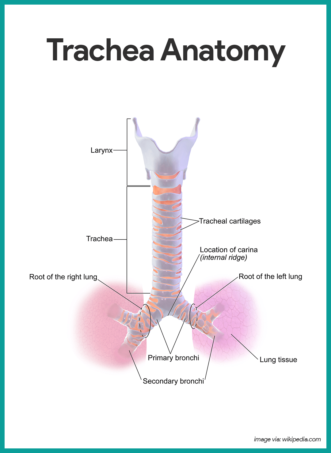
- Length. Air entering the trachea or windpipe from the larynx travels down its length (10 to 12 cm or about 4 inches) to the level of the fifth thoracic vertebra , which is approximately midchest.
- Structure. The trachea is fairly rigid because its walls are reinforced with C-shaped rings of hyaline cartilage; the open parts of the rings abut the esophagus and allow it to expand anteriorly when we swallow a large piece of food, while the solid portions support the trachea walls and keep it patent, or open, in spite of the pressure changes that occur during breathing.
- Cilia. The trachea is lined with ciliated mucosa that beat continuously and in a direction opposite to that of the incoming air as they propel mucus, loaded with dust particles and other debris away from the lungs to the throat, where it can be swallowed or spat out.
- Structure. The right and left main (primary) bronchi are formed by the division of the trachea.
- Location. Each main bronchus runs obliquely before it plunges into the medial depression of the lung on its own side.
- Size. The right main bronchus is wider, shorter, and straighter than the left.
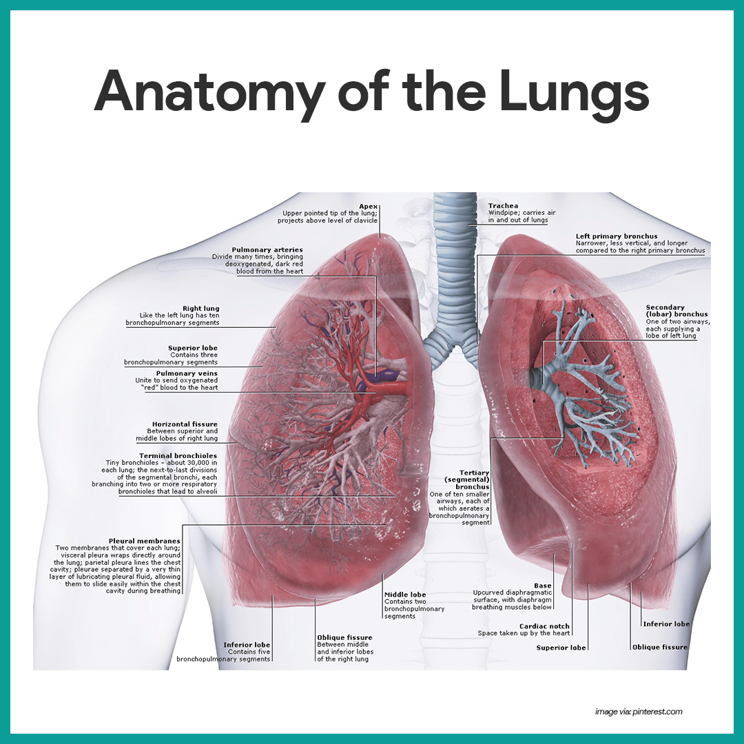
- Location. The lungs occupy the entire thoracic cavity except for the most central area, the mediastinum , which houses the heart, the great blood vessels, bronchi, esophagus, and other organs.
- Apex. The narrow, superior portion of each lung, the apex, is just deep into the clavicle .
- Base. The broad lung area resting on the diaphragm is the base.
- Division. Each lung is divided into lobes by fissures; the left lung has two lobes , and the right lung has three .
- Pleura. The surface of each lung is covered with a visceral serosa called the pulmonary , or visceral pleura, and the walls of the thoracic cavity are lined by the parietal pleura .
- Pleural fluid. The pleural membranes produce pleural fluid, a slippery serous secretion that allows the lungs to glide easily over the thorax wall during breathing movements and causes the two pleural layers to cling together.
- Pleural space. The lungs are held tightly to the thorax wall, and the pleural space is more of a potential space than an actual one.
- Bronchioles . The smallest of the conducting passageways are the bronchioles.
- Alveoli. The terminal bronchioles lead to the respiratory zone structures, even smaller conduits that eventually terminate in alveoli or air sacs.
- Respiratory zone. The respiratory zone, which includes the respiratory bronchioles, alveolar ducts, alveolar sacs, and alveoli, is the only site of gas exchange .
- Conducting zone structures. All other respiratory passages are conducting zone structures that serve as conduits to and from the respiratory zone.
- Stroma. The balance of the lung tissue, its stroma, is mainly elastic connective tissue that allows the lungs to recoil passively as we exhale.
- Wall structure. The walls of the alveoli are composed largely of a single, thin layer of squamous epithelial cells.
- Alveolar pores. Alveolar pores connect neighboring air sacs and provide alternative routes for air to reach alveoli whose feeder bronchioles have been clogged by mucus or otherwise blocked.
- Respiratory membrane. Together, the alveolar and capillary walls, their fused basement membranes, and occasional elastic fibers construct the respiratory membrane (air-blood barrier), which has gas (air) flowing past on one side and blood flowing past on the other.
- Alveolar macrophages . Remarkably efficient alveolar macrophages sometimes called “dust cells” , wander in and out of the alveoli picking up bacteria, carbon particles, and other debris.
- Cuboidal cells. Also scattered amid the epithelial cells that form most of the alveolar walls are chunky cuboidal cells, which produce a lipid (fat) molecule called surfactant , which coats the gas-exposed alveolar surfaces and is very important in lung function.
Physiology of the Respiratory System
The major function of the respiratory system is to supply the body with oxygen and to dispose of carbon dioxide. To do this, at least four distinct events, collectively called respiration, must occur.
- Pulmonary ventilation . Air must move into and out of the lungs so that gasses in the air sacs are continuously refreshed, and this process is commonly called breathing.
- External respiration. Gas exchange between the pulmonary blood and alveoli must take place.
- Respiratory gas transport. Oxygen and carbon dioxide must be transported to and from the lungs and tissue cells of the body via the bloodstream.
- Internal respiration. At systemic capillaries, gas exchanges must be made between the blood and tissue cells.
- Rule. Volume changes lead to pressure changes, which lead to the flow of gasses to equalize pressure.
- Inspiration. Air is flowing into the lungs; the chest is expanded laterally, the rib cage is elevated, and the diaphragm is depressed and flattened; lungs are stretched to the larger thoracic volume, causing the intrapulmonary pressure to fall and air to flow into the lungs.
- Expiration. Air is leaving the lungs; the chest is depressed and the lateral dimension is reduced, the rib cage is descended, and the diaphragm is elevated and dome-shaped; lungs recoil to a smaller volume, intrapulmonary pressure rises, and air flows out of the lung.
- Intrapulmonary volume. Intrapulmonary volume is the volume within the lungs.
- Intrapleural pressure. The normal pressure within the pleural space, the intrapleural pressure, is always negative, and this is the major factor preventing the collapse of the lungs.
- Nonrespiratory air movements. Nonrespiratory movements are a result of reflex activity, but some may be produced voluntarily such as coughing , sneezing, crying, laughing, hiccups, and yawning.
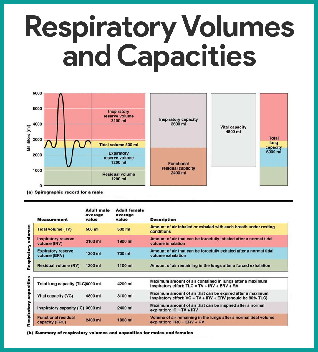
- Tidal volume. Normal quiet breathing moves approximately 500 ml of air into and out of the lungs with each breath.
- Inspiratory reserve volume. The amount of air that can be taken in forcibly over the tidal volume is the inspiratory reserve volume, which is normally between 2100 ml to 3200 ml.
- Expiratory reserve volume. The amount of air that can be forcibly exhaled after a tidal expiration, the expiratory reserve volume, is approximately 1200 ml.
- Residual volume. Even after the most strenuous expiration, about 1200 ml of air still remains in the lungs and it cannot be voluntarily expelled; this is called residual volume, and it is important because it allows gas exchange to go on continuously even between breaths and helps to keep the alveoli inflated.
- Vital capacity. The total amount of exchangeable air is typically around 4800 ml in healthy young men, and this respiratory capacity is the vital capacity, which is the sum of the tidal volume, inspiratory reserve volume, and expiratory reserve volume.
- Dead space volume. Much of the air that enters the respiratory tract remains in the conducting zone passageways and never reaches the alveoli; this is called the dead space volume and during a normal tidal breath, it amounts to about 150 ml.
- Functional volume. The functional volume, which is the air that actually reaches the respiratory zone and contributes to gas exchange , is about 350 ml.
- Spirometer. Respiratory capacities are measured with a spirometer, wherein as a person breathes, the volumes of air exhaled can be read on an indicator, which shows the changes in air volume inside the apparatus.
- Bronchial sounds. Bronchial sounds are produced by air rushing through the large respiratory passageways (trachea and bronchi).
- Vesicular breathing sounds. Vesicular breathing sounds occur as air fills the alveoli, and they are soft and resemble a muffled breeze.
- External respiration. External respiration or pulmonary gas exchange involves oxygen being loaded and carbon dioxide being unloaded from the blood.
- Internal respiration. In internal respiration or systemic capillary gas exchange , oxygen is unloaded and carbon dioxide is loaded into the blood.
- Gas transport. Oxygen is transported in the blood in two ways: most attaches to hemoglobin molecules inside the RBCs to form oxyhemoglobin, or a very small amount of oxygen is carried dissolved in the plasma ; while carbon dioxide is transported in plasma as bicarbonate ion, or a smaller amount (between 20 to 30 percent of the transported carbon dioxide) is carried inside the RBCs bound to hemoglobin.
Neural Regulation
- Phrenic and intercostal nerves . These two nerves regulate the activity of the respiratory muscles, the diaphragm, and external intercostals.
- Medulla and pons . Neural centers that control respiratory rhythm and depth are located mainly in the medulla and pons; the medulla, which sets the basic rhythm of breathing, contains a pacemaker , or self-exciting inspiratory center, and an expiratory center that inhibits the pacemaker in a rhythmic way; pons centers appear to smooth out the basic rhythm of inspiration and expiration set by the medulla.
- Eupnea. The normal respiratory rate is referred to as eupnea, and it is maintained at a rate of 12 to 15 respirations/minute .
- Hyperpnea. During exercise, we breathe more vigorously and deeply because the brain centers send more impulses to the respiratory muscles, and this respiratory pattern is called hyperpnea.
Non-neural Factors Influencing Respiratory Rate and Depth
- Physical factors. Although the medulla’s respiratory centers set the basic rhythm of breathing, there is no question that physical factors such as talking, coughing, and exercising can modify both the rate and depth of breathing, as well as an increased body temperature, which increases the rate of breathing.
- Volition (conscious control). Voluntary control of breathing is limited, and the respiratory centers will simply ignore messages from the cortex (our wishes) when the oxygen supply in the blood is getting low or blood pH is falling .
- Emotional factors. Emotional factors also modify the rate and depth of breathing through reflexes initiated by emotional stimuli acting through centers in the hypothalamus .
- Chemical factors. The most important factors that modify respiratory rate and depth are chemical- the levels of carbon dioxide and oxygen in the blood; increased levels of carbon dioxide and decreased blood pH are the most important stimuli leading to an increase in the rate and depth of breathing, while a decrease in oxygen levels become important stimuli when the levels are dangerously low.
- Hyperventilation. Hyperventilation blows off more carbon dioxide and decreases the amount of carbonic acid, which returns blood pH to the normal range when carbon dioxide or other sources of acids begin to accumulate in the blood.
- Hypoventilation. Hypoventilation or extremely slow or shallow breathing allows carbon dioxide to accumulate in the blood and brings blood pH back into normal range when blood starts to become slightly alkaline.
Respiratory efficiency is reduced with age. They are unable to compensate for increased oxygen need and are significantly increasing the amount of air inspired. Therefore, difficulty in breathing is usually common especially during activities. Expiratory muscles become weaker so their cough efficiency is reduced and the amount of air left in the lungs is increased. Health promotion teaching can include smoking cessation, preventing respiratory infections through handwashing , and ensuring up to date influenza and pneumonia vaccinations.
Craving more insights? Dive into these related materials to enhance your study journey!
- Anatomy and Physiology Nursing Test Banks . This nursing test bank includes questions about Anatomy and Physiology and its related concepts such as: structure and functions of the human body, nursing care management of patients with conditions related to the different body systems.
15 thoughts on “Respiratory System Anatomy and Physiology”
Hello, My name is Sharon and I think I just struck gold! This web site is just what I need to study, learn, and understand the Human Body. Thank you so much.
Thank you so much. you just made my day. The information was comprehensive and comes as an easy resource. Violet.
Thanks very much, I was satisfied with the answers
Thanks very much. I am satisfied with the notes
Thank you so much the notes are perfect and useful to my studies
Very helpful and easy to understand. Thank you so much.
It is fantastic and very helpfull and thanks a lot
This is very helpful, thank you.
You’re welcome! Happy to hear that you found the respiratory system anatomy and physiology material helpful. If there’s anything more you’d like to dive into or any questions you have, feel free to reach out. Always here to help with your learning journey.
Thank you so much
Anatomy and physiology is a subject to learn the figure the rate of human body as well. Thanks
I need McQ for this note please
Please check out the “see also” section for the quizzes.
Very helpful content especially for me as an RN student
HELLO , MY NAMEN IS DESTRI I’m come from indonesia i think is very information about nurse very important ,thank u very much guys …
reagard me ,
Leave a Comment Cancel reply

Respiratory System
Preparing for the MCAT requires a deep understanding of the respiratory system, a fundamental component of the Organ Systems foundation. Mastery of lung functions, gas exchange, and respiratory physiology provides critical insights into how the body maintains oxygen and carbon dioxide levels, essential for achieving a high MCAT score.
Learning Objective
In studying the “Respiratory System” for the MCAT , you should aim to understand the anatomy and physiology of respiratory structures, including the lungs, airways, and diaphragm. Learn the mechanisms of gas exchange, ventilation, and oxygen transport. Explore how the body regulates breathing and maintains acid-base balance. Additionally, delve into the pathophysiology of common respiratory conditions such as asthma, COPD, and pneumonia. Apply this knowledge to interpret diagnostic results, such as pulmonary function tests and arterial blood gases, in MCAT practice passages, focusing on how respiratory health is integral to overall systemic function.
Understanding Respiratory System Structure
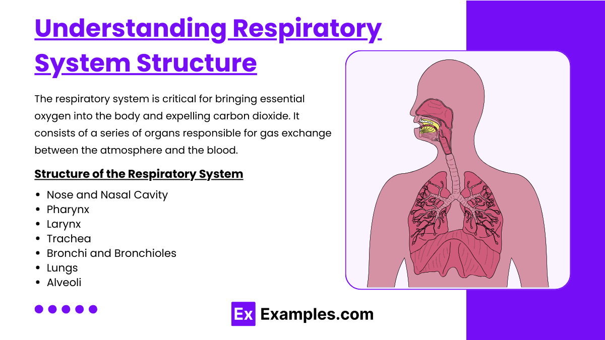
The respiratory system is a network of organs and tissues responsible for gas exchange between the body and the environment. Its primary function is to deliver oxygen to the blood and remove carbon dioxide. The system includes the nasal cavity, pharynx, larynx, trachea, bronchi, and lungs. Within the lungs, tiny air sacs called alveoli facilitate the exchange of gases with the bloodstream. The respiratory system also helps regulate blood pH, filters inhaled particles, and protects the airways with mucus and cilia. It plays a vital role in maintaining cellular respiration and overall metabolic processes. Here’s an overview of its structure:
Structure of the Respiratory System
- The main external opening for air, the nose is lined with hair and mucus to filter dust and pathogens. The nasal cavity also warms and moistens incoming air.
- A muscular tube that serves both respiratory and digestive systems, connecting the nasal cavity and mouth to the larynx and esophagus. It helps in the process of swallowing and provides a pathway for the movement of air.
- Located at the entrance to the trachea and functions in voice production. The larynx contains the vocal cords and works as a valve that closes off the trachea during swallowing, preventing food from entering the airway.
- A rigid tube leading from the larynx to the bronchi, it provides a clear airway for air to enter and exit the lungs. The trachea is lined with cilia and mucus to trap and expel contaminants.
- The trachea divides into two primary bronchi, one entering each lung. These bronchi branch into smaller bronchioles within the lung tissue, further dividing into even smaller passages that lead to the alveoli.
- Two large respiratory organs located in the thorax. They house the bronchi, bronchioles, and alveoli. The right lung is typically larger and divided into three lobes, while the left lung has two lobes.
- Tiny sac-like structures at the end of the bronchioles where gas exchange occurs. Each alveolus is surrounded by a network of capillaries where oxygen enters the blood and carbon dioxide is removed.
Function of the Respiratory System
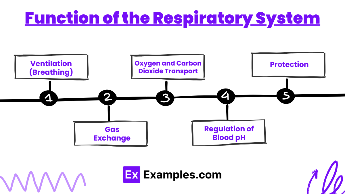
- Involves the movement of air in and out of the lungs through the act of inhalation and exhalation. This process is driven by the diaphragm and the intercostal muscles.
- Occurs in the alveoli. Oxygen from inhaled air diffuses into the capillaries, while carbon dioxide from the blood diffuses into the alveoli to be exhaled.
- Blood transports oxygen from the lungs to the cells of the body and brings carbon dioxide back to the lungs to be exhaled.
- The respiratory system helps maintain the acid-base balance of the body by regulating the levels of carbon dioxide in the blood, which influences blood pH.
- Provides defense against pathogens and particulates through the filtering action of nose hairs, mucus, and cilia. Additionally, coughing and sneezing are reflexes that help clear the airways of irritants.
Pathophysiology of Respiratory Disorders
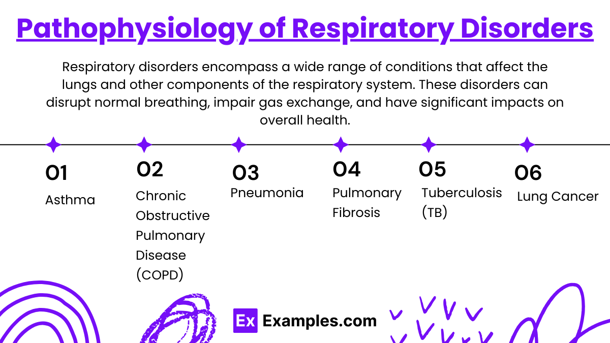
Respiratory disorders encompass a wide range of conditions that affect the lungs and other components of the respiratory system. These disorders can disrupt normal breathing, impair gas exchange, and have significant impacts on overall health. Here’s an overview of the pathophysiology behind some common respiratory disorders:
- Pathophysiology : Asthma is characterized by chronic inflammation of the airways, leading to episodes of wheezing, breathlessness, chest tightness, and coughing. During an asthma attack, the airways become hyperresponsive, leading to bronchoconstriction, mucosal edema, and increased mucus production, all of which narrow the airways and obstruct airflow.

2. Chronic Obstructive Pulmonary Disease (COPD)
- Pathophysiology : COPD includes conditions such as chronic bronchitis and emphysema, often caused by long-term exposure to irritating gases or particulate matter, most commonly from cigarette smoke. Chronic bronchitis involves inflammation of the lining of the bronchial tubes, leading to persistent cough and mucus production. Emphysema involves damage to the alveoli (air sacs in the lungs), reducing the surface area available for gas exchange and causing air to become trapped in the lungs, which impairs breathing.
3. Pneumonia
- Pathophysiology : Pneumonia is an infection that inflames the air sacs in one or both lungs. The air sacs may fill with fluid or pus, causing symptoms such as cough with phlegm or pus, fever, chills, and difficulty breathing. The infection can be bacterial, viral, or fungal.
4. Pulmonary Fibrosis
- Pathophysiology : Pulmonary fibrosis involves the progressive scarring of lung tissue, which leads to a continuous decline in lung function. The thickening and stiffening of tissue make it difficult to breathe and reduce oxygen transfer from the lungs to the bloodstream.
5. Tuberculosis (TB)
- Pathophysiology : Caused by the bacterium Mycobacterium tuberculosis , TB primarily affects the lungs but can also affect other parts of the body. The infection leads to the formation of granulomas—small nodules of immune cells—in the lungs, which can cause tissue damage and reduce lung function.
6. Lung Cancer
- Pathophysiology : Lung cancer originates in the lung tissues, typically in the cells lining the air passages. It is often associated with smoking, but it can also occur in non-smokers. The disease interferes with normal respiratory function primarily through the growth of tumors that block air passages and through the systemic effects of cancer.
Example 1: Gas Exchange During Exercise
- Scenario : Increased oxygen demand during vigorous exercise.
- Process : During exercise, the respiratory rate and depth of breathing increase to enhance the oxygen intake and carbon dioxide expulsion. This ensures that more oxygen reaches the muscles and more CO2 is removed from the body, meeting the increased metabolic demands.
Example 2: Asthma Attack
- Scenario : Exposure to allergens leading to an asthma attack.
- Process : In asthma, exposure to specific allergens can trigger an inflammatory response that leads to bronchoconstriction, airway edema, and increased mucus production. This results in difficulty breathing, wheezing, and coughing. Treatment typically involves bronchodilators which relax the airway muscles.
Example 3: COPD and Airflow Limitation
- Scenario : Chronic exposure to cigarette smoke leading to COPD.
- Process : In COPD, prolonged exposure to irritants such as cigarette smoke causes chronic inflammation in the airways, destruction of alveolar walls, and narrowing of the airways, which impairs airflow both in and out of the lungs, leading to shortness of breath, especially during physical exertion.
Example 4: Carbon Monoxide Poisoning
- Scenario : Inhalation of carbon monoxide (CO) in a closed garage.
- Process : CO competes with oxygen for binding sites on hemoglobin, forming carboxyhemoglobin, which reduces the blood’s oxygen-carrying capacity. This leads to tissue hypoxia and can result in symptoms ranging from headaches and dizziness to death, depending on the exposure level.
Example 5: Acute Respiratory Acidosis
- Scenario : Respiratory failure due to severe pneumonia.
- Process : Pneumonia can impair gas exchange by filling the alveoli with fluid or pus, reducing the lung’s ability to oxygenate blood and remove carbon dioxide. This buildup of CO2 leads to respiratory acidosis, where the blood becomes too acidic, potentially requiring mechanical ventilation to correct.
Practice Questions
Question 1: mechanism of breathing.
What primarily drives the inhalation process during normal quiet breathing?
A) Contraction of the abdominal muscles B) Relaxation of the diaphragm C) Contraction of the diaphragm D) Contraction of the pectoral muscles
Correct Answer: C) Contraction of the diaphragm
Explanation: During normal quiet breathing, inhalation is primarily driven by the contraction of the diaphragm. When the diaphragm contracts, it moves downward, increasing the volume of the thoracic cavity and reducing the pressure inside the lungs relative to the atmosphere, causing air to flow into the lungs.
Question 2: Gas Exchange
Which gas has the highest partial pressure in the alveoli of the lungs?
A) Oxygen (O2) B) Carbon dioxide (CO2) C) Nitrogen (N2) D) Water vapor (H2O)
Correct Answer: A) Oxygen (O2)
Explanation: In the alveoli of the lungs, oxygen (O2) typically has the highest partial pressure compared to other gases present in inspired air. This high partial pressure gradient between the alveoli and the blood drives the diffusion of oxygen into the blood, facilitating its transport to tissues throughout the body.
Question 3: Respiratory Adjustments in Exercise
During vigorous exercise, what adjustment does the respiratory system primarily make to meet increased oxygen demand?
A) Decrease in respiratory rate B) Increase in tidal volume and respiratory rate C) Increase in carbon dioxide output only D) Decrease in tidal volume
Correct Answer: B) Increase in tidal volume and respiratory rate
Explanation: During vigorous exercise, the respiratory system adjusts by increasing both tidal volume (the amount of air inhaled and exhaled in a single breath) and respiratory rate (the number of breaths taken per minute). This adjustment maximizes the amount of air exchanged during breathing and improves the body’s ability to increase oxygen intake and carbon dioxide expulsion to meet the elevated metabolic demands.
Sciencing_Icons_Science SCIENCE
Sciencing_icons_biology biology, sciencing_icons_cells cells, sciencing_icons_molecular molecular, sciencing_icons_microorganisms microorganisms, sciencing_icons_genetics genetics, sciencing_icons_human body human body, sciencing_icons_ecology ecology, sciencing_icons_chemistry chemistry, sciencing_icons_atomic & molecular structure atomic & molecular structure, sciencing_icons_bonds bonds, sciencing_icons_reactions reactions, sciencing_icons_stoichiometry stoichiometry, sciencing_icons_solutions solutions, sciencing_icons_acids & bases acids & bases, sciencing_icons_thermodynamics thermodynamics, sciencing_icons_organic chemistry organic chemistry, sciencing_icons_physics physics, sciencing_icons_fundamentals-physics fundamentals, sciencing_icons_electronics electronics, sciencing_icons_waves waves, sciencing_icons_energy energy, sciencing_icons_fluid fluid, sciencing_icons_astronomy astronomy, sciencing_icons_geology geology, sciencing_icons_fundamentals-geology fundamentals, sciencing_icons_minerals & rocks minerals & rocks, sciencing_icons_earth scructure earth structure, sciencing_icons_fossils fossils, sciencing_icons_natural disasters natural disasters, sciencing_icons_nature nature, sciencing_icons_ecosystems ecosystems, sciencing_icons_environment environment, sciencing_icons_insects insects, sciencing_icons_plants & mushrooms plants & mushrooms, sciencing_icons_animals animals, sciencing_icons_math math, sciencing_icons_arithmetic arithmetic, sciencing_icons_addition & subtraction addition & subtraction, sciencing_icons_multiplication & division multiplication & division, sciencing_icons_decimals decimals, sciencing_icons_fractions fractions, sciencing_icons_conversions conversions, sciencing_icons_algebra algebra, sciencing_icons_working with units working with units, sciencing_icons_equations & expressions equations & expressions, sciencing_icons_ratios & proportions ratios & proportions, sciencing_icons_inequalities inequalities, sciencing_icons_exponents & logarithms exponents & logarithms, sciencing_icons_factorization factorization, sciencing_icons_functions functions, sciencing_icons_linear equations linear equations, sciencing_icons_graphs graphs, sciencing_icons_quadratics quadratics, sciencing_icons_polynomials polynomials, sciencing_icons_geometry geometry, sciencing_icons_fundamentals-geometry fundamentals, sciencing_icons_cartesian cartesian, sciencing_icons_circles circles, sciencing_icons_solids solids, sciencing_icons_trigonometry trigonometry, sciencing_icons_probability-statistics probability & statistics, sciencing_icons_mean-median-mode mean/median/mode, sciencing_icons_independent-dependent variables independent/dependent variables, sciencing_icons_deviation deviation, sciencing_icons_correlation correlation, sciencing_icons_sampling sampling, sciencing_icons_distributions distributions, sciencing_icons_probability probability, sciencing_icons_calculus calculus, sciencing_icons_differentiation-integration differentiation/integration, sciencing_icons_application application, sciencing_icons_projects projects, sciencing_icons_news news.
- Share Tweet Email Print
- Home ⋅
- Science ⋅
- Biology ⋅
Classroom Activities on the Respiratory System

Classroom Activities for Scientific Notation
The respiratory system transports oxygen through the blood to all the major organs in the human body. Through breathing, the lungs pull oxygen into the body and expel carbon dioxide. Red blood cells transport oxygen to all the cells around the body.
Oxygen is essential for cell growth and reproduction. Humans breathe about 20 times a minute and much more during physical exertion.
Respiratory system activities and respiratory system games (high school, middle school, and college based) make these complex systems easier to understand.
Physical Demonstration
Students are assigned one of three roles -- lungs, oxygen and carbon dioxide . Children assigned to be the lungs hold hands to form two circles with an opening at the top. As the lungs inhale, children step out to widen the circle. Simultaneously, the students representing oxygen enter the lungs through the opening at the top, then pass into the bloodstream under the joined hands in the circle.
As the lungs "exhale," the students representing carbon dioxide enter the circle under the joined hands. The children in the circle step closer together, forcing the carbon dioxide out of the openings at the top of the circles. This physical demonstration helps children understand the breathing process.
Respiratory System Activities to Test Lung Capacity
Students work in pairs to measure lung capacity. Using a balloon and string, each child breathes one breath as hard as they can into the balloon. The other child measures the circumference of the balloon using the string.
The class discusses that the air in the balloon represents the amount of air that used to be in the students' lungs. They discuss the differences in the lung capacity between the students and come up with various hypotheses on why lung capacity varies.
In this activity, children observe breathing patterns at rest and after exercise. Students sit quietly for 30 seconds and reflect on their breathing. Students discuss their breathing rate and how they feel.
Then the children get up and do jumping jacks for 30 seconds. Immediately after the jumping jacks, the students reflect on their breathing. Class discussion focuses on whether the children are breathing faster after exercise and why this is (the body needs to take in more oxygen).
To make it a bit more complex for older students, have them take specific measurements and analyze the data. These measurements could include:
- Breaths per minute (at rest and after exercise)
- Pulse
- Number of jumping jacks in 30 seconds
- Lung capacity of each student
Have the students craft a hypothesis , choose what data they collect and have them analyze the data via graphs, equations and comparison of data points.
Worksheet Activities
Children learn key words and concepts about the respiratory system through word searches, puzzles and crosswords. Puzzle activities can be created where the children must put the actions associated with breathing in the correct order (for example, inhalation, oxygen enters the lungs, exhalation, carbon dioxide leaves the lungs).
To make the puzzles more durable, put each action on a piece of cardboard and laminate it. Children can also label drawings of the respiratory system, including lungs and red blood cells . You can make your own worksheets and puzzles or obtain them from the Internet.
Respiratory system games, high school level in particular, can get students engaged with the lesson. You can even have the students create their own game, which will get them to truly understand the information and craft a usable activity.
Online Dissection and Lung Models
Lung models help students better visualize the entire respiratory system . Look for an online dissection and/or lung model system that's interactive.
There may even be dissection and respiratory system games (high school based, middle school based, etc) on these online resources that allow students to match parts of the system for points, quiz themselves and more.
Related Articles
Fun science activities for force & motion, excretory system science project ideas, how to build a 3d model of the solar system, how to make a 3d model of the muscular system for a..., how to measure air pollution for a science fair project, science fair projects for lung capacity, rainforest ecosystem school projects, how to make a 3d model of the respiratory system, phases of the moon activities for kids, creative ideas to make the solar system, color-mixing paint activities for preschoolers, science projects: how to make a skeleton, fun outdoor math activities, activities for rational counting for preschool, dragonfly learning activities for preschool, what are the types of technology in a mathematics classroom, fun games & activities for field day, how to teach first grade math subtraction tables, how to make a model of the solar system for the fifth....
- KidsHealth in the Classroom: Human Body Series:Respiratory System
- Henry County Schools: My Body, the Inside Story: Respiratory System Instructional Activities
- The Canadian Lung Association: Inside the Human Body: The Respiratory System
About the Author
For more than a decade, Tia Benjamin has been writing organizational policies, procedures and management training programs. A C-level executive, she has more than 15 years experience in human resources and management. Benjamin obtained a Bachelor of Science in social psychology from the University of Kent, England, as well as a Master of Business Administration from San Diego State University.
Photo Credits
thorax x-ray of the lungs image by JoLin from Fotolia.com
Find Your Next Great Science Fair Project! GO

Want to create or adapt books like this? Learn more about how Pressbooks supports open publishing practices.
21 The Respiratory System
Dialogue cards, question sets.
Major Respiratory System Organs
Respiratory Epithelium
Regions of the Pharynx
Respiratory Membrane and Alveoli
The Process of Breathing
Ventilation Control Centers
Interactive Activities for Human Anatomy and Physiology Copyright © by Open Education Lab is licensed under a Creative Commons Attribution-NonCommercial-ShareAlike 4.0 International License , except where otherwise noted.
Share This Book
upper respiratory system 1
There is a printable worksheet available for download here so you can take the quiz with pen and paper.
The scorecard of a champion
100% needed
Other Games of Interest

Latest Quiz Activities
- soyoiyo played the game 6 hours ago
upper respiratory system 1 — Quiz Information
This is an online quiz called upper respiratory system 1
You can use it as upper respiratory system 1 practice, completely free to play.


- Types of Bones
- Axial Skeleton
- Appendicular Skeleton
- Skeletal Pathologies
- Muscle Tissue Types
- Muscle Attachments & Actions
- Muscle Contractions
- Muscular Pathologies
- Functions of Blood
- Blood Vessels
- Circulation
- Circulatory Pathologies
- Respiratory Functions
- Upper Respiratory System
- Lower Respiratory System
- Respiratory Pathologies
- Oral Cavity
- Alimentary Canal
- Accessory Organs
- Absorption & Elimination
- Digestive Pathologies
- Lymphatic Structures
- Immune System
- Urinary System Structures
- Urine Formation
- Urine Storage & Elimination
- Urinary Pathologies
- Female Reproductive System
- Male Reproductive System
- Reproductive Process
- Endocrine Glands
Welcome to the Visible Body Learn Site
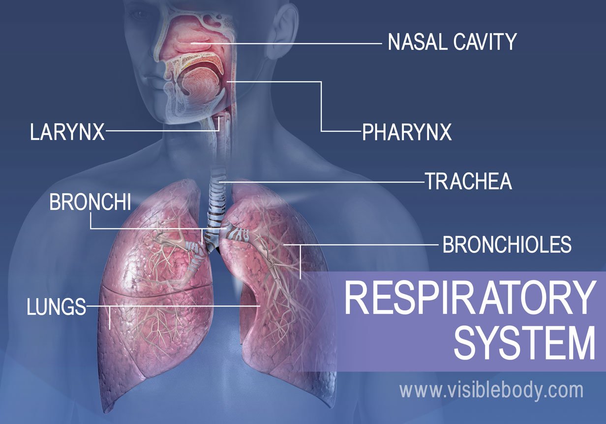
Top 5 Functions of the Respiratory System: A Look Inside Key Respiratory Activities
Through breathing, inhalation and exhalation, the respiratory system facilitates the exchange of gases between the air and the blood and between the blood and the body’s cells. The respiratory system also helps us to smell and create sound. The following are the five key functions of the respiratory system.
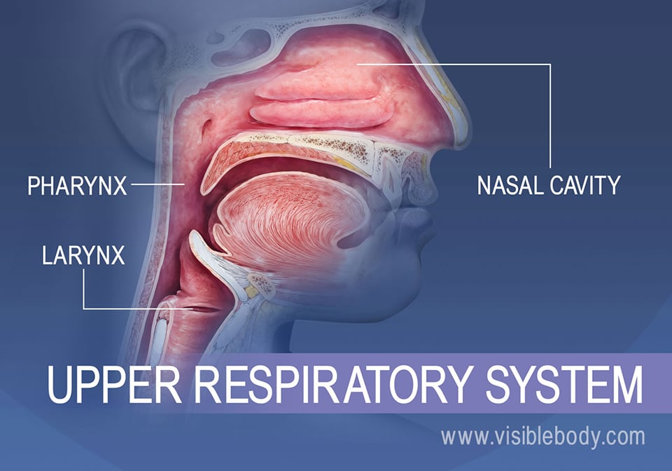
Breathing In and Speaking Out: How the Structures of the Upper Respiratory System Work
The structures of the upper respiratory system, or respiratory tract, allow us to breathe and speak.
- The nose and nasal cavities provide airways for respiration.
- The paranasal sinuses surround the nasal cavities.
- The pharynx connects the nasal and oral cavities to the larynx and esophagus.
- The larynx and vocal cords allow us to breathe and talk and sing.
- Structures that produce sound depend on the hyoid bone.
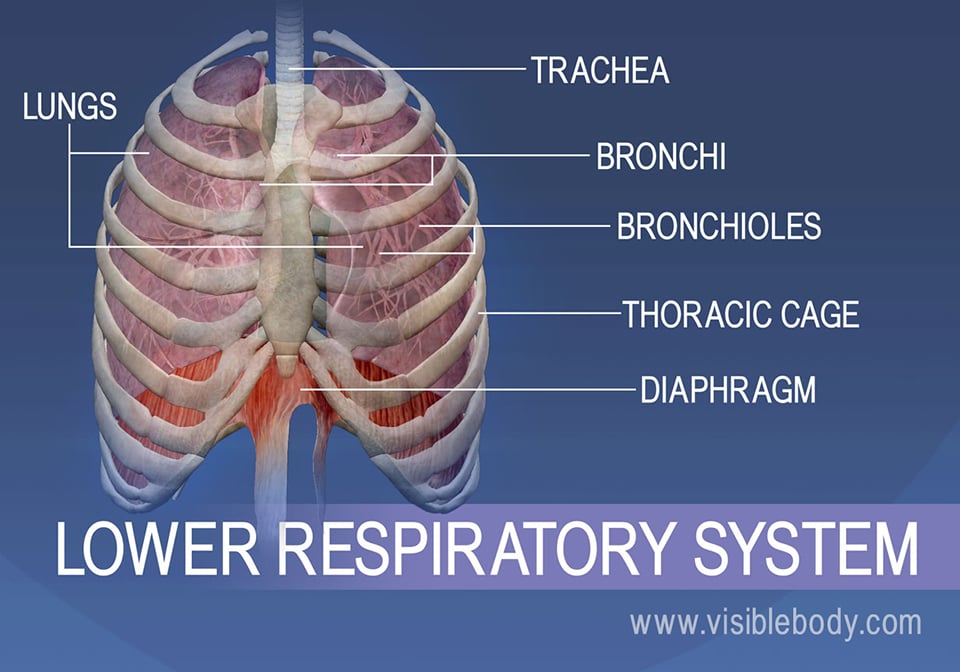
Drawing In and Processing Air: Functions of the Trachea, Bronchi, Lungs, and Alveoli
- Trachea: the main airway to the lungs
- Bronchi: passageways that bring air in and out of the lungs
- Lungs: structures responsible for gas exchange between the air we breathe and our bodies
- Alveoli: microscopic air sacs that are the site of external respiration
- Diaphragm: the muscle that is key to the physical process of breathing
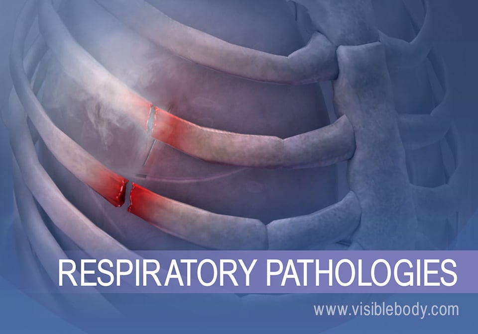
Common Respiratory Diseases and Disorders: COPD, Asthma, Sinusitis, Influenza, and Pneumothorax
- Most respiratory diseases and disorders can be described as either infectious or chronic.
- Inflamed airways become irritated during inhalation during an asthma attack.
- Sinusitis is the inflammation of mucous membranes in the nasal sinuses.
- The flu virus can pass through the air from one person to another.
- Chest trauma can cause pneumothorax, a collapsed lung.
For students
For instructors
Get our awesome anatomy emails!
When you select "Subscribe" you will start receiving our email newsletter. Use the links at the bottom of any email to manage the type of emails you receive or to unsubscribe. See our privacy policy for additional details.
- User Agreement
- Permissions
Respiratory System (Anatomy)
Definition: The respiratory system (also referred to as the ventilator system) is a complex biological system comprised of several organs that facilitate the inhalation and exhalation of oxygen and carbon dioxide in living organisms (or, in other words, breathing). Most of the organs of the respiratory system help to distribute air, but only the tiny, grape-like alveoli and the alveolar ducts are responsible for actual gas exchange.
In addition to air distribution and gas exchange, the respiratory system filters warm and humidify the air we breathe. Organs in the respiratory system also play a role in speech and the sense of smell. It also helps the body maintain homeostasis, or balance among the many elements of the body’s internal environment.
In most fish, and a number of other aquatic animals (both vertebrates and invertebrates) the respiratory system consists of gills, which are either partially or completely external organs, bathed in the watery environment. Other animals, such as insects, have respiratory systems with very simple anatomical features, and in amphibians, even the skin plays a vital role in gas exchange. Plants also have respiratory systems but the directionality of gas exchange can be opposite to that in animals. The respiratory system in plants includes anatomical features such as stomata, that are found in various parts of the plant.
A properly functioning respiratory system is a vital part of our good health. Respiratory infections can be acute and sometimes life-threatening. They can also be chronic, in which case they place tremendous long-term stress on the immune system, endocrine system, HPA axis, and much more.
Anatomy of the Respiratory System: In humans and other mammals, the anatomy of a typical respiratory system is the respiratory tract. The tract is divided into an upper and a lower respiratory tract. The upper tract includes the nose, nasal cavities, sinuses, pharynx and the part of the larynx above the vocal folds. The lower tract includes the lower part of the larynx, the trachea, bronchi, bronchioles, and the alveoli.
The respiratory system is divided into two main components:
Upper respiratory tract: Composed of the nose, the pharynx, and the larynx, the organs of the upper respiratory tract are located outside the chest cavity.
- Nasal cavity: Inside the nose, the sticky mucous membrane lining the nasal cavity traps dust particles, and tiny hairs called cilia help move them to the nose to be sneezed or blown out.
- Sinuses: These air-filled spaces alongside the nose help make the skull lighter.
- Pharynx: Both food and air pass through the pharynx before reaching their appropriate destinations. The pharynx also plays a role in speech.
- Larynx: The larynx is essential to human speech.
Lower respiratory tract: Composed of the trachea, the lungs, and all segments of the bronchial tree (including the alveoli), the organs of the lower respiratory tract are located inside the chest cavity.
- Trachea: Located just below the larynx, the trachea is the main airway to the lungs.
- Lungs: Together the lungs form one of the body’s largest organs. They’re responsible for providing oxygen to capillaries and exhaling carbon dioxide.
- Bronchi: The bronchi branch from the trachea into each lung and create the network of intricate passages that supply the lungs with air.
- Diaphragm: The diaphragm is the main respiratory muscle that contracts and relaxes to allow air into the lungs.
The major organs of the respiratory system function primarily to provide oxygen to body tissues for cellular respiration, remove the waste product carbon dioxide, and help to maintain acid-base balance. Portions of the respiratory system are also used for non-vital functions, such as sensing odors, speech production, and for straining, such as during childbirth or coughing.
The alveoli are the dead end terminals of the “tree”, meaning that any air that enters them has to exit via the same route. A system such as this creates dead space, a volume of air (about 150 ml in the adult human) that fills the airways after exhalation and is breathed back into the alveoli before the environmental air reaches them. At the end of inhalation, the airways are filled with environmental air, which is exhaled without coming in contact with the gas exchanger.
Functionally, the respiratory system can be divided into a conducting zone and a respiratory zone. The conducting zone of the respiratory system includes the organs and structures not directly involved in gas exchange. The gas exchange occurs in the respiratory zone.
- Conducting Zone – The major functions of the conducting zone are to provide a route for incoming and outgoing air, remove debris and pathogens from the incoming air, and warm and humidify the incoming air. Several structures within the conducting zone perform other functions as well. The epithelium of the nasal passages, for example, is essential to sensing odors, and the bronchial epithelium that lines the lungs can metabolize some airborne carcinogens.
- Respiratory Zone – In contrast to the conducting zone, the respiratory zone includes structures that are directly involved in gas exchange. The respiratory zone begins where the terminal bronchioles join a respiratory bronchiole, the smallest type of bronchiole, which then leads to an alveolar duct, opening into a cluster of alveoli.
Homeostatic Control of Respiration: The last physiological role of the respiratory system is the homeostatic control of respiration or, in other words, the body’s ability to maintain a steady breathing rate. This is termed eupnea. This state should remain constant until the body has a demand for increased oxygen and carbon dioxide levels due to increased exertion, most likely caused by physical activity. When this happens, chemoreceptors will pick up on the increased partial pressure of the oxygen and carbon dioxide and send triggers to the brain. The brain will then signal the respiratory center to make adjustments to the breathing rate and depth in order to face the increased demands.
Clinical significance of the Respiratory system:
Disorders of the respiratory system can be classified into several general groups:
- Airway obstructive conditions (e.g., emphysema, bronchitis, asthma)
- Pulmonary restrictive conditions (e.g., fibrosis, sarcoidosis, alveolar damage, pleural effusion)
- Vascular diseases (e.g., pulmonary edema, pulmonary embolism, pulmonary hypertension)
- Infectious, environmental and other “diseases” (e.g., pneumonia, tuberculosis, asbestosis, particulate pollutants)
- Primary cancers (e.g. bronchial carcinoma, mesothelioma)
- Secondary cancers (e.g. cancers that originated elsewhere in the body, but have seeded themselves in the lungs)
- Insufficient surfactant (e.g. respiratory distress syndrome in pre-term babies).
Disorders of the respiratory system are usually treated by a pulmonologist and respiratory therapist.
Information Source:
- opentextbc.ca
- healthline.com
- adrenalfatiguesolution.com
Roundworm Protein can Unlock the Cellular ‘Fountain of Youth’
Aquatic toxicology – a multidisciplinary field which integrates toxicology, hypermagnesemia, recombinant dna (rdna), a beautiful bird “tawny eagle”, instagram may soon let you post from desktop, bonding in metals, inventory reconciliation management, cross-cutting breakdowns in alzheimer’s disease revealed by new technologies, submitting application for leave of absents from school by student, latest post, cadmium chloride, improving eye tracking for evaluating brain abnormalities, psychedelics stimulate cells in the hippocampus to alleviate anxiety, sodium iodide – an ionic compound, sodium iodate (naio3), adding depth to the connection between brain structure and ideology.

IMAGES
VIDEO
COMMENTS
bronchioles. The _________ zone includes the respiratory bronchioles, alveolar ducts, alveolar sacs, and alveoli and is where gas exchange occurs. respiratory. The process of moving air in and out of the lungs is called: pulminary ventilation. The most important stimulus for breathing in a healthy person is the body's need to rid itself of the ...
pulmonary ventilation. respiratory system. consists of a series of passages that brings outside air in contact with special structures that lie close to blood capillaries. external respiration. moves oxygen from the air into the blood. internal respiration. moves oxygen from the blood to the tissues. inspiration. air into lungs, inhalation.
Respiratory Tract. The respiratory tract in humans is made up of the following parts: External nostrils - For the intake of air. Nasal chamber - which is lined with hair and mucus to filter the air from dust and dirt. Pharynx - It is a passage behind the nasal chamber and serves as the common passageway for both air and food.
The respiratory system organs oversee the gas exchanges that occur between the blood and the external environment. Passageway. Passageways that allow air to reach the lungs. Humidifier. Purify, humidify, and warm incoming air. Anatomy of the Respiratory System. The organs of the respiratory system include the nose, pharynx, larynx, trachea ...
Respiratory system (Systema respiratorum) The respiratory system, also called the pulmonary system, consists of several organs that function as a whole to oxygenate the body through the process of respiration (breathing).This process involves inhaling air and conducting it to the lungs where gas exchange occurs, in which oxygen is extracted from the air, and carbon dioxide expelled from the body.
The respiratory system, which includes air passages, pulmonary vessels, the lungs, and breathing muscles, aids the body in the exchange of gases between the air and blood, and between the blood ...
The respiratory system is a network of organs and tissues responsible for gas exchange between the body and the environment. Its primary function is to deliver oxygen to the blood and remove carbon dioxide. The system includes the nasal cavity, pharynx, larynx, trachea, bronchi, and lungs. Within the lungs, tiny air sacs called alveoli ...
Human respiratory system, the system in humans that takes up oxygen and expels carbon dioxide. The major organs of the respiratory system include the nose, pharynx, larynx, trachea, bronchi, lungs, and diaphragm. Learn about the anatomy and function of the respiratory system in this article.
The respiratory system transports oxygen through the blood to all the major organs in the human body. Through breathing, the lungs pull oxygen into the body and expel carbon dioxide. Red blood cells transport oxygen to all the cells around the body. Oxygen is essential for cell growth and reproduction. Humans breathe about 20 times a minute and ...
This is respiratory acidosis. If large amounts of CO 2 are blown off as in hyperventilation then the pH of the blood rises and this is respiratory alkalosis. Remember that the normal range of plasma pH is 7.35-7. The respiratory system/blood plasma buffer works to keep the pH in this range.
Major Respiratory System Organs. Respiratory Epithelium. Regions of the Pharynx. Respiratory Membrane and Alveoli. The Process of Breathing. Ventilation Control Centers. Previous: Lymphatic and Immune Systems. Next: The Digestive System. License.
This online quiz is called upper respiratory system 1 . It was created by member Hiro Flavored Pepsi and has 10 questions.
The structures of the upper respiratory system, or respiratory tract, allow us to breathe and speak. The nose and nasal cavities provide airways for respiration. The paranasal sinuses surround the nasal cavities. The pharynx connects the nasal and oral cavities to the larynx and esophagus. The larynx and vocal cords allow us to breathe and talk ...
Created by. Tyerra_Chapman. Study with Quizlet and memorize flashcards containing terms like Traps inhaled particles and moistens the inhaled air, Transport the mucus to the pharynx where it can be swallowed so the trapped particles can be destroyed in the stomach, it warms the incoming air that flows across the mucosal surface and more.
Respiratory System (Anatomy) Definition: The respiratory system (also referred to as the ventilator system) is a complex biological system comprised of several organs that facilitate the inhalation and exhalation of oxygen and carbon dioxide in living organisms (or, in other words, breathing). Most of the organs of the respiratory system help to distribute air, but only the tiny, grape-like ...
The upper respiratory region consists of the nose, nasal cavity, sinuses, pharynx, and the region above the vocal cords in the larynx. The lower respiratory region consists of the larynx, trachea, bronchi, and lungs. «Labeled.». Review the anatomy of the upper respiratory area and drag and drop the correct term by the proper anatomical structure.
32484. Removal of lung, pneumonectomy. 32440. Diagnostic thoracoscopy with biopsy of the mediastinal space. 32606. Thoracic sympathectomy via thoracoscope. 32664. Study with Quizlet and memorize flashcards containing terms like Transpalatine repair of choanal atresia, Initial posterior control of nasal hemorrhage with posterior cautery ...
The respiratory system's main purpose is to provide Oxygen to the blood so it can be transported around the body. The respiratory system is made up of the nasal cavity, the two lungs, the throat and the larynx, bronchi and trachea. We wanted to investigate the effect that exercise has on our breathing rates. To do this we nominated one person ...
Assignment 3 - Respiratory System. Share. Get a hint. Tuberculosis. Click the card to flip 👆. Contagious disease caused by airborne bacteria. - seen with a 2 view chest x-ray with an apical lordotic chest x-ray. Click the card to flip 👆. 1 / 15.