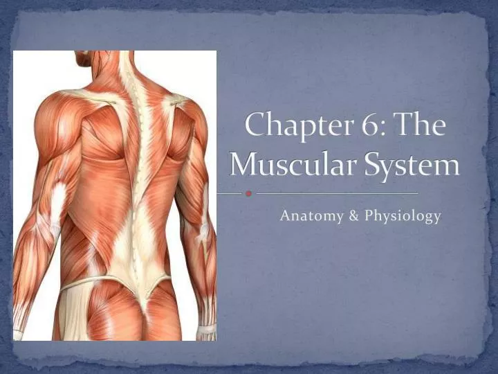
The Muscular System

PowerPoint ® Lecture Slide Presentation

by Patty Bostwick-Taylor, �Florence-Darlington Technical College

Copyright © 2009 Pearson Education, Inc., publishing as Benjamin Cummings

  • Overview of Muscle Tissue
  • Muscles are responsible for all types of body movement
  • Three basic muscle types are found in the body
  • Skeletal muscle
  • Cardiac muscle
  • Smooth muscle
  • Muscle Types
  • Skeletal and smooth muscle cells are elongated (muscle cell = muscle fiber)
  • Contraction of muscles is due to the movement of microfilaments
  • All muscles share some terminology
  • Prefixes myo and mys refer to “muscle”
  • Prefix sarco refers to “flesh”
  • Comparison of Skeletal, Cardiac, �and Smooth Muscles
  • Skeletal Muscle Characteristics
  • Most are attached by tendons to bones
  • Cells are multinucleate
  • Striated—have visible banding
  • Voluntary—subject to conscious control
  • Connective Tissue Wrappings of Skeletal Muscle
  • Cells are surrounded and bundled by connective tissue
  • Endomysium—encloses a single muscle fiber
  • Perimysium—wraps around a fascicle (bundle) of muscle fibers
  • Epimysium—covers the entire skeletal muscle
  • Fascia—on the outside of the epimysium

Skeletal Muscle Attachments

  • Epimysium blends into a connective tissue attachment
  • Tendons—cord-like structures
  • Mostly collagen fibers
  • Often cross a joint due to toughness and small size
  • Aponeuroses—sheet-like structures
  • Attach muscles indirectly to bones, cartilages, or connective tissue coverings
  • Sites of muscle attachment
  • Connective tissue coverings

Smooth Muscle Characteristics

  • Lacks striations
  • Spindle-shaped cells
  • Single nucleus
  • Involuntary—no conscious control
  • Found mainly in the walls of hollow organs

Figure 6.2a

  • Cardiac Muscle Characteristics
  • Usually has a single nucleus
  • Branching cells
  • Joined to another muscle cell at an intercalated disc
  • Involuntary
  • Found only in the heart
  • Skeletal Muscle Functions
  • Produce movement
  • Maintain posture
  • Stabilize joints
  • Generate heat

Microscopic Anatomy of Skeletal Muscle

  • Sarcolemma—specialized plasma membrane
  • Myofibrils—long organelles inside muscle cell
  • Sarcoplasmic reticulum—specialized smooth endoplasmic reticulum
  • Myofibrils are aligned to give distinct bands
  • I band = light band
  • Contains only thin filaments (actin)
  • A band = dark band
  • Contains the entire length of the thick filaments (myosin)
  • Sarcomere—contractile unit of a muscle fiber
  • Organization of the sarcomere
  • Myofilaments
  • Thick filaments = myosin filaments
  • Thin filaments = actin filaments
  • Composed of the protein myosin
  • Has ATPase enzymes
  • Myosin filaments have heads (extensions, or cross bridges)
  • Myosin and actin overlap somewhat
  • Composed of the protein actin
  • Anchored to the Z disc
  • At rest, there is a bare zone that lacks actin filaments called the H zone
  • Sarcoplasmic reticulum (SR)
  • Stores and releases calcium
  • Surrounds the myofibril
  • Skeletal Muscle Activity

Stimulation and Contraction of Single Skeletal Muscle Cells

  • Excitability (also called responsiveness or irritability)—ability to receive and respond to a stimulus
  • Contractility—ability to shorten when an adequate stimulus is received
  • Extensibility—ability of muscle cells to be stretched
  • Elasticity—ability to recoil and resume resting length after stretching

The Nerve Stimulus and Action Potential

  • Skeletal muscles must be stimulated by a motor neuron (nerve cell) to contract
  • Motor unit—one motor neuron and all the skeletal muscle cells stimulated by that neuron

Figure 6.4b

  • Neuromuscular junction
  • Association site of axon terminal of the motor neuron and muscle
  • Synaptic cleft
  • Gap between nerve and muscle
  • Nerve and muscle do not make contact
  • Area between nerve and muscle is filled with interstitial fluid

Transmission of Nerve Impulse to Muscle

  • Neurotransmitter—chemical released by nerve upon arrival of nerve impulse
  • The neurotransmitter for skeletal muscle is acetylcholine (ACh)
  • Acetylcholine attaches to receptors on the sarcolemma
  • Sarcolemma becomes permeable to sodium (Na+)
  • Sodium rushes into the cell generating an action potential
  • Once started, muscle contraction cannot be stopped
  • The Sliding Filament Theory �of Muscle Contraction
  • Activation by nerve causes myosin heads (cross bridges) to attach to binding sites on the thin filament
  • Myosin heads then bind to the next site of the thin filament and pull them toward the center of the sarcomere
  • This continued action causes a sliding of the myosin along the actin
  • The result is that the muscle is shortened (contracted)

The Sliding Filament Theory of �Muscle Contraction

Figure 6.7a–b

The Sliding Filament Theory

Figure 6.8a

Figure 6.8b

Figure 6.8c

  • Contraction of Skeletal Muscle as a Whole
  • Muscle fiber contraction is “all or none”
  • Within a skeletal muscle, not all fibers may be stimulated during the same interval
  • Different combinations of muscle fiber contractions may give differing responses
  • Graded responses—different degrees of skeletal muscle shortening

Contraction of Skeletal Muscle

  • Graded responses can be produced by changing…
  • The frequency of muscle stimulation
  • The number of muscle cells being stimulated at one time

Types of Graded Responses

  • Single, brief contraction
  • Not a normal muscle function
  • Tetanus (summing of contractions)
  • One contraction is immediately followed by another
  • The muscle does not completely �return to a resting state
  • The effects are added
  • Unfused (incomplete) tetanus
  • Some relaxation occurs between contractions
  • The results are summed
  • Fused (complete) tetanus
  • No evidence of relaxation before the following contractions
  • The result is a smooth, sustained muscle contraction
  • Muscle Response to Strong Stimuli
  • Muscle force depends upon the number of fibers stimulated
  • More fibers stimulation results in stronger muscle contraction
  • Muscles can continue to contract unless they run out of energy

Energy for Muscle Contraction

  • Initially, muscles use stored ATP for energy
  • ATP bonds are broken to release energy
  • Only 4–6 seconds worth of ATP is stored by muscles
  • After this initial time, other pathways must be utilized to produce ATP
  • Direct phosphorylation of ADP by creatine phosphate (CP)
  • Muscle cells store CP
  • CP is a high-energy molecule
  • After ATP is depleted, ADP is left
  • CP transfers high-energy phosphate to ADP, to regenerate ATP
  • CP supplies are exhausted in less than 15 seconds
  • Aerobic respiration
  • Occurs at rest and during light to moderate exercise
  • A series of metabolic pathways occur in the mitochondria & uses oxygen
  • Glucose is broken down to carbon dioxide and water, releasing energy (36 ATP)
  • Slow process requiring continuous O 2 & nutrient fuels
  • Anaerobic glycolysis and lactic acid formation
  • Reaction that breaks down glucose without oxygen
  • Occurs in cytosol
  • Glucose is broken down to pyruvic acid to produce 2 ATP
  • Pyruvic acid is converted to lactic acid
  • This reaction is not as efficient but is fast
  • 30-60 sec of activity
  • Huge amounts of glucose are needed
  • Lactic acid produces muscle fatigue
  • Muscle Fatigue and Oxygen Deficit
  • When a muscle is fatigued, it is unable to contract even with a stimulus
  • Common cause for muscle fatigue is oxygen debt
  • Oxygen must be “repaid” to tissue to remove oxygen deficit
  • Oxygen is required to get rid of accumulated lactic acid, and make ATP and CP reserves
  • Increasing acidity (from lactic acid) and lack of ATP causes the muscle to contract less
  • Types of Muscle Contractions
  • Isotonic contractions (same tone/tension)
  • Myofilaments are able to slide past each other during contractions
  • The muscle shortens and movement occurs
  • Isometric contractions (same measurement/length)
  • Tension in the muscles increases
  • The muscle is unable to shorten or produce movement
  • Muscle Tone
  • Some fibers are contracted even in a relaxed muscle
  • Different fibers contract at different times to provide muscle tone
  • The process of stimulating various fibers is under involuntary control
  • Homeostatic imbalance:
  • Loss of tone/paralysis
  • Caused by nerve damage; muscle is no longer stimulated
  • Results in flaccid (soft/flabby) muscle
  • Atrophy will begin (waste away)

Effect of Exercise on Muscles

  • Exercise increases muscle size, strength, and endurance
  • Aerobic (endurance) exercise (biking, jogging) results in stronger, more flexible muscles with greater resistance to fatigue
  • Increases blood supply, increase in number of mitochondria, & ability to store O 2
  • Makes body metabolism more efficient
  • Improves digestion, coordination, skeleton strength, heart & lung efficiency
  • No significant increase in muscle size
  • Resistance (isometric) exercise (weight lifting) increases muscle size and strength
  • Enlargement of individual muscle cells (make more filaments)
  • Increase in amount of connective tissue

Figure 6.11

Five Golden Rules of Skeletal Muscle Activity

  • Muscle Movements, Types and Names
  • Movement is attained due to a muscle moving an attached bone
  • Muscles are attached to at least two points
  • Origin: Attachment to an immoveable bone
  • Insertion: Attachment to a movable bone
  • Common Body Movements
  • Decreases the angle of the joint
  • Brings two bones closer together
  • Typical of hinge joints like knee and elbow
  • Opposite of flexion
  • Increases angle between two bones

Types of Ordinary Body Movements

  • Movement of a bone around its longitudinal axis
  • Common in ball-and-socket joints
  • Example is when you move atlas around the dens of axis (shake your head “no”)
  • Movement of a limb away from the midline
  • Opposite of abduction
  • Movement of a limb toward the midline
  • Circumduction
  • Combination of flexion, extension, abduction, and adduction

Special Movements

  • Dorsiflexion
  • Lifting the foot so that the superior surface approaches the shin
  • Plantar flexion
  • Depressing the foot (pointing the toes)
  • Turn sole of foot medially
  • Turn sole of foot laterally
  • Forearm rotates laterally so palm faces anteriorly
  • Forearm rotates medially so palm faces posteriorly
  • Move thumb to touch the tips of other fingers on the same hand
  • Interactions of Skeletal Muscles in the Body
  • Prime mover—muscle with the major responsibility for a certain movement
  • Example: biceps brachii
  • Antagonist—muscle that opposes or reverses a prime mover
  • Triceps brachii
  • Synergist—muscle that aids a prime mover in a movement and helps prevent rotation
  • Wrist muscles when you make a fist
  • Fixator—stabilizes the origin of a prime mover
  • Muscles anchoring to scapulae and thorax for posture

Naming Skeletal Muscles

  • By direction of muscle fibers
  • Example: Rectus (straight)
  • By relative size of the muscle
  • Example: Maximus (largest)
  • By location of the muscle
  • Example: Temporalis (temporal bone)
  • By number of origins
  • Example: Triceps (three heads)
  • By location of the muscle’s origin and insertion
  • Example: sternocleidomastoid (sternum, clavicle, mastoid process)
  • By shape of the muscle
  • Example: Deltoid (triangular)
  • By action of the muscle
  • Example: Flexor and extensor (flexes or extends a bone)
  • Arrangement of Fascicles

Figure 6.14

  • Gross Anatomy of Skeletal Muscles

Head and Neck Muscles

  • Facial muscles (facial expression)
  • Occipitofrontalis/Epicranius – 2 muscles + an aponeurosis
  • Frontalis—elevates eyebrows & wrinkles forehead
  • Occipitalis – draws scalp backward
  • Orbicularis oculi—closes eyes, squints, blinks, winks
  • Orbicularis oris—closes mouth and protrudes the lips
  • Buccinator—pulls cheeks against teeth when chewing
  • Zygomaticus major—elevates corners of the mouth
  • Depressor anguli oris – depresses corners of the mouth
  • Platysma – tenses skin of neck, depresses lower lip
  • Chewing muscles (muscles of mastication)
  • Masseter—closes the jaw and elevates mandible
  • Temporalis—synergist of the masseter, closes jaw; elevates mandible
  • Medial & lateral pterygoid – elevate mandible and moves mandible laterally
  • Neck muscles
  • Platysma—tenses skin of neck; depresses lower lip
  • Sternocleidomastoid—flexes the neck, rotates the head
  • Torticollis (wryneck) – injury of one of a baby’s sternocleidomatoid muscles during difficult birth; may result in spasms
  • Anterior Neck Muscles
  • Suprahyoid muscles – elevate hyoid
  • Infrahyoid muscles – depress hyoid

Muscles of Trunk, Shoulder, Arm

Anterior muscles

  • Pectoralis major—adducts and flexes the humerus
  • Pectoralis minor – pulls scapula downward and forward on thoracic wall
  • Serratus anterior – same as pectoralis minor
  • Teres major – extends and adducts humerus
  • Intercostal muscles
  • External intercostals—elevate rib cage during inhalation
  • Internal intercostals—depress the rib cage during exhalation (especially when done so forcibly)
  • Diaphragm – contraction pulls central portion down, increasing thorax volume
  • Muscles of the abdominal girdle
  • Rectus abdominis—flexes vertebral column and compresses abdominal contents (defecation, childbirth, forced breathing)
  • External and internal obliques—flex vertebral column; rotate trunk and bend it laterally
  • Transversus abdominis—compresses abdominal contents

Posterior muscles

  • Trapezius—elevates, depresses, adducts, stabilizes, and rotates the scapula
  • Latissimus dorsi—extends and adducts the humerus
  • Rhomboids – elevate and retract scapula
  • Levator scapulae – elevates scapula
  • Rotator cuff muscles – collectively abduct and rotate humerus
  • Erector spinae—extend the vertebral column
  • Quadratus lumborum—extends lumbar vertebral column, laterally flexes vertebral column
  • Deltoid—abducts, flexes, and extends the humerus
  • Muscles of the Upper Limb
  • Biceps brachii—supinates forearm, flexes (flexor) elbow
  • Brachialis—elbow flexion
  • Brachioradialis—weak muscle, flexes elbow
  • Triceps brachii—elbow extension (extensor) (antagonist to biceps brachii)

ANTERIOR Surface of Forearm

  • Pronators: Pronator teres & Pronator quadratus
  • Flexors: Flexor retinaculum, Flexor carpi radialis (flexes wrist, abducts hand), Palmaris longus (flexes wrist), Flexor carpi ulnaris (flexes wrist, adducts hand), Flexor digitorum superficialis (flexes wrist & digist 2-5), Flexor digitorum profundus, Flexor pollicis longus (flexes wrist & digits 2-5)

POSTERIOR Surface of Forearm

  • Supinators: Supinator
  • Extensors: Extensor retinaculum, Extensor carpi radialis longus (extends wrist, abducts hand), extensor carpi radialis brevis (extends wrist, abducts hand), Extensor digitorum (extends wrist, extends digits 2-5), Extensor carpi ulnaris (extends wrist, adducts hand), Extensor pollicis longus (extends thumb) Extensor pollicis brevis (extends thumb), Abductor pollicis longus (adbducts thumb)

Muscles of the Lower Limb

  • Gluteus maximus (B) – hip extension/extends thigh
  • Gluteus medius (A) – hip abduction/abducts thigh, steadies pelvis when walking
  • Gluteus minimus (C) – abducts thigh
  • Iliopsoas—hip/thigh flexion, keeps the upper body from falling backward when standing erect
  • Adductor muscles—flex thigh/hip; adduct the thighs
  • Tensor fascia latae – connects to iliotibial band
  • Gracilis – flexes leg/knee joint; adducts thigh
  • Pectineus – flexes thigh/ hip; adducts thigh
  • Muscles causing movement at the knee joint
  • Hamstring group—thigh extension and leg flexion
  • Biceps femoris
  • Semimembranosus
  • Semitendinosus
  • Sartorius—flexes the thigh
  • Quadriceps group—extends the leg/knee joint
  • Rectus femoris
  • Vastus muscles (three: medialis, intermedius, lateralis)
  • Muscles causing movement at ankle and foot
  • Tibialis anterior—dorsiflexion and foot inversion
  • Tibialis posterior – plantar flex foot; invert foot
  • Extensor digitorum longus—toe extension (2-5) and dorsiflexion of the foot
  • Extensor digitorum brevis – extends toes 2-4
  • Extensor hallucis longus – extends big toe; dorsiflexes foot
  • Extensor hallucis brevis – extends great toe
  • Fibularis muscles—plantar flexion, everts the foot
  • Soleus—plantar flexion
  • Gastrocnemius – plantar flexes foot and flexes knee joint and leg
  • Peroneus longus & brevis – plantar flex & evert foot
  • Popliteus – plantar flex foot; flex leg/knee
  • Plataris – plantar flex foot; flex leg/knee
  • Flexor hallucis longus – plantar flex foot; flex great toe
  • Flexor digitorum longus – flex toes 2-5

Superficial Muscles: Anterior

Figure 6.21

Superficial Muscles: Posterior

Figure 6.22

Superficial Anterior Muscles of the Body

Table 6.3 (1 of 3)

Table 6.3 (2 of 3)

Table 6.3 (3 of 3)

Superficial Posterior Muscles of the Body

Table 6.4 (1 of 3)

Table 6.4 (2 of 3)

Table 6.4 (3 of 3)

  • Intramuscular Injection Sites

Figure 6.18, 6.19b, d

Developmental Aspects of the Muscular System

  • Fetal Development
  • Muscle tissue laid down in segments (like earthworm)
  • Each segment invaded by nerves
  • First fetal movements (felt by mom) occur by 16 weeks
  • Infant/Toddler Development
  • Muscle control develops along with nervous system
  • Moves in a cephalic/caudal and proximal/distal direction
  • Gross movements 1 st , fine movements 2 nd
  • Midadolescence
  • Peak level of development of natural control
  • Athletic training creates optimal precision
  • Increased amount of connective tissue
  • Decreased amount of muscle tissue (stringy)
  • Decreased body weight
  • Decreased muscle strength (by ~50% at 80 years of age)
  • Can be delayed with regular exercise

Homeostatic imbalance

  • Muscular Dystrophy: group of inherited muscle-destroying diseases affecting specific muscle groups
  • Muscles enlarge (fat & connective tissue deposits), but muscle fibers degenerate and atrophy (AKA pseudohypertrophic)
  • Dystrophin (muscle protein) is lacking; helps maintain the sarcolemma
  • No cure; but gene therapy trials are showing promise in mice

Duchenne’s muscular dystrophy

  • Most common & most serious
  • Usually affects boys (X-linked recessive); females usually carriers vs. diseased
  • Diagnosed between 2-7 years of age
  • Begins in extremities and works superiorly
  • Usually fatal by their 20’s due to respiratory failure

Posture changes during progression of Duchenne muscular dystrophy.

  • Myasthenia gravis (muscle + weakness + heavy): generalized muscle weakness and fatigability in adulthood
  • Drooping eyelids, difficulty in swallowing and talking
  • Shortage of ACh receptors @ NMJ
  • Probably an autoimmune disease b/c those affected have antibodies to ACh receptors
  • ACh receptors appear damaged/diseased with progression of disease
  • Muscles weaken due to lack of stimulation
  • Death occurs due to respiratory failure

Muscular System Advanced Anatomy Presentation PPT With Student Summary Notes

Show preview image 1

What educators are saying

Also included in.

presentation muscular system ppt

Description

A comprehensive 62-slide PPT presentation (with 5 pages of student notes) that covers the anatomy, structure, function, and disorders of the muscular system . Topics include: functions of the muscular system, types of muscle - skeletal, smooth and cardiac, muscle anatomy, superficial muscles of the body - anterior and posterior, origin and insertion, agonist and antagonists, synergist and fixators, types of muscle contraction, actin and myosin, the sarcomere, sliding filament model, muscle fatigue, and effects of age & exercise on muscles, as well as diseases and disorders of the muscular system. Also includes 5 pages of student notes and 5 pages of answer keys (see below).

Designed for high school level biology as well as AP anatomy courses. Perfectly suitable for the Science Olympiad anatomy event at the (B) and (C) levels. Simply pick and choose your level of detail and hide the other slides!

PPT Contents:

  • What is the muscular system
  • Main functions of the skeletal system (e.g. movement, posture, etc.)
  • Directional movements of skeletal muscles (e.g. abduction, adduction, etc.)
  • Tendons and aponeuroses
  • Origin and insertion
  • Agonist, antagonists, and prime movers
  • Synergists and fixators
  • Methods of naming skeletal muscles (e.g. location, position, action, etc.)
  • Latin roots for naming skeletal muscles (e.g. longus, brevis, etc.)
  • Superficial anterior and posterior muscles of the body (2 diagrams)
  • Summary table of 61 major skeletal muscles (includes: origin, insertion & action)
  • What is muscle tissue
  • Main characteristics of muscle tissue ( e.g. excitability, contractility, etc.)
  • Types of muscle tissue (skeletal, cardiac, smooth)
  • Skeletal muscle structure
  • Muscle fiber structure
  • Sarcomere structure and function
  • Actin and myosin
  • Sliding filament model
  • Special topic: effects of aging on muscles (e.g. sarcopenia, hypotrophy, etc.)
  • Special topic: effects of exercise on muscles (e.g. hypertrophy, endurance, etc.)
  • Diseases and disorder of the muscular system

Includes Student Summary Notes (10 Pages Total)

  • This PPT includes a 5-page student summary notes document taken from my Introductory Series PPT on the same topic
  • Includes a corresponding 5-page answer key for teachers
  • Summary notes features diagram labeling, short answer, fill in the blanks, and summary table questions
  • Questions are directly tied to each PPT topic in chronological order

Relevant NGSS Disciplinary Core Idea(s) Addressed by This Product:

NGSS HS-LS1 - From Molecules to Organisms: Structures and Processes

  • NGSS HS-LS1.A - Life Science: Structures and Processes

You Might Also Like the Following Related Unit Resources:

BUNDLE - Muscular System Mini Unit

PPT - Muscular System (Advanced) - With Student Notes

Unit Overview & Key Words - Muscular System (Advanced)

Study Sheet - Muscular System

Worksheet - Muscular System Medical Terminology

Worksheet - What are Muscles, Skeletal Muscles of the Body (HS-LS1)

Worksheet - What is Muscle Tissue Structure and Function (HS-LS1)

Quiz - Muscles of the Body (HS-LS1)

Quiz - Structure and Function of Muscles (HS-LS1)

You Might Also Like the Following Anatomy PPT Products:

Introductory PPT Series - Designed for Middle School & Early High School

BUNDLE - Anatomy PowerPoints (Introductory Series)

PPT - Organ Systems (Introduction) - With Student Notes

PPT - Circulatory System (Introduction) - With Student Notes

PPT - Digestive System (Introduction) - With Student Notes

PPT - Immune System (Introduction) - With Student Notes

PPT - Integumentary System (Introduction) - With Student Notes

PPT - Muscular System (Introduction) - With Student Notes

PPT - Nervous System (Introduction) - With Student Notes

PPT - Respiratory System (Introduction) - With Student Notes

PPT - Reproductive System (Introduction) - With Student Notes

PPT - Reproductive Health (Introduction) - With Student Notes

PPT - Sensory System (Introduction) - With Student Notes

PPT - Skeletal System (Introduction) - With Student Notes

Advanced PPT Series - Designed for High School, AP & Science Olympiad

BUNDLE - Anatomy PPTs (Advanced Series)

PPT - Circulatory System (Advanced) - With Student Notes

PPT - Digestive System (Advanced) - With Student Notes

PPT - Endocrine System (Advanced) - With Student Notes

PPT - Immune System (Advanced) - With Student Notes

PPT - Integumentary System (Advanced) - With Student Notes

PPT - Lymphatic System (Advanced) - With Student Notes

PPT - Microbiology, Pathogens, Chain of Infection - With Student Notes

PPT - Nervous System (Advanced) - With Student Notes

PPT - Sensory System (Advanced) - With Student Notes

PPT - Skeletal System - (Advanced) - With Student Notes

PPT - Respiratory System (Advanced) - With Student Notes

PPT - Urinary System (Advanced) - With Student Notes

PPT - Digestive System (Advanced) (In Development)

PPT - Infection (Advanced) - Pathogens & Chain of Infection

PPT - Respiratory System (Advanced) (In Development)

PPT - Urinary System (Advanced)

Connect with More Science With Mr. Enns Resources:

Be sure to follow my TpT store by clicking on the red Follow Me next to my seller picture to receive notifications of new products and upcoming sales. 

Terms of Use:

Copyright © Douglas Enns . All rights reserved by author. This product is to be used by the original downloader only. Copying for more than one teacher, classroom, department, school, or school system is prohibited. This product may not be distributed or displayed digitally for public view. Failure to comply is a copyright infringement and a violation of the Digital Millennium Copyright Act (DMCA). Clipart and elements found in this document are copyrighted and cannot be extracted and used outside of this file without permission or license. Intended for classroom and personal use ONLY.

Questions & Answers

Science with mr enns.

  • We're hiring
  • Help & FAQ
  • Privacy policy
  • Student privacy
  • Terms of service
  • Tell us what you think

Got any suggestions?

We want to hear from you! Send us a message and help improve Slidesgo

Top searches

Trending searches

presentation muscular system ppt

infertility

30 templates

presentation muscular system ppt

linguistics

89 templates

presentation muscular system ppt

15 templates

presentation muscular system ppt

28 templates

presentation muscular system ppt

public health

35 templates

presentation muscular system ppt

holy spirit

38 templates

Physiotherapy Major for College: Muscular System

Physiotherapy major for college: muscular system presentation, free google slides theme, powerpoint template, and canva presentation template.

There’s like 600 muscles in the human body. This sounds like a lot to teach, however you will find that it’s not that complicated or boring with the right resources! This template provides visuals and simple explanations to help students understand the various muscles, their muscle actions, and major injuries associated with them. Organize muscles in different groups and explain all about them with this soft and clear template with many visual illustrations. With this template, your physiotherapy classes will be more interactive and informative than ever before!

Features of this template

  • 100% editable and easy to modify
  • 35 different slides to impress your audience
  • Contains easy-to-edit graphics such as graphs, maps, tables, timelines and mockups
  • Includes 500+ icons and Flaticon’s extension for customizing your slides
  • Designed to be used in Google Slides, Canva, and Microsoft PowerPoint
  • 16:9 widescreen format suitable for all types of screens
  • Includes information about fonts, colors, and credits of the resources used

How can I use the template?

Am I free to use the templates?

How to attribute?

Attribution required If you are a free user, you must attribute Slidesgo by keeping the slide where the credits appear. How to attribute?

Related posts on our blog.

How to Add, Duplicate, Move, Delete or Hide Slides in Google Slides | Quick Tips & Tutorial for your presentations

How to Add, Duplicate, Move, Delete or Hide Slides in Google Slides

How to Change Layouts in PowerPoint | Quick Tips & Tutorial for your presentations

How to Change Layouts in PowerPoint

How to Change the Slide Size in Google Slides | Quick Tips & Tutorial for your presentations

How to Change the Slide Size in Google Slides

Related presentations.

The Anatomy and Physiology of the Muscular System presentation template

Premium template

Unlock this template and gain unlimited access

Muscular System Infographics presentation template

Register for free and start editing online

PPT Star

Muscular System Presentation Template

Download this template as well as our other 21,747 templates for only $99.

Muscular System Presentation Template, Master Slide

Slide: 1 / 20

Muscular System Presentation Template Special Features

Make your next PowerPoint, Google Slides, or Keynote presentation more effective with our professionally designed Muscular System template. Improve the way you present seminars, webinars and lectures. Make more exciting sales presentations, trade show displays and product promotions, or use them in any educational setting for more impact and greater retention.

Muscular System presentation template includes built-in layouts and stunning backgrounds to make your presentation a winner. The attention we pay to the finest detail make this presentation template truly world-class.

We take pride in employing features most companies skip because it's "too much work." This Muscular System presentation theme is carefully considered and implemented to maximize the beauty, consistency, clarity and -- most importantly -- audience impact of your presentation.

Intuitive and Easy To Use

Enhanced colors, stunning imagery, professional typography, make life easy, 17 professional pre-made slides, make life even easier.

All our presentation templates are saved in POT, PPT, and PPTX formats to make them compatible with your presentation software like Office for Mac, Keynote, Google Docs, OpenOffice, and others. Some MS PowerPoint features may not be supported by your presentation software. Please read more about Microsoft PowerPoint compatibility in your software documentation.

  • All Themes (16676)
  • Abstract/Textures (2449)
  • Agriculture (338)
  • America (206)
  • Animals and Pets (344)
  • Animated (32)
  • Art & Entertainment (802)
  • Business (1951)
  • Business Concepts (3754)
  • Careers/Industry (2977)
  • Cars and Transportation (482)
  • Computers (284)
  • Construction (976)
  • Consulting (2852)
  • Education & Training (2803)
  • Financial/Accounting (903)
  • Flags/International (297)
  • Food & Beverage (754)
  • General (12382)
  • Global (704)
  • Health and Recreation (306)
  • Holiday/Special Occasion (1167)
  • Legal (219)
  • Medical (1236)
  • Military (137)
  • Nature & Environment (1665)
  • People (1868)
  • Politics and Government (234)
  • Real Estate (283)
  • Religious/Spiritual (480)
  • Sports (732)
  • Technology and Science (2159)
  • Telecommunication (497)
  • Utilities/Industrial (407)

With the all-inclusive price of $99 you are only paying $.01 per template!

Join our 81,532 happy customers and get complete access to our 21,747 templates, 100% satisfaction guaranteed or your money back.

sign up today

muscular and skeletal system

Muscular and Skeletal System

Jul 30, 2014

420 likes | 709 Views

Muscular and Skeletal System. Powerpoint #2 Unit 8 – Chapters 35/36 Working together to create movement . Skeletal System. Structures : Bones Cartilage Ligaments Tendons. Skeletal System. Function: Supports body Protects internal organs Allows for movement

Share Presentation

  • muscle contraction
  • internal organs
  • muscle fibers
  • tiny contractile myosin bridges
  • connective tissue tendons
  • connective tissue

marek

Presentation Transcript

Muscular and Skeletal System Powerpoint #2 Unit 8 – Chapters 35/36 Working together to create movement

Skeletal System Structures: • Bones • Cartilage • Ligaments • Tendons

Skeletal System Function: • Supports body • Protects internal organs • Allows for movement • Stores mineral reserves • Provides a site for blood cell formation

How many bones in a human Skeleton? • 206 • How many in babies? ~ 300 • What are bones? • A solid network of living cells and protein fibers that are surrounded by deposits of calcium salts

Bones: 3 Parts • Spongy bone • Not soft or spongy • Very strong • Structure resembles the supporting structure of bridges. • Strong but lightweight

Compact bone: • Very dense (no spaces like spongy bone) • Outer portion of bone • Contains Haversian canal for veins and arteries to run through

Haversian Canals

Bone Marrow: • Soft tissue • Found in bone cavities • Yellow Marrow: fat cells • Red marrow: • makes red blood cells, platelets and most white blood cells • ~1/2 red marrow turns into yellow in adults • If severe blood loss, some yellow can turn back to red

Development of bone: • Cartilage: Tough, elastic, connective tissue • Found in: ears, between bones, larynx, and other various places.

Development of Bone • http://www.youtube.com/watch?v=78RBpWSOl08 • Ossification: cartilage replaced by bone • Osteoblasts:cells that build bone • Osteoclasts:cells that break down bone • growth Plates: found in most long bones (leg and arm bones) cartilage continues to grow here until completely replaced by bone during adolescence

Types of Joints • Ball and socket

Types of Joints • Hinge

Types of Joints • Saddle

Types of Joints • Pivot

Structure of Joints • Ligaments: Connect Bone to Bone

Anterior Cruciate Ligament

Structure of Joints • Bursa:

Muscle Structure and Function

Skeletal muscle Cardiac muscle Smooth muscle Types of Muscle • The human body is comprised of 324 muscles • Muscle makes up 30-35% (in women) and 42-47% (in men) of body mass. Three types of muscle:

A. Skeletal (Striated) Muscle • Connects the various parts of the skeleton through one or more connective tissue tendons • During muscle contraction, skeletal muscle shortens and moves various parts of the skeleton • Activated through signals carried to the muscles via nerves voluntary control • Repeated activation of a skeletal muscle can lead to fatigue • Can have many nuclei

Skeletal Muscles work in PAIRSBending or straightening of elbow requires the coordinated interplay of the biceps and triceps muscles

B. Smooth Muscle • Located in the blood vessels, the respiratory tract, the iris of the eye, the gastro-intestinal tract • The contractions are slow and uniform • Is fatigue resistant • Activation is involuntary • Has one nucleus

C. Cardiac Muscle • Has characteristics of both skeletal and smooth muscle • Functions to provide the contractile activity of the heart • Is very fatigue resistant • Activation of cardiac muscle is involuntary (like smooth muscle) • Can have 2 nuclei, usually has 1

Components of skeletal muscle myofibril muscle fiber muscle fiber bundle

Muscle Fibers • Cylinder-shaped cells that make up skeletal muscle • Each myofibril is made up of a number of myofilaments • Diameterof fiber(0.05-0.10 mm) Lengthof fiber(appr. 15 cm) • Each fibercontains contractile machinery and cell organelles • Group of fiberactivated via same nerve: motor unit • Each fiberhas capillaries that supply nutrients and eliminate waste • Divided into functional units called sarcomeres

High microscope magnification of sarcomereswithin a myofibril

Muscle Contraction • Organized in series (attached end to end) • Two types of protein myofilaments: - Actin:thin filament - Myosin: thick filament • Projecting from each myosin are tiny contractile myosin bridges

Muscle Contraction • During muscle contraction the myofilaments myosin and actin slide toward each other and overlap. This shortens the sacromere and the entire muscle. Muscle cells are "shocked" by nerve impulses from motor neurons.

(a) At rest Muscle Contraction The filaments slide together because myosin attaches to actinand pulls on it. Myosin head (H) attaches to actin filament (A), forming a cross bridge. After the cross bridge is formed the myosin head bends, pulling on the actin filaments and causing them to slide: Muscle contraction is a little like climbing a rope. The cross bridge cycle is: grab -> pull -> release, repeated over and over b) Contraction

Tendons • Connect Muscle to Bone

Tendon Ruptures

  • More by User

Skeletal and Muscular System

Skeletal and Muscular System

Skeletal and Muscular System. Jeopardy Test Review Game. How many bones are in the human body?. 206. If you get a sunburn, what type of tissue have you injured?. epithelial. Name 4 bones that are found in the axial skeleton. Skull, vertebrae, ribs, sternum.

1.5k views • 103 slides

Skeletal and Muscular Systems

Skeletal and Muscular Systems

Functions_of_the_Skeletal_System.asf. Skeletal and Muscular Systems. by. Janet Baity. The 5 Functions:. 1. Skeleton gives us structure and support. 2. Skeleton protects the internal organs. 3. Skeleton provides a place for muscles to attach.

267 views • 15 slides

Skeletal and Muscular System

Skeletal and Muscular System. 1. Functions of the Skeletal System. Read page 38 of the blue human body book. Record the five functions on the back of your skeletal system sheet. Click on picture to check answer . 2. Parts of the Skeletal System.

379 views • 19 slides

Muscular-Skeletal

Muscular-Skeletal

Muscular-Skeletal. Lecture 3. Amputation. Pg 1080. Amputation. “removal of a body part, often an extremity”. Amputation. Common Causes Peripheral Vascular Disease (PVD) Trauma Osteomyelitis Tumor. osteosarcoma. Which of the following type of amputation is the most common?

1.32k views • 112 slides

Skeletal System & Muscular System Review

Skeletal System & Muscular System Review

Skeletal System & Muscular System Review. Muscular System. Bundle of muscle tissue held together by connective tissue Skeletal, cardiac, smooth Muscle strain Abduction Gastrocnemius Circumduction Tendons Deltoid Deltoid, Gluteus maximus , Quadriceps Rest, pain med. Muscular System.

225 views • 8 slides

Skeletal, Muscular, and Integumentary System

Skeletal, Muscular, and Integumentary System

Chapter 45. Skeletal, Muscular, and Integumentary System. Intercellular Junctions . A tight junction completely encircles an epithelial cell near its apex (top) and joins it tightly to the neighboring cells. Like a six pack, each cell have complementary groves and ridges to make it zipper like.

941 views • 66 slides

THE SKELETAL/ MUSCULAR SYSTEM

THE SKELETAL/ MUSCULAR SYSTEM

THE SKELETAL/ MUSCULAR SYSTEM. Maame Kwarkye Joon Bak Ana Recio Dalia Simmons. SKELETAL and MUSCULAR SYSTEM. MAIN FUNCTIONS: SUPPORT PROTECTION STORAGE BLOOD CELL FORMATION

704 views • 33 slides

Muscular/Skeletal System

Muscular/Skeletal System

Muscular/Skeletal System. By: Matthew Burton . What is the Muscular/Skeletal System?. Muscular/Skeletal System- helps keep you up right and moving so your not a piece of goop on the ground . Diseases of Muscular/Skeletal System. Rheumatoid Arthritis- attacks cell lining of your joint

504 views • 6 slides

Skeletal, Muscular, and Nervous System

Skeletal, Muscular, and Nervous System

Skeletal, Muscular, and Nervous System. Chapter 15 Lesson 1.

1.62k views • 100 slides

Skeletal and Muscular System

Skeletal and Muscular System. Skeletal System. Axial Skeleton – supports the central axis of the body Skull Clavicle Ribs and Sternum Vertebrae Appendicular Skeleton Arms Legs. Skeletal System. Skull – protects the brain Clavicle – hold the upper extremities of the hand

298 views • 13 slides

Skeletal and Muscular.

Skeletal and Muscular.

Skeletal and Muscular. Brittni Parrish. Osteoporosis. What is it? Osteoporosis is a disease in which bones over time become fragile and are more prone to breaking. Signs and Symptoms. The only sign or symptom of osteoporosis is the breaking of a bone. Causes.

282 views • 20 slides

Skeletal and Muscular systems

Skeletal and Muscular systems

Skeletal and Muscular systems. By Sandy. Vocabulary. SKELETAL AND MUSCULAR SYSTEM Skeletal system; the organ system including the bones that protects the body and gives it structure Muscular system the organ system that includes the muscles and allows the body to move

312 views • 4 slides

Muscular and Skeletal System

Muscular and Skeletal System. Hufflepuff Beider Danae Danny Eymi Kelsey Joshua Jose. Muscular system. Your bones support your body but your muscles are organ that work together to more your body.

437 views • 19 slides

Skeletal and Muscular System

Skeletal and Muscular System. By Vignesh Vudatha and Oliver Wiegel. Motion is caused by the various forms of sensory inputs to the nervous system. These inputs cause muscles to work against the skeleton to facilitate movement of the body. What are the functions of a skeleton?. Support.

381 views • 22 slides

Transition Lecture: Skeletal System Review/Muscular System

Transition Lecture: Skeletal System Review/Muscular System

Special features of bone tissue Few cells with large amounts of intercellular material =Matrix. Special features of muscle tissue. Contract Extend Excitable Elastic. Transition Lecture: Skeletal System Review/Muscular System. Special features of bone tissue Matrix = minerals and salt

990 views • 86 slides

Muscular-Skeletal System

Muscular-Skeletal System

Muscular-Skeletal System. Physiological Characteristics Irritability (excitability): react to stimuli (electrical stimulation) Chemical reaction creates muscle contraction Contractility: increase tension Shorter and thicker Extensibility: stretched beyond resting length

150 views • 8 slides

Muscular Skeletal system

Muscular Skeletal system

Muscular Skeletal system. By: Rudolph S., Jun Qi, Justin Jacky, Benny. Introduction. The major parts of the muscular skeletal system are Cardiac Muscle Skeletal muscle Smooth muscle. Major parts and Functions.

274 views • 9 slides

Skeletal and Muscular System

Skeletal and Muscular System. Skeletal System. Interacts with muscular system for movement of the body Coordinated by nervous system. What is Cartilage?. Cartilage is connective tissue that is more flexible than bone Cushions bones. Cartilage. Bones. 1. Supports body 2. Protects Organs

525 views • 42 slides

Skeletal and Muscular Systems

Skeletal and Muscular Systems . 8 th Grade Growth and Development Lesson 1. The Skeletal System. Body System : a group of organs that work together to perform an important function.

179 views • 17 slides

Skeletal and muscular systems

Skeletal and muscular systems

Skeletal and muscular systems. Module 2- Principles of Biomechanics. Skeleton. Axial Skeleton Skull, vertebral column, ribs, sternum Appendicular Skeleton Shoulder girdle, arms, hips, legs. Spine. 7 Cervical Vertebrae 12 Thoracic Vertebrae 5 Lumbar Vertebrae 5 Sacral Vertebrae. Joints.

64 views • 6 slides

The  Musculo -skeletal system (muscular)

The Musculo -skeletal system (muscular)

The Musculo -skeletal system (muscular). 1.1: The structure and functions of the musculo -skeletal systems Classification of muscles types Locations and role of voluntary muscle types Antagonstic muscle pairs Characteristics of muscle types

280 views • 23 slides

Skeletal and Muscular System

Skeletal and Muscular System. Support and Locomotion. Function : to protect, hold up body and move it from place to place Definitions: Motile - capable of locomotion Locomotion - being able to move one's self from place to place Sessile - stationary, staying in one place.

420 views • 39 slides

IMAGES

  1. PPT

    presentation muscular system ppt

  2. PPT

    presentation muscular system ppt

  3. PPT

    presentation muscular system ppt

  4. PPT

    presentation muscular system ppt

  5. PPT

    presentation muscular system ppt

  6. PPT

    presentation muscular system ppt

VIDEO

  1. Muscular System

COMMENTS

  1. The Muscular System

    12. SR is an elaborate, smooth endoplasmic reticulum - runs longitudinally and surrounds each myofibril - Form chambers called terminal cisternae on either side of the T- tubules A single T-tubule and the 2 terminal cisternae form a triad SR stores Ca++ when muscle not contracting - When stimulated, calcium released into sarcoplasm - SR membrane has Ca++ pumps that function to pump ...

  2. Ch. 7

    Contractility. Muscles can shorten with force causing movement of structures to which they're attached; lengthen passively (gravity or force of opposing muscle) Excitability. Muscles respond to stimuli (usually nerves cause contraction of skeletal muscle) Extensibility. Skeletal muscle can be stretched to resting length and a little beyond.

  3. The Anatomy and Physiology of the Muscular System Presentation

    The human body is fascinating—isn't it amazing how many muscles make up our body? It's time to talk about the anatomy and physiology of the muscular system! This template perfect for high school anatomy classes will allow you to explain different muscles of the body. If you notice, the included illustrations are very detailed.

  4. Ch 08 Muscular System

    1 Muscular System 2 Types of Muscle Skeletal - striated & voluntary Smooth - involuntary Cardiac - heart The word "striated" means striped. Skeletal muscle appears striped under a microscope.

  5. The muscular system powerpoint

    The muscular system powerpoint - Download as a PDF or view online for free. The muscular system powerpoint - Download as a PDF or view online for free. Submit Search. Upload. ... Presentation for Bella Mahl 2024-03-28-24-MW-Overview-Bella.pptx.

  6. PDF THE MUSCULAR SYSTEM

    A typical myofiber is 2-3 centimeters ( 3/4-1 1/5 in) long and 0.05millimeters (1/500 inch) in diameter and is composed of narrower structures - myofibrils. These contain thick and thin myofilaments made up mainly of the proteins actin and myosin. Numerous capillaries keep the muscle supplied with the oxygen and glucose needed to fuel ...

  7. Ch. 6 Lecture

    Effect of Exercise on Muscles. Exercise increases muscle size, strength, and endurance. Aerobic (endurance) exercise (biking, jogging) results in stronger, more flexible muscles with greater resistance to fatigue. Increases blood supply, increase in number of mitochondria, & ability to store O2.

  8. Muscular System Infographics

    Premium Google Slides theme and PowerPoint template. The muscular system is the one that allows us to be mobile, active and healthy. Explain what this important system of the human body consists of with this template that contains 30 different infographics that you can customize to use as you need. They are designed in red, black and white ...

  9. PPT PowerPoint Presentation

    PowerPoint Presentation. Muscular System. Poudre High School. Human Anatomy/Physiology. Mr. Bradley. The Muscular System Characteristics of Muscles Skeletal Muscle striated, voluntary Multi-nucleated - fibers in bundles 1-40 mm long, 10-100 microns thick 42% of male body weight, 36% in females General 1. sarcoplasm - cytoplasm of muscle ...

  10. PPT

    Presentation Transcript. Chapter 6: The Muscular System Anatomy & Physiology. Five Golden Rules of Skeletal Muscle Activity. Muscles and Body Movements • Movement can occur because muscles attach to our bones • Muscles are attached to at least two points known as: • Origin • Attachment to a moveable bone • Insertion • Attachment to ...

  11. PPT

    990 likes | 1.95k Views. Anatomy of the Muscular System. Anatomy & Physiology. Skeletal Muscle Structure. Endomysium : connective tissue membrane that surrounds individual muscle fibers or cells Fascicles : groups of skeletal muscle fibers Perimysium : connective tissue membrane surrounding fascicles. Download Presentation.

  12. PPT

    Presentation Transcript. Muscular System Chapter 16 (pgs 310-324) Muscular System • Muscles responsible for all types of body movement • 3 basic muscle types found in body: • Cardiac muscle • Smooth muscle • Skeletal muscle. Muscle Types. Cardiac Muscle • Only found in heart • Striated and branched • Involuntary • Membranes of ...

  13. The muscular system

    10 likes • 2,180 views. Mickelder Kercy. This PowerPoint presentation covers the anatomy, physiology and common disorders of the muscular system. Health & Medicine. 1 of 62. Download Now. Download to read offline. The muscular system - Download as a PDF or view online for free.

  14. Muscular System PPT

    Muscular System PPT. Chapter 10 ppt.ppt — Microsoft PowerPoint presentation, 23526 kB (24090624 bytes)

  15. Muscular System Advanced Anatomy Presentation PPT With Student Summary

    Description. A comprehensive 62-slide PPT presentation (with 5 pages of student notes) that covers the anatomy, structure, function, and disorders of the muscular system. Topics include: functions of the muscular system, types of muscle - skeletal, smooth and cardiac, muscle anatomy, superficial muscles of the body - anterior and posterior ...

  16. Physiotherapy Major: Muscular System

    Physiotherapy Major for College: Muscular System Presentation . Education . Free Google Slides theme, PowerPoint template, and Canva presentation template . There's like 600 muscles in the human body. This sounds like a lot to teach, however you will find that it's not that complicated or boring with the right resources! ...

  17. PPT

    Jul 15, 2014. 510 likes | 739 Views. Muscular System. The Skeleton and its joints support, protect, and provide flexibility for the body, but the Skeleton CANNOT Move Itself. 2. That job is performed by the Muscle Tissue that makes up the MUSCULAR SYSTEM . Download Presentation. protein filaments thick. body parts.

  18. Introduction to Anatomy (Muscular System)

    Types Muscles Skeletal muscles are. Structure of a. Properties of skeletal. •Raphe is between. Aponeuroses - thin. Different forms Rhomboid. Strap muscles. Fusiform Bipennate; Lumbricoid Rectus Flat Unipennate Multipennate. Introduction to Anatomy (Muscular System) - Download as a PDF or view online for free.

  19. Muscular System Presentation Template for PowerPoint, Google Slides

    Muscular System Presentation Template Special Features. Make your next PowerPoint, Google Slides, or Keynote presentation more effective with our professionally designed Muscular System template. Improve the way you present seminars, webinars and lectures. Make more exciting sales presentations, trade show displays and product promotions, or ...

  20. PPT

    This ppt covers some of the essential topics under the muscular system namely; 1. The three types of Skeletal Muscles 2. Functions of Skeletal Muscles 3. The Skeletal Muscle Contraction 4. Different Shapes of Skeletal Muscles 5. Functional Groups of Muscles 6.

  21. PPT

    Presentation Transcript. Muscular and Skeletal System Powerpoint #2 Unit 8 - Chapters 35/36 Working together to create movement. Skeletal System Structures: • Bones • Cartilage • Ligaments • Tendons. Skeletal System Function: • Supports body • Protects internal organs • Allows for movement • Stores mineral reserves ...